
Recommended
More Related Content
What's hot
What's hot (20)
Similar to Trapezius Muscle
Similar to Trapezius Muscle (20)
Recently uploaded
Recently uploaded (20)
Trapezius Muscle
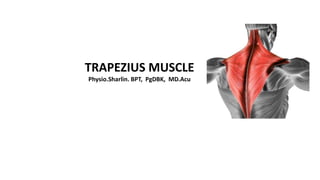
- 1. TRAPEZIUS MUSCLE Physio.Sharlin. BPT, PgDBK, MD.Acu
- 2. The trapezius muscle is a large, triangular, paired muscle located on the posterior aspect of the neck and thorax. When viewed together, this pair forms a diamond or trapezoid shape The trapezius has many attachment points, extending from the skull and vertebral column to the shoulder girdle. The trapezius belongs to the superficial layer of the extrinsic muscles of the back, along with latissimus dorsi, rhomboid major and minor, and levator scapulae muscles. The trapezius is largely involved in movements of the shoulder girdle, and is therefore functionally considered as a muscle of the upper limb rather than of the back.
- 3. Trapezius muscle has 3 parts 1. Upper trapezius / Descending part 2. Middle trapezius / Transverse part 3. Lower trapezius / Ascending part DESCENDING PART Origin: medial third of the superior nuchal line, external occipital protuberance, nuchal ligament Insertion: lateral third of clavicle Blood supply: transverse muscular branches arising from the occipital artery (branch of the external carotid), which passes along the deep surface of the muscle. TRANSVERSE PART Origin: spinous processes and supraspinous ligaments of vertebrae T1-T4 (or C7-T3) Insertion: medial acromial margin, superior crest of spine of scapula Blood supply: supplied by the superficial cervical artery, or by a branch from the transverse cervical artery.
- 4. ASCENDING PART Origin: spinous processes and supraspinous ligaments of vertebrae T4-T12 Insertion: medial end of scapular spine Blood supply:supplied by muscular branches of the dorsal scapular artery, which arises from the subclavian artery. Innervation The trapezius muscle is the only muscle of the upper limb that does not receive its innervation from the brachial plexus. Instead, motor innervation to the trapezius is conveyed by the accessory nerve (CN XI) as well as the anterior rami of the C3 and C4 spinal nerves. Function The main function of the trapezius is stabilizing the scapula in its anatomical place, as well as controlling it during movements of the shoulder and upper limb. The throwing action is a common manoeuvre in which the trapezius muscle is active, along with the deltoid muscle and the rotator cuff muscles. In addition, the trapezius is also involved in the movements of the head and neck.
- 5. Descending (upper) fibers The descending (upper) fibers act with the levator scapulae muscle to produce an elevation of the scapula at the scapulothoracic joint. In the same manner, they also maintain the level of the shoulders against gravity e.g. when a weight is being carried in the hand. When the muscle is acting unilaterally, the descending fibres produce an ipsilateral lateral flexion of the head and neck by acting on the atlanto-occipital joint. Unilateral contraction may also result in contralateral rotation of the head at the atlantoaxial joint. Bilateral contraction of the descending part of trapezius (i.e. when both left and right muscles contract) causes an extension of the head and neck. Ascending (lower) fibers The ascending (lower) fibers are responsible for depression of the medial part of the scapula, and thus lowering the shoulder. This action is especially important for activities in which the shoulders are lowered against resistance, for example when using the hands to help oneself up from a seated position. Together with the descending part, the ascending fibers also produce a rotation of the scapula around an axis which runs anteroposteriorly through the base of the scapular spine.
- 6. Transverse (middle) fibers The transverse (middle) fibers act together with the rhomboids to produce a retraction of the scapula, by pulling it towards the midline. The trapezius muscle is also responsible for upward rotation of the scapula, along with the serratus anterior muscle. This allows us to raise our arm above our heads beyond the level of the shoulder. Clinical notes The function of the trapezius muscle can be tested by placing a hand on the patient’s shoulder and assessing their ability to elevate or ‘shrug’ the shoulder against resistance. This test, coupled with the functionality test for sternocleidomastoid muscle, can be used to assess damage of the accessory nerve. Weakness of the trapezius muscle with intact functioning of the sternocleidomastoid muscle would indicate damage to the accessory nerve at a more distal point, such as in the posterior triangle of the neck. Weakness of both the trapezius and sternocleidomastoid muscles would allude to damage closer to where the accessory nerve exits the base of the skull. As the trapezius muscle has an extensive vascular supply, it can be used as a site for musculocutaneous tissue flap harvesting for reconstructive purposes in other areas of the body, such as for breast reconstruction.
- 7. Strengthening Exercise 1.Shoulder shrug 2.Barbell deadlift 3.Dumbell lift 4. Shoulder bracing