
Recommended
More Related Content
What's hot
What's hot (20)
Similar to Growth and Development of mandible.pptx
Similar to Growth and Development of mandible.pptx (20)
More from SadhuAbhijeet
More from SadhuAbhijeet (11)
Recently uploaded
Recently uploaded (20)
Growth and Development of mandible.pptx
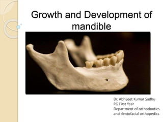
- 1. Growth and Development of mandible Dr. Abhijeet Kumar Sadhu PG First Year Department of orthodontics and dentofacial orthopedics
- 2. Contents 1. Prenatal Development 2. Postnatal Development The ramus Lingual Tuberosity Ramus To Corpus Remodelling Coronoid Process The Mandibular Condyle The Human Chin 3. Anomalies Of Development Mandibular Dysostosis Agnathia Microglossia Macrognathia
- 3. Prenatal Development 1. Neural Crest Cells 2. Mandibular nerve 3. Osteogenic membrane 36 to 38 days. 4. Meckel’s Cartilage 41st to 45th days
- 4. 5. Ossification Center - 6th week 6. Meckel’s cartilage disappears - 24th week 7. lamellar bone - 5th month
- 5. 8. Secondary accessory cartilages - 10th and 14th weeks 9. Condylar Secondary Cartilage - 10th week
- 7. The Ramus 1. Function 2. principal vectors of mandibular "growth“ and Displacement 3. Remodeling conversion
- 8. 4. Ramus Uprighting - Remodeling Rotation 5. Reason - middle cranial fossa enlarges 6. Lingual Tuberosity a’ b'
- 10. 3. 4. Prominence - Lingual fossa 5. Relocates slightly lateraly
- 11. Ramus to Corpus Remodelling Conversion 1. Ramus alignes with corpus 2. As a whole - Not straight line backward growth as mentioned earlier
- 12. Coronoid process 1. Propeller like twist 2. Grows superiorly
- 13. 3. 4.
- 14. 6. Effect on ramus 7. Lower part of Ramus bellow the coronoid
- 15. 8. Antegonial notch 9. Relationship between Ramus corpus Angle And Antgonial notch
- 16. The Mandibular Condyle 1. Pacesetting Master centre 2. Grows facing direct pressure
- 17. 3. Secondary Cartilage - multidirectional capacity for growth
- 18. 5. Condylar Neck
- 19. The Condylar Question 1. What is the physical force that produces the primary forward and downward displacement of the mandible
- 20. The Human Chin 1. During vertical nasomaxillary growth the descent of the maxillary arch
- 21. Anomalies Of Development 1. MANDIBULAR DYSOSTOSIS Characterized by defect of structures arising from first and second brachial arches Autosomal dominant Gene for this mapped to chromosome 5 Mandible is under developed with retruded chin. Cleft palate > 1/3 of cases. Respiratory and feeding difficulties in infants due to hypoplasia of the nasopharynx, oropharynx TREATMENT: Mild cases: no treatment. Severe cases: Cosmetic surgery Combined orthodontic therapy along with orthognathic surgery.
- 22. 2. Agnathia Characterized by hypoplasia or absence of mandible. More commonly, only a portion of jaw is missing. Partial absence of mandible is more common. Bilateral agenesis of condyles and ramus have also been reported. In case of unilateral absence of mandibular ramus, ears may be deformed or absent. This is believed to be due to failure of migration of neural crest mesenchyme into maxillary prominence at the fourth to fifth week post conception. Poor prognosis considered to be lethal.
- 23. 4. Micrognathia Means small jaw, either the maxilla or mandible may be involved. Some cases may produce illusion of micrognathia due to abnormal positioning or abnormal relation of one jaw to the other. Two types – Congenital - Caused due to abnormalities like pierre robin syndrome Acquired - Since normal growth of mandible depend normally on developing condyle , condylar ankylosis may result in deficient mandible
- 24. 4. Macrognathia MACROGNATHIA - It is often associated with other conditions like: • Paget’s disease of bone –excessive bone resorption and haphazard bone growth. • Acromegaly- is a hormonal disorder that develops when your pituitary gland produces too much growth hormone during adulthood. When you have too much growth hormone, your bones increase in size
- 25. References 1. Enlow, D.H. and Hans, M.G. (2008) Essentials of facial growth. Ann Arbor, MI: Distributed by Needham Press. 2. Sperber, G.H. (2001) Craniofacial Development. Hamilton, Ont.: B.C. Decker. 3. Shafer, W.G. et al. (2006) A textbook of oral pathology. New Delhi: Elsevier.
Editor's Notes
- The cartilages and bones of the mandibular skeleton form from embryonic neural crest cells that originate in the mid- and hindbrain regions of the neural folds. These cells migrate ventrally to form the mandibular (and maxillary) facial prominences, where they differentiate into bones and connective tissues. The first structure to develop in the region of the lower jaw is the mandibular division of the trigeminal nerve that precedes the ectomesenchymal condensation forming the first (mandibular) pharyngeal arch. The prior presence of the nerve has been postulated as requisite for inducing osteogenesis by the production of neurotrophic factors. The mandible is derived from ossification of an osteogenic membrane formed from ectomesenchymal condensation at 36 to 38 days post conception. The resulting intramembranous bone lies lateral to Meckel’s cartilage of the first (mandibular) pharyngeal arch 4. The cartilage skeleton of the first arch, known as Meckel’s cartilage. Arises at the 41st to 45th days post conception and provides a template for subsequent development of the mandible; however, most of its cartilage substance disappears in the formed mandible
- 5. A single ossification center for each half of the mandible arises in the 6th week post conception (the mandible and the clavicle are the first bones to begin to ossify) in the region of the bifurcation of the inferior alveolar nerve and artery into mental and incisive branches. . The ossifying membrane is lateral to Meckel’s cartilage and its accompanying neurovascular bundle. From the primary center ossification spreads upwards below and around the inferior alveolar nerve and its incisive branch, to form a trough for the developing teeth. The spread of the intramembranous ossification dorsally and ventrally forms the body and ramus of the mandible. Meckel’s cartilage becomes surrounded and invaded by bone. Ossification stops dorsally at the site that will become the mandibular lingula, where Meckel’s cartilage continues into the middle ear. The prior presence of the neurovascular bundle ensures the formation of the mandibular foramen and canal and the mental foramen. 6.Meckel’s cartilage lacks the enzyme phosphatase found in ossifying cartilages, thus precluding its ossification; Bone ALP is a major regulator of bone mineralization. It hydrolyzes inorganic pyrophosphate which is a naturally occurring inhibitor of mineralization, almost all of Meckel’s cartilage disappears by the 24th week after conception. 7. lamellar bone, and typical haversian systems are already present at the 5th month post conception. This remodeling occurs earlier than it occurs in other bones, and is thought to be a response to early intense sucking and swallowing, which stress the mandible.
- 8. Secondary accessory cartilages appear between the 10th and 14th weeks post conception to form the head of the condyle, part of the coronoid process, and the mental protuberance. The secondary cartilage of the coronoid process develops within the temporalis muscle, as its predecessor. The coronoid accessory cartilage becomes incorporated into the expanding intramembranous bone of the ramus and disappears before birth. 9. The condylar secondary cartilage appears during the 10th week post conception as a cone-shaped structure in the ramal region. This condylar cartilage is the primordium of the future condyle. 14th week, the first evidence of endochondral bone appears in the condyle region much of the cone-shaped cartilage is replaced with bone, but its upper end persists into adulthood, acting as both growth and articular cartilage. Changes in mandibular position and form are related to the direction and amount of condylar growth. The condylar growth rate increases at puberty, peaks between 121⁄2 and 14 years of age, and normally ceases at about 20 years of age. However, the continuing presence of the cartilage provides a potential for continued growth
- Although the mandible appears as a single bone in the adult, it is developmentally and functionally divisible into several skeletal subunits. The basal bone of the body forms one unit, to which are attached the alveolar, coronoid, angular, and condylar processes and the chin The growth pattern of each of these skeletal subunits is influenced by a functional matrix that acts upon the bone the teeth act as a functional matrix for the alveolar unit the action of the temporalis muscle influences the coronoid process the masseter and medial pterygoid muscles act upon the angle and ramus of the mandible the lateral pterygoid has some influence on the condylar process the mandible does not simply "grow" as pictured in Figure A. It "remodels“ and is simultaneously "displaced" as "forward and downward" movement proceeds from the temporomandibular interface.
- It is important because (1) it positions the lower arch in occlusion with the upper, and (2) it is continuously adaptive to the multitude of changing craniofacial conditions. the principal vectors of mandibular "growth" are posterior and superior. The ramus is thereby remodeled in a generally posterosuperior manner while the mandible as a whole becomes displaced anteriorly and inferiorly This allows posterior lengthening of the corpus and dental arch. The posterior development of the mandibular bony arch simultaneously proceeds into the region that was previously occupied by the ramus. This requires a remodeling conversion from what used to be ramus into what then becomes mandibular corpus. That is, the whole ramus becomes relocated posteriorly by resorptive and depository remodeling, and the former anterior part of the ramus is structurally altered into an addition to the corpus, which thereby becomes lengthened by this remodeling process. The overall extent of ramus movement amounts to several centimeters, not merely the width of the molar. 4..
- 4. The ramus normally becomes more vertically aligned during its development this is accomplished by greater amounts of bone additions on the inferior part of the posterior border than on the superior part. A correspondingly greater amount of matching resorption on the anterior border takes place inferiorly than superiorly. A "remodeling" rotation of ramus alignment thus occurs. Condylar growth becomes directed in a more vertical course along with the rest of the ramus. 5. The skeletal dimension of the pharynx is established by the size of the middle cranial fossa because the floor of this basicranial fossa is the roof of the pharyngeal compartment, and ramus must span the anteropsterior dimension of pharynx. pharynx (and middle cranial fossa) enlarges horizontally from a to a'. The ramus enlarges, correspondingly, from b to b' to match it. It also lengthens vertically, however. Angle c is thereby reduced to c' in order to accommodate the vertical nasomaxillary growth also taking place at the same time The "gonial angle" thus must undergo change (close) in order to prevent change in the occlusal relationship between the maxillary and mandibular arches. 6. The remodeling movement of the ramus in a backward direction has usually been pictured as essentially a two-dimensional process This is not merely an incomplete explanation; it is inaccurate as well. The problem is that some of the key anatomic parts that participate in the relocation and remodeling process of the ramus and corpus cannot be seen or represented in conventional two-dimensional headfilms and tracings. Among these is the lingual tuberosity
- 1.It ia a irrregular area of bony prominence at the distal termination of mylohyoid line. 2. This is an important structure because it is the direct anatomic equivalent of the maxillary tuberosity . Just as the maxillary tuberosity is a major site of growth for the upper bony arch, so is the lingual tuberosity a major site of growth for the mandible. Yet, this structure is not even included in the basic vocabulary of cephalometries. The reason, simply, is that it is not recognizable in the headfilm. It is major growth and remodeling site but it also the effective boundary between the two basic parts of the mandible: the ramus and the corpus.
- 3. The lingual tuberosity grows posteriorly by deposits on its posterior facing surface. lingual tuberosity protrudes noticeably in a lingual (medial) direction and it lies well toward the midline from the ramus. 4. The prominence of the tuberosity is augmented by the presence of a large resorptive field just below it. This resorptive field produces a sizable depression The Lingual Fossa. The combination of periosteal resorption in the fossa and deposition on the medial-facing surface of the tuberosity itself greatly accentuates the contours of both regions 5. The tuberosity remodels (relocates) in an almost directly posterior direction with a slight lateral shift because most of the bilateral growth of the basicranium has occurred by about the second and third years this calls for a key remodeling movement to accommodate the more narrow arch
- 1.As this takes place, that part of the ramus just behind the tuberosity remodels medially by deposits on the lingual side of the ramus thus, resultant lingual shift of the anterior part of the ramus to become added to the corpus 2.The growth thus is not straight line backward growth process with anterior resorption and posterior resorption. the growth direction follows the x arrows, rather than the straightline axis shown by the y arrows. As pointed out above, because the bicondylar dimension is established much earlier in childhood, bilateral growth separation between the right and left condyles is minimal beyond the early childhood years
- The coronoid process has a propeller-like twist, so that its lingual side faces three general directions all at once: posteriorly, superiorly, and medially. When bone is added onto the lingual side of the coronoid process, its growth thereby proceeds superiorly, and this part of the ramus thereby becomes increased in vertical dimension each coronoid process lengthens vertically, even though additions are made on the medial (lingual surface). This is an example of enlows enlarging v principle, where v is oriented vertically. growth movement and enlargement proceeds by additions of bone on the inside with removal from the outside. These same deposits of bone on the lingual side also bring about a posterior direction of growth movement, because this surface also faces posteriorly, A backward movement of the two coronoid processes is the result This is also an example of the expanding V principle , with the V oriented horizontally
- 3. These same deposits of bone on the lingual side also function to carry the base of the coronoid process and the anterior part of the ramus in a medial direction in order to add this part to the lengthening corpus. 4.Medial bone deposition also relocates the coronoid posteriorly . So, the area occupied by the anterior part of the early childhood ramus in 1 is re lo c a t e d and its former location becomes remodeled into the posterior part of the corpus in 2.
- 6. the buccal side of the coronoid process has a resorptive type of periosteal surface. And most of the superior part of the ramus, including the whole area just below the mandibular (sigmoid) notch, grows superiorly by depositon on the lingual side and resorption from the buccal side. 7. The lower part of the ramus bellow the coronoid process also has a twisted contour. Its buccal side faces posteriorly toward the direction of backward growth and thus, has a depository type of surface.
- 8. Antegonial gonial notch is the depression located on the inferior edge of the mandible at the ramus-corpus junction. This forms as there is single field of surface resorption on the inferior aspect of ramus as the ramus relocates posteriorly. 9. A mandible characteristically has a less prominent antegonial notch (b) if the angle between the ramus and corpus becomes closed. Much more prominent antegonial notch (a) if it becomes opened
- Historically, the condyle has been regarded as a kind of cornucopia from which the whole mandible itself pours forth. The condyle was believed to be the ultimate determinant that, essentially, establishes the mandibular rate of growth, the amount of growth, growth direction, overall mandibular size, and overall mandibular shape. It is no longer believed to represent a pacesetting "master center" with all other regional growth fields subordinate to and dependent on it for direct control. What, then, it must be asked, could be even more important than serving as a master center 2. Cartilage is a special non-vascular tissue and is involved because variable levels of compression occur at its articular contact with the temporal bone of the basicranium There are no capillaries in cartilage to press closed by a compressive surface force. In addition, importantly, the intercellular matrix of cartilage is, is turgid and unyielding to surface pressure . An endochondral growth mechanism is required for this part of the mandible because the condyle grows in a direction toward its articulation into the face of direct pressure. An intramembranous type of growth could not operate, because the intramembranous mode of osteogenesis is not pressure adapted and has a low threshold for compressive forces. the endochondral bone tissue (b) formed in association with the condylar cartilage (a) is laid down only in the medullary portion of the condyle. The enclosing bony cortices (c) are produced by periosteal-endosteal osteogenic activity; these vascular membranes are not subject to the compressive forces of articulation, but, rather, are essentially tension related because of muscle and connective tissue attachments. The real functional significance of the condylar cartilage thus involves an avascular and matrix-firm adaptation for regional pressure and movable articulation
- 3. The condylar cartilage is a secondary type of cartilage, which means that it does not develop by differentiation from the established primary cartilages of the fetal skull. It is not structurally comparable to a long bone's cartilaginous epiphyseal plate. have a special multidirectional capacity for growth and remodeling in selective response to varied mandibular displacement movements and rotations. Unlike characteristic linearly oriented direction of chondroblast proliferation, of long bone epiphyseal plates A unique capsular layer of poorly vascularized connective tissue covers the articular surface of the condyle Just deep to it is a special layer of prechondroblast cells. This is the predominant site for cellular proliferation. new developing cartilage. endochondral replacement by bone Unlike the arrangement in typical primary growth cartilage (i.e., epiphyseal plates of long bones), these various zones do not have linear columns of daughter cells. This is a notable histologic difference between primary and secondary types of growth cartilage It also means, importantly, that the cap of condylar cartilage has multidirectional proliferative capacity. Depending on where in the condylar cartilage that mitotic divisions occur, that part of the condyle (and ramus) thereby proliferates more vertically or more posteriorly, or virtually any point between, in response to maxillary counterpart to achieve proper occlusion
- 5. The lingual and buccal sides of the neck characteristically have resorptive surfaces. Thus, the condyle is quite broad and the neck is narrow. The neck is progressively relocated into areas previously held by the much wider condyle, and it is sequentially derived from the condyle as the condyle moves in a superoposterior course. This is done by periosteal resorption combined with endosteal deposition. This is another example of the V principle, with the V-shaped cone of the condylar neck growing toward its wide end. the endosteal surface of the neck actually faces the growth direction; the periosteal side points away from the course of growth
- 1. It was pointed out that functional mandibles totally lacking condyles exist in nature where condyle and part of the condylar neck are congenitally missing. the mandible functions (albeit with distress) in masticatory movements even though it lacks an articulation. (Mandibulofacial dysostosis (Treacher Collins syndrome, Oculoauriculovertebral syndrome (Goldenhar syndrome), hurler's syndrome, Proteus syndrome) First, the condyles may not play the kingpin role of a "master center" pacesetting the growth processes in the other parts of the mandible . Second, the whole mandible can become displaced anteriorly and inferiorly into its functional position without a "push" against the basicranium. These observations led to a consideration "functional matrix“"the origin, development and maintenance of all skeletal units are secondary, compensatory and mechanically obligatory responses to operationally prior demands of related functional matrices.“ Thus, as the mandible is displaced away from its basicranial articular contact, with the growth expansion of the soft tissue matrix associated with it.the condyle and whole ramus secondarily (but virtually simultaneously) remodels toward it.
- The vertical drift of the mandibular teeth, the anterior mandibular teeth simultaneously drift lingually and superiorly. The remodeling process that brings this about (F ig. 4-18) involves periosteal resorption on the labial bony cortex (a), deposition on the alveolar surface of the labial cortex (b), resorption on the alveolar surface of the lingual cortex (c), and deposition on the lingual side of the lingual cortex (d). At the same time, bone is progressively added onto the external surface of the mandibular basal bone area, including the mental protuberance (chin). The reversal between these two growth fields usually occurs at the point where the concave surface contour becomes convex. The result of this two-way growth process is a progressively enlarging mental protuberance – Chin.