
Recommended
More Related Content
What's hot
What's hot (20)
Similar to spinal cord and spinal nerves.ppt
Similar to spinal cord and spinal nerves.ppt (20)
Recently uploaded
Recently uploaded (20)
spinal cord and spinal nerves.ppt
- 2. Enteric Nervous System • GIT( Gut) Central Nervous System (CNS) • Brain • Spinal cord Peripheral Nervous System (PNS) • Cranial nerves (12 pairs) • Spinal nerves (31 pairs)
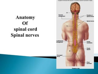
- 3. Spinal cord Lower elongated part of central nervous system Cylindrical in shape Extends from upper border of the 1st cervical vertebrae to the lower border of 1st lumbar vertebrae L1 in adult L3 (newborn)
- 4. Conus medullaris: tapered inferior end. Cauda equina: origins of spinal nerves extending inferiorly from lumbosacral enlargement and conus medullaris. Filum terminale median ligament of pia mater extending from conus medularis to first coccygeal vertebra.
- 5. Conus Medullaris Cauda Equina (horse’s tail) L5 L4 L3 L2 Filum terminale
- 6. Provide physical stability and shock absorption Three layers ◦ Dura mater ◦ Arachnoid mater ◦ Pia mater Spaces ◦ Epidural: anesthesia injected. Contains blood vessels, areolar connective tissue and fat. ◦ Subdural: serous fluid ◦ Subarachnoid: CSF and blood vessels within web-like strands of arachnoid tissue.
- 7. The adult spinal cord terminates at the level of the first lumbar vertebra (L1) Lumber puncture in adult is done in between L3-L4.
- 8. 8 White matter = myelinated processes (white in color) Gray matter = nerve cell bodies, dendrites, axon terminals, bundles of unmyelinated axons and neuroglia (gray color)
- 9. H-shaped pillar with anterior & posterior gray horns United by gray commissure containing the central canal Lateral gray column (horn) present in thoracic & upper lumbar segments Amount of gray matter related to the amount of muscle innervated Consists of nerve cells, neuroglia, blood vessels
- 10. Cervical Thoracic Lumbar Sacral Regional Differences Cervical enlargement Lumbar enlargement Lateral horn
- 11. • fiber tracts for transmission of information • Ascending (sensory) tracts • Descending (motor) tracts White Matter Posterior funiculus Lateral funiculus Anterior funiculus
- 12. 12- 12
- 13. 12- 13
- 15. 12- 15
- 16. The basic classifications of a spinal cord injury are: Tetraplegia (Quadriplegia) Injury to the spinal cord in the cervical region with associated loss of muscle strength in all four extremities. Paraplegia :- Injury to the spinal cord in the thoracic, lumbar, or sacral segments, including the cauda equina and conus medullaris, are associated with loss of muscle strength in the lower extremities.
- 17. Vessels of the spinal cord Anterior spinal vessels Posterior spinal vessels Radicular vessels
- 18. Anterior spinal artery (Medial Medullary syndrome) Supply anterior ⅔ of spinal cord Posterior spinal arterie Arise from vertebral artery or (PICA) Supply posterior ⅓ of spinal cord
- 20. 8 pairs of cervical spinal nerves; *C1-C8 12 pairs of thoracic spinal nerves; T1-T12 5 pairs of lumbar spinal nerves; L1-L5 5 pairs of sacral spinal nerves; S1-S5 1 pair of coccygeal spinal nerves; C0
- 21. Thirty-one pairs of spinal nerves First pair exit vertebral column between skull and atlas. Last four pair exit via the sacral foramina Others exit through inter-vertebral foramina 12- 21
- 22. Spinal nerves indicated by capital letter and number Dermatomal map: skin area supplied with sensory innervation by spinal nerves 12- 22
- 23. 12- 23
- 24. Complex network of ventral primary divisions of spinal nerves Four large plexuses Ventral rami of C1-C4= cervical plexus Ventral rami of C5-T1= brachial plexus Ventral rami of L1-L4= lumbar plexus Ventral rami of L4-S4= sacral plexus
- 25. S4-S5; coccygeal nerve Muscles of pelvic floor Sensory information from skin over coccyx
- 26. C1-C4 Innervates superficial neck structures, skin of neck, posterior portion of head Ansa cervicalis: loop between C2 and C3 Phrenic nerve ◦ From C3-C5 (cervical and brachial plexuses) ◦ Innervate diaphragm 12- 26
- 28. Nerve plexus of C5- C8 and T1 Five ventral rami form three trunks that separate into six divisions then form cords that give rise to: Branches/nerves ◦ Axillary ◦ Radial ◦ Musculocutaneous ◦ Ulnar ◦ Median ◦ Smaller nerves such as pectoral, long thoracic, thoracodorsal, subscapular, suprascapular 12- 28
- 29. Laterally rotate arm-teres minor Abducts arm- deltoid Skin: inferior lateral shoulder 12- 29
- 30. Movements at elbow and wrist, thumb movements Skin- posterior surface of arm and forearm, lateral 2/3 of dorsum of hand 12- 30
- 31. Movements at shoulder, elbow and wrist. Skin- lateral surface of forearm 12- 31
- 32. Movements at wrist, fingers, hand Skin- medial 1/3 of hand, little finger, and medial ½ of ring finger 12- 32
- 33. Movement of hand, wrist, fingers, thumb Skin- Lateral 2/3 palm, thumb, index and middle fingers; lateral ½ of ring finger and dorsal tips of same fingers 12- 33
- 35. Lumbar plexus: ventral rami of L1-L4 Sacral plexus: ventral rami of L4-S4 Usually considered together because of their close relationship Four major nerves exit and enter lower limb ◦ Obturator ◦ Femoral ◦ Tibial ◦ Common fibular (peroneal) 12- 35
- 36. Adduction of the thigh and knee Skin- superior middle side of thigh 12- 36
- 37. Movements of hip and knee: iliopsoas, sartorius, quadriceps femoris Skin- anterior and lateral thigh; medial leg and foot 12- 37
- 38. The two nerves together referred to as the sciatic (ischiadic) nerve Tibial ◦ Movement of hip, knee, foot, toes ◦ Skin: none ◦ Branches are medial and lateral plantar nerves, sural nerve 12- 38
- 39. Common fibular ◦ Anterior and lateral muscles of the leg and foot ◦ Skin distribution: lateral and anterior leg and dorsum of the foot. ◦ Branches are deep and superficial fibular (peroneal) nerves 12- 39
- 40. General disorders ◦ Anesthesia: loss of sensation Hyperesthesia: increased sensitivity to pain, pressure, light Paresthesia: tingling, prickling, burning Neuralgia: nerve inflammation causing stabbing pain Sciatica: pain radiating down back of thigh and leg Infections ◦ Herpes: skin lesions ◦ Shingles or herpes zoster: adult disease of chickenpox ◦ Poliomyelitis: infantile paralysis ◦ Anesthetic leprosy: bacterial infection of peripheral nerves Genetic and autoimmune disorders ◦ Myasthenia gravis: results in fatigue and muscular weakness due to inadequate ACh receptors
- 41. THANK YOU