More Related Content
Similar to alzheimer disease 2009 mucke.pdf
Similar to alzheimer disease 2009 mucke.pdf (20)
alzheimer disease 2009 mucke.pdf
- 1. Lots of people are forgetful. Are
there any particular warning signs
of Alzheimer’s disease?
Most people’s memory declines a little with
age, so the line between normal age-related
forgetfulness and the earliest signs of Alzhe-
imer’s disease (AD) can be fine — so fine that
a category of ‘mild cognitive impairment’,
or MCI, has been created, in part to avoid
diagnosing AD in people with more benign
memory impairments. However, many people
with MCI progress to AD. Typically, AD shows
itself as a gradual loss of episodic memory (for
instance, forgetting that a conversation took
place the day before). This is often more appar-
ent to others than to the patient. But AD can
also present as word-finding difficulties, get-
ting lost in familiar neighbourhoods, or more
complex behavioural changes, sometimes
brought on suddenly by a change in environ-
ment (such as hospitalization).
How is AD diagnosed?
Diagnosing AD with 100% certainty requires a
detailedpost-mortemmicroscopicexamination
of the brain. But nowadays, AD can be diag-
nosed with more than 95% accuracy in living
patients by using a combination of tools. These
include taking a careful history from patients
and their families, and assessing cognitive func-
tionbyneuropsychologicaltests.Othercausesof
dementia must be ruled out, such as low thyroid
function,vitamindeficiencies,infections,cancer
and depression. It’s also crucial to differentiate
AD from other neurodegenerative dementias,
includingfrontotemporaldementia,Lewy-body
dementia and Creutzfeldt–Jakob disease. Brain
imaging and tests of cerebrospinal fluid (CSF)
can help to distinguish AD from these condi-
tions. Patients with AD typically show shrink-
age of brain regions involved in learning and
memory on magnetic resonance images, as well
as decreased glucose metabolism and increased
uptake of radioligands that detect abnormal
protein deposits (amyloid) on positron emis-
sion tomography scans. CSF abnormalities
include low levels of amyloid-β (Aβ) peptides
and increased levels of the protein tau.
How big a problem is the disease?
Very big — in large part because people are
living longer, and ageing is a major risk fac-
tor. The Alzheimer’s Association estimates
that, without better ways to prevent the dis-
ease, the number of people with AD could
rise from around 5 million in the United
States today to between 11 million and 16 mil-
lion, and from about 26 million to more than
100 million worldwide, by 2050. This could
severely strain health-care systems because the
disease is so persistent, disabling and costly.
What are the causes of AD?
There are many. A lot of evidence suggests that
neurodegenerative diseases, including AD, stem
from the abnormal accumulation of harmful
proteins in the nervous system (Fig. 1). In AD,
these include Aβ peptides, the lipid-carrier pro-
tein apolipoprotein E (apoE), the microtubule-
associated protein tau, and the presynaptic
protein α-synuclein, which is also involved in
NEUROSCIENCE
Alzheimer’sdisease
Lennart Mucke
The neurodegenerative disorder Alzheimer’s disease is becoming more prevalent in ageing populations
worldwide. The identification of effective treatments will require a better understanding of the
physiological mechanisms involved, and innovative approaches to drug development and evaluation.
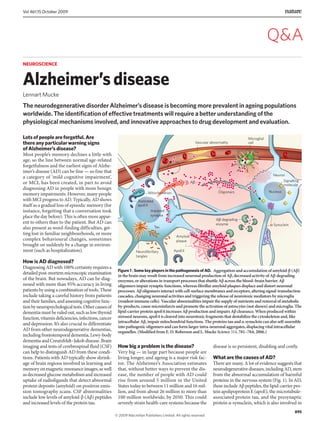
Figure 1 | Some key players in the pathogenesis of AD. Aggregation and accumulation of amyloid-β (Aβ)
in the brain may result from increased neuronal production of Aβ, decreased activity of Aβ-degrading
enzymes, or alterations in transport processes that shuttle Aβ across the blood–brain barrier. Aβ
oligomers impair synaptic functions, whereas fibrillar amyloid plaques displace and distort neuronal
processes. Aβ oligomers interact with cell-surface membranes and receptors, altering signal-transduction
cascades, changing neuronal activities and triggering the release of neurotoxic mediators by microglia
(resident immune cells). Vascular abnormalities impair the supply of nutrients and removal of metabolic
by-products, cause microinfarcts and promote the activation of astrocytes (not shown) and microglia. The
lipid-carrier protein apoE4 increases Aβ production and impairs Aβ clearance. When produced within
stressed neurons, apoE4 is cleaved into neurotoxic fragments that destabilize the cytoskeleton and, like
intracellular Aβ, impair mitochondrial functions. The proteins tau and α-synuclein can also self-assemble
into pathogenic oligomers and can form larger intra-neuronal aggregates, displacing vital intracellular
organelles. (Modified from E. D. Roberson and L. Mucke Science 314, 781–784; 2006.)
AC
Oligomers
Amyloid
plaque
Signalling
molecule
ApoE4
B-Synuclein
Truncated
apoE4
Mitochondrion
Neurofibrillary
tangles
Tau
Impaired
synapse
Nucleus
Neuron
AC-degrading
enzyme
Vascular abnormality
Microglial
cell
895
Vol 461|15 October 2009
Q&A
© 2009 Macmillan Publishers Limited. All rights reserved
- 2. Parkinson’sdisease.AllofusmakeAβpeptidein
the brain and other organs — it’s released from
theamyloidprecursorprotein(APP)aftercleav-
age by β-secretase and γ-secretase enzymes. But
Aβ is usually quickly removed from our brains
by clearance mechanisms. When its concentra-
tion is increased by overproduction or defective
clearance, Aβ self-aggregates into assemblies
ranging from oligomers to protofibrils, fibrils
and amyloid plaques. Tau and α-synuclein can
also self-aggregate into oligomers and into
larger inclusions in neurons, known as neuro-
fibrillary tangles and Lewy bodies, respectively.
By definition, all patients with AD have many
plaques and tangles; most patients also have
Lewy bodies.
How do these changes cause cognitive
decline?
This is a hotly debated issue. Most probably,
Aβ and tau cause faulty neural-network activ-
ity and impair synapses between neurons that
form and maintain microcircuits supporting
learning, memory and other cognitive func-
tions. Ultimately, vulnerable groups of neurons
atrophy and die in a process that may involve
excitotoxicity (overstimulation of neurotrans-
mitter receptors on neuronal surface mem-
branes), collapse of calcium homeostasis,
inflammation, and depletion of energy and
growth factors. A form of apolipoprotein E,
apoE4, contributes to the abnormal accumula-
tion of Aβ and tau, but probably also damages
mitochondria and the cellular cytoskeleton.
Aβ, tau, apoE and α-synuclein interact with
many other molecules and modulate diverse
signalling cascades that regulate neuronal
activity and survival. Genetically modified
rodents and other experimental models are
being used to tease out this complexity and
to determine which biochemical cascades
have the greatest impact on the initiation and
progression of the disease.
Can AD be inherited?
Yes. A small number of patients (probably
fewer than 1%) have early-onset AD because
they have inherited autosomal dominant muta-
tions in genes whose protein products — APP,
presenilin 1 (PS1) or PS2 — are involved in the
production of Aβ peptides. Presenilin is the
enzymatic centre of the γ-secretase complex.
The most powerful genetic risk factor for the
more common forms of AD is the APOE ε4
gene, which encodes the apoE4 lipid carrier.
The more common apoE3 and the rare apoE2
forms of apoE are relatively protective against
AD. More than 60% of Caucasian patients
with AD carry at least one APOE ε4 gene.
Certain variants of genes encoding another
lipid carrier, clusterin (apoJ), the intracellular
trafficking protein PICALM, or complement
component (3b/4b) receptor 1 also modu-
late AD risk, possibly by affecting Aβ levels,
synaptic functions or inflammation.
What about non-genetic causes?
The risk of AD may be increased by a low level
of education, severe head injury, cerebrovascu-
lar disease, diabetes and obesity. But it remains
uncertain whether avoiding these risk factors
can significantly lower one’s chances of getting
the disease, especially in people with genetic
risks for AD. It is likely that AD-predisposing
genes interact with other disease genes and
environmental factors. An otherwise healthy
person may get AD early in life simply because
they’ve inherited an aggressive PS1 mutation.
Another may get AD because they’ve inherited
two APOE ε4 genes, and yet another because
they’ve inherited one or more minor risk genes,
but are also overweight and diabetic.
What has ageing got to do with it?
Ageing is the most important risk factor for
AD. Even aggressive autosomal dominant AD
mutations typically don’t lead to obvious defi-
cits until the fourth or fifth decade of life. Sev-
eral mechanisms may protect the young brain
against AD, including higher levels of growth
factors, better energy metabolism and more
efficient mechanisms for clearing misfolded
proteins and repairing cells. Failure of these
protective mechanisms may contribute to the
development of AD. Ageing also increases the
prevalence of obesity, diabetes and athero-
sclerosis, which may promote AD through
metabolic or vascular mechanisms. Inflamma-
tion could be the common denominator here,
as the inflammatory activity of immune cells,
particularly macrophages and microglia, and
of astrocytes, increases with ageing. Some of
these activities are probably beneficial, whereas
others may promote or allow the development
of ageing-related disorders such as AD.
Arethereanyavailabletreatments?
Medicines currently prescribed for AD fall
into three groups: inhibitors of acetylcho-
linesterase; an antagonist of a receptor for the
neurotransmitter glutamate; and drugs from
the psychiatric toolbox to control depression
and behavioural abnormalities. The neuro-
transmitter acetylcholine is depleted in AD
brains, and inhibition of acetylcholineste-
rase, its degrading enzyme, aims to improve
cholinergic neurotransmission. Excitotoxic-
ity resulting from overstimulation of NMDA-
type glutamate receptors may contribute to
AD, providing a rationale for blocking these
receptors. Several clinical trials have shown
beneficial effects for inhibitors of acetylcho-
linesterase or NMDA receptors, although the
benefits were typically small, and these drugs
don’t seem to arrest or reverse AD.
How about diet and lifestyle changes?
It’s often suggested that adopting a healthy
diet and lifestyle to avoid high cholesterol and
high blood pressure may help because of the
potential contribution of vascular disease to
AD. Regular physical exercise also increases
growth factors in memory centres of the brain.
Social engagement and mental activity have
also been linked to lower AD risk in epidemi-
ological studies. In mouse models, increased
activity and environmental enrichment pre-
vent or delay the development of AD-like
signs. But the control groups in many of these
mouse studies were kept in rather impover-
ished conditions, which may have exaggerated
the benefits of the ‘enriched’ conditions.
Are there any other treatment options?
In my opinion, one of the most productive
things to do is for patients and their relatives to
enrol in carefully controlled prospective clinical
trials. There is an urgent need to increase the
proportion of patients with AD and of healthy
elderly people who participate in these trials.
By contrast, dietary fads and unproven over-
the-counter drugs and herbs should be discour-
aged. The claims to fame for these compounds
are notoriously transient. They’ve also added a
troublesome burden of confounding variables
(‘noise’) among trial subjects and complicatethe
task of designing informative clinical trials.
Why have so many drug trials failed?
For several reasons. In some cases, the trial
may reveal that the drug target does not have a
crucial pathogenic role. In other cases, the drug
may block a truly pathogenic pathway, but the
overall impact may be negligible because other
Cognitively
normal
Cognitively
normal
Alzheimer’s
disease
Figure 2 | The challenge of finding AD biomarkers.
Two normal controls (top and bottom rows) and
an age-matched AD patient (middle row) were
given an intravenous injection of the radioligand
PIB, which binds to fibrillar Aβ deposits. PIB
retention in the brain is detected by positron
emission tomography. Low levels of PIB binding
(cooler colours) are seen in most cognitively
normal people (top) and high levels of PIB
binding (warmer colours) in people with AD
(middle). But some cognitively normal people
also have high levels of PIB binding (bottom),
suggesting that the presence of amyloid plaques is
not sufficient to cause cognitive deficits. Whether
cognitively normal people with high levels of PIB
binding will develop AD later on is unknown.
Images courtesy of Gil Rabinovici (University of
California, San Francisco) and William Jagust
(Lawrence Berkeley National Laboratory).
896
NATURE|Vol 461|15 October 2009
NEWS & VIEWS Q&A
© 2009 Macmillan Publishers Limited. All rights reserved
- 3. branches of the multifactorial pathogenic cas-
cade are untouched. For example, in a recent
trial of an anti-Aβ agent, APOE ε4 gene carriers
had more side effects and may have benefited
less than non-carriers. Assessing whether the
drug affects the most relevant target can also
be challenging. Considerable evidence suggests
that small Aβ oligomers cause more damage
to synaptic and cognitive functions than larger
amyloid plaques. Plaque loads can be estimated
by radiological imaging, but brain levels of Aβ
oligomers can’t be reliably measured in living
patients, making it unclear whether anti-Aβ
treatments in clinical trials actually lower levels
of the harmful Aβ oligomers. Treatment failure
may also be the result of ‘too little, too late’. AD
probably develops insidiously over many years,
if not decades. Some of my colleagues believe
that even so-called early clinical stages of AD
reflect advanced-stage brain failure that may
be impossible to reverse.
Is there any chance of disease reversal?
This depends, in part, on the ‘plasticity’ of the
brain, which is much greater than that of other
organs, although AD-associated factors such
as Aβ and apoE4 may impair these adaptive
mechanisms, adding insult to injury. The flip
side of this coin is that removing these factors
might unleash powerful repair mechanisms
that could fix or help circumvent broken neu-
ral circuits so that functional recovery may be
possible. Many people have shown an impres-
sive recovery of neurological functions after
extensive loss of nerve cells from other causes.
The test will be to see if the AD-damaged brain
is capable of similar feats when all inhibitors of
effective regeneration have been eliminated.
Is stem-cell therapy an option?
The idea behind using stem cells is that these
cells might be used to replace destroyed neu-
rons. But AD poses particular challenges in this
regard, as it affects diverse types of neuron in
different brain regions. For now, it’s unclear if
stem cells can be induced to differentiate into
all these cell types and if the resulting neurons
would effectively integrate into broken circuits,
particularly in a hostile environment full of
harmful proteins and inflammatory mediators.
Again,regenerationandrepairmightbeassisted
by removing these hostile factors. Where stem
cells could yield more immediate rewards is as
models for studying the heterogeneity of AD. It
isnowpossibletoestablishpluripotentstem-cell
lines from skin cells of individual patients and
todifferentiatethemintoneuronsorotherbrain
cells. Comparing these cellular models might
lead to the identification of patient-specific
pathogenic pathways and modifier genes.
And prevention — is this feasible?
Preventative treatments would probably have
to be started years, if not decades, before the
first symptoms of AD appear. Treating people
for such long periods would require drugs with
minimal side effects and the ability to identify
people with significant risk factors early on. We
still do not have reliable early biomarkers for
AD, although progress has been made (Fig. 2).
AnAlzheimer’sDiseaseNeuroimagingInitiative
is under way to determine if measuring changes
in brain volume over time, glucose metabolism
and amyloid deposition in the brain, and levels
of Aβ and tau in the CSF, can identify people
at high risk of developing the disease. Pro-
teomics profiling of blood plasma has yielded
protein ‘fingerprints’ that might be diagnostic
and possibly even predictive of AD. Although
whole-genome sequencing as a routine screen-
ing method is still quite a way off, it is relatively
straightforward to screen for the known auto-
somal dominant AD mutations in the APP, PS1
and PS2 genes, and for the APOE ε4 gene.
So should everyone get genetic testing?
This depends on many factors, including one’s
family history, outlook on life and the desire
to secure certain types of insurance. If early-
onset AD runs in the family and one is con-
templating having children, genetic testing
for autosomal dominant AD mutations may
be appropriate. In general, genetic testing
for AD should be undertaken only with the
advice of a physician and a genetic counsellor
who are experienced in helping people weigh
up all the risks and benefits. Many clinicians
advise against genotyping for APOE ε4 and
other susceptibility genes because these genes
are primarily risk factors, and some carriers
never develop AD. The lack of established pre-
ventative treatments also diminishes the value
of knowing one’s risks, although greater public
awareness of AD risks might help to intensify
the fight against this devastating condition.
Is there reason for hope?
Indeed there is. As we gain a greater under-
standing of the mechanisms of AD, drugs can
be aimed at its root causes (not just at its symp-
toms). Several drugs with disease-modifying
potential are already in advanced clinical trials
(Table 1), and more are in the pipeline. Large-
scale risk-factor profiling using genomic and
proteomicscreensmaymake it possibletoiden-
tify subgroups of patients who stand to benefit
from particular drugs or drug combinations.
Zeroing in on the most responsive patient pop-
ulationscould makeclinical trialsmoreeffective
and guide long-term prevention strategies. ■
Lennart Mucke is at the Gladstone Institute of
Neurological Disease and the Department of
Neurology, University of California,
San Francisco, California 94158, USA.
e-mail: lmucke@gladstone.ucsf.edu
FURTHER READING
Alzheimer’s Association 2009 Alzheimer’s Disease
Facts and Figures (2009).
Chin, J., Roberson, E. D. & Mucke, L. in Learning and
Memory: A Comprehensive Reference Vol. 4 (ed. Byrne,
J. H.) Ch. 15 (Academic, 2008).
Mahley,R.W.,Weisgraber,K.H.&Huang,Y.
ApolipoproteinE4:Acausativefactorandtherapeutic
targetinneuropathology,includingAlzheimer’sdisease.
Proc.NatlAcad.Sci.USA103, 5644–5651(2006).
Roberson, E. D. & Mucke, L. 100 years and counting:
Prospects for defeating Alzheimer’s disease. Science
314, 781–784 (2006).
www.alzforum.org
www.clinicaltrials.gov
The author declares competing financial interests.
See online article for details.
See online at go.nature.com/phyLwm for more on
neuroscience.
TABLE 1 | ONGOING CLINICAL TRIALS FOR TREATING ALZHEIMER’S DISEASE
Approach or drug Proposed mechanism of action Phase
β-Secretase inhibition Decreases formation of Aβ from amyloid precursor protein II
γ-Secretase inhibition Decreases formation of Aβ from amyloid precursor protein II/III
Active immunization with Aβ
peptides
Generates anti-Aβ antibodies that interact with Aβ and remove it
from the brain by uncertain downstream mechanisms
II
Passive immunization with
anti-Aβ antibodies
The antibodies interact with Aβ and remove it from the brain by
uncertain downstream mechanisms
III
Intravenous pooled
immunoglobulins
May enhance clearance of Aβ and other harmful proteins from the
brain; may decrease harmful inflammatory processes
III
Scyllo-inositol Decreases formation and stability of pathogenic Aβ assemblies II
Latrepirdine Prevents mitochondrial dysfunction III
Inhibition of receptor for
advanced glycation
end products (RAGE)
Blocks stimulation of the cell-surface receptor RAGE, which binds
Aβ, decreasing Aβ levels in the brain and preventing Aβ from
activating pathogenic pathways
II
Stimulation of insulin signalling Prevents hyperglycaemia; may overcome insulin resistance
in the brain
II
Selective oestrogen-receptor
modulator
Promotes neuroprotective effects of oestrogen without eliciting
its harmful effects
II
Neurotrophic and
neuroprotective agents
Stimulate neurotrophic and antioxidant pathways or pathways
that protect against excitotoxicity
II
The above selection focuses on potentially disease-modifying strategies and is based on a review of websites, oral reports at scientific
meetings, and discussions with Paul Aisen (University of California, San Diego) and Laurie Ryan (National Institute on Aging).
Phase II and phase III trials assess the safety and efficacy of new treatments; phase III trials involve many more subjects, are conducted in
multiple centres, and are required for drug approval by regulatory agencies.
897
NATURE|Vol 461|15 October 2009 NEWS & VIEWS Q&A
© 2009 Macmillan Publishers Limited. All rights reserved