How to Integrate Perioperative Immunotherapy Into Multimodal Treatment Plans to Improve Outcomes in Resectable NSCLC
•
0 likes•46 views

Co-Chairs, Nasser Altorki, MD, and Jonathan D. Spicer, MD, PhD, FRCSC, prepared useful Practice Aids pertaining to NSCLC for this CME/MOC activity titled “How to Integrate Perioperative Immunotherapy Into Multimodal Treatment Plans to Improve Outcomes in Resectable NSCLC.” For the full presentation, downloadable Practice Aids, and complete CME/MOC information, and to apply for credit, please visit us at https://bit.ly/3xb6WS1. CME/MOC credit will be available until June 14, 2023.

Recommended
Recommended
More Related Content
Similar to How to Integrate Perioperative Immunotherapy Into Multimodal Treatment Plans to Improve Outcomes in Resectable NSCLC
Similar to How to Integrate Perioperative Immunotherapy Into Multimodal Treatment Plans to Improve Outcomes in Resectable NSCLC (20)
More from PVI, PeerView Institute for Medical Education
More from PVI, PeerView Institute for Medical Education (20)
Recently uploaded
Recently uploaded (20)
How to Integrate Perioperative Immunotherapy Into Multimodal Treatment Plans to Improve Outcomes in Resectable NSCLC
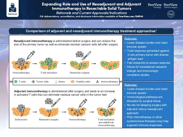
- 1. Expanding Role and Use of Neoadjuvant and Adjuvant Immunotherapy in Resectable Solid Tumors Rationale and Current Approvals/Indications Full abbreviations, accreditation, and disclosure information available at PeerView.com/GWF40 Neoadjuvant immunotherapy is administered before surgery and can reduce the size of the primary tumor as well as eliminate residual cancers cells left after surgery Rationale: • Lower disease burden and intact immune system • T-cell response generated against in situ primary tumor with diverse antigen load • Fast endpoints to assess response • Allows for translational research: biologic and immunologic correlative studies Rationale: • Lower disease burden and intact immune system • Immunological pathways are disrupted by surgical stress • No risk for delaying surgery with adjuvant versus neoadjuvant approach • Prior chemotherapy or other systemic/local therapies may help augment immune responses Adjuvant immunotherapy is administered after surgery and leads to an increase in activated T cells that can eliminate residual cancer cells in the tumor bed Comparison of adjuvant and neoadjuvant immunotherapy treatment approaches1 Immunotherapy T cells T-cell activation Resection surgery Solid tumor Resection surgery Immunotherapy T-cell activation and additional immunotherapy Tumor cells Artery Healthy cells Immunotherapy
- 2. Expanding Role and Use of Neoadjuvant and Adjuvant Immunotherapy in Resectable Solid Tumors Rationale and Current Approvals/Indications Full abbreviations, accreditation, and disclosure information available at PeerView.com/GWF40 Overall survival (OS) is the gold-standard outcome measure for phase 3 trials, but the protracted length of these clinical trials in resectable cancers makes this research daunting and expensive One strategy to expedite clinical trials, including those assessing immunotherapies in early-stage cancer settings, is the use of newer, innovative surrogate measurements for endpoints Because pathologic response reflects a therapy’s ability to eradicate tumor cells more directly than radiographic evaluation using RECIST criteria, it may better correlate with clinically meaningful outcomes Several trials in different tumor types have correlated the novel endpoints with more traditional endpoints such as OS, DFS, and RFS, but additional confirmatory studies are needed Many adjuvant approvals have been based on disease-free survival (DFS) Trials in neoadjuvant settings provide an opportunity to assess pathologic response as an early surrogate marker for survival outcomes, and pathologic response criteria such as major pathological response (MPR) and pathologic complete response (pCR) have been assessed in neoadjuvant immunotherapy trials; there are various definitions, but generally: MPR: ≤10% of viable tumor in the treated tumor bed pCR: complete absence of viable tumor in the treated tumor bed Recently, immune-related pathologic response criteria (irPRC) have also been developed with the aim of assessing the full spectrum of response to immunotherapy in resection specimens Different scoring systems exist or are in development for evaluating pathologic response in various tumor types (eg, melanoma, lung cancer, bladder cancer) Relevant endpoints for neoadjuvant and adjuvant immunotherapy clinical trials2-8
- 3. Expanding Role and Use of Neoadjuvant and Adjuvant Immunotherapy in Resectable Solid Tumors Rationale and Current Approvals/Indications Full abbreviations, accreditation, and disclosure information available at PeerView.com/GWF40 Adjuvant treatment of patients with urothelial carcinoma who are at high risk of recurrence after undergoing radical resection Adjuvant treatment of patients with completely resected esophageal or gastroesophageal junction cancer with residual pathologic disease who have received neoadjuvant chemoradiotherapy Adjuvant treatment of patients with melanoma with lymph node involvement or metastatic disease who have undergone complete resection Current perioperative approvals and indications of immunotherapies in solid tumors9 Nivolumab Adjuvant treatment of patients with cutaneous melanoma with pathologic involvement of regional lymph nodes of more than 1 mm who have undergone complete resection, including total lymphadenectomy Ipilimumab Newest Represents the first FDA approval for neoadjuvant therapy for early-stage NSCLC Neoadjuvant treatment of patients with resectable non–small cell lung cancer with platinum-doublet chemotherapy
- 4. Expanding Role and Use of Neoadjuvant and Adjuvant Immunotherapy in Resectable Solid Tumors Rationale and Current Approvals/Indications Full abbreviations, accreditation, and disclosure information available at PeerView.com/GWF40 Neoadjuvant treatment of high-risk, early-stage triple-negative breast cancer with chemotherapy adjuvant treatment after surgery as a single agent Adjuvant treatment of patients with renal cell carcinoma at intermediate-to-high or high risk of recurrence following nephrectomy, or following nephrectomy and resection of metastatic lesions Adjuvant treatment of patients with stage IIB, IIC, or III melanoma following complete resection Patients with BCG-unresponsive, high-risk, non–muscle invasive bladder cancer with carcinoma in situ with or without papillary tumors who are ineligible for or have elected not to undergo cystectomy Current perioperative approvals and indications of immunotherapies in solid tumors9 Pembrolizumab Adjuvant treatment following resection and platinum-based chemotherapy in patients with stage II to IIIA non–small cell lung cancer whose tumors have PD-L1 expression on ≥1% of tumor cells Atezolizumab 1. Krishnamoorthy M et al. J Natl Cancer Inst. 2021;113:823-832. 2. Topalian SL et al. Science 2020;367:eaax0182. 3. Benitez JC et al. Clin Cancer Res. 2020;26:5068-5077. 4. Bilusic M, Gulley JL. J Natl Cancer Inst. 2021;113:799-800. 5. Krishnamoorthy M et al. J Natl Cancer Inst. 2021;113:823- 832. 6. O’Donnell JS et al. Clin Cancer Res. 2019;25:5743-5751. 7. Hellmann MD et al. Lancet Oncol. 2014;15:e42-50. 8. Cottrell TR et al. Ann Oncol. 2018;29:1853-1860. 9. https://www.fda.gov/drugs/resources-information-approved-drugs/oncology-cancer-hematologic-malignancies- approval-notifications.
- 5. IASLC Multidisciplinary Recommendations for Pathologic Assessment of Lung Cancer Specimens Following Neoadjuvant Therapy1 Full abbreviations, accreditation, and disclosure information available at PeerView.com/GWF40 Recommendation 1 The term “tumor bed” is the area where the original pretreatment tumor was considered to be located. It can be challenging to determine whether necrosis and stromal inflammation and/or fibrosis are due to regression secondary to neoadjuvant therapy, native tumor characteristics, or a combination. For this reason we favor the term tumor bed. It is suggested to simply describe the major components of the tumor bed as (1) viable tumor, (2) necrosis, or (3) stroma (which can include inflammation or fibrosis). See page 3 for an example. Recommendation 2 It is essential that information be provided from the surgical team to the pathology laboratory on whether the patient received neoadjuvant therapy in order for this specimen processing protocol to be followed. If there is more than one tumor in the specimen, it is of critical importance to also provide this information. It is good clinical practice to correctly label the specimen with the lobe(s) resected and to clarify any issues that may be needed for pathologic staging such as the pericardium, diaphragm, or chest wall. Recommendation 3 Lung cancer resection specimens following neoadjuvant therapy should be sampled to optimize comprehensive gross and histologic assessment of the lung tumor bed for pathologic response. The tumor should be cut in its greatest dimension to maximize the tumor bed cross section. In cases where identification and/or orientation of the tumor are difficult, review of the preoperative CT scan can be helpful. Tumors 3 cm or less in size should be completely sampled. For larger tumors greater than 3 cm, the tumor should be cut across in serial sections 0.5 cm thick, and after gross inspection, the most representative cross section showing viable tumor should be sampled. At least one cross section of the entire tumor (0.5 cm thick) with a gross photograph and histologic mapping should be made. Histologic sections at the tumor periphery should include 1 cm of adjacent lung parenchyma. Pathologic response cannot be assessed in small biopsies; a resection specimen is required. Recommendation 4 To determine the border of the tumor bed, the edge of the tumor needs to be distinguished from the surrounding non-neoplastic lung parenchyma. This can be facilitated by review of the gross specimen and the histologic slides from the periphery of the tumor bed. Recommendation 5 Determination of the pathologic response to therapy should be made after review of all H&E slides of tumor by estimating the percentages of (1) viable tumor, (2) necrosis, and (3) stroma, which includes both fibrosis and inflammation, so each of these three components add up to 100%. Each component should be assessed in 10% increments unless the amount is below 5% when an estimate of single percentages should be recorded. While this is primarily done by review of histologic sections of the tumor bed, correlation with the gross findings, in some cases facilitated by a gross photograph, may be important in markedly necrotic and/or cavitated tumors where it is not possible to reflect this change in histologic sections. Note: Although it may be useful to record the amount of each of these components on each individual histologic slide, it needs to be kept in mind that the amount of tumor bed varies on each slide, so these percentages cannot be summed and averaged as if they were in equal amounts. This is a semiquantitative process. There is no validated quantitative method that is available that can be implemented in a timely fashion for clinical decisions. A proposed synoptic template for reporting pathologic findings for resected lung cancers following neoadjuvant therapy is summarized on page 2.
- 6. IASLC Multidisciplinary Recommendations for Pathologic Assessment of Lung Cancer Specimens Following Neoadjuvant Therapy1 Full abbreviations, accreditation, and disclosure information available at PeerView.com/GWF40 Recommended Synoptic Template for Recording Lung Cancers Following Neoadjuvant Therapy Primary tumor Type of neoadjuvant therapy • No known presurgical therapy: _____ • Type of neoadjuvant therapy – Chemotherapy: ____________________ – Radiotherapy: ____________________ – Immunotherapy (please specify): ____________________ – TKI (please specify): ____________________ – Other (please specify): ____________________ Treatment effect in primary tumor • Percentage of viable tumor (record in 10% increments except below 10%; then record single digits between 1%-5%): _____ • No residual viable tumor identified: _____ • Percentage of necrosis: _____ • Percentage of stroma (includes fibrosis and inflammation): _____ Grade of inflammation (choose the appropriate grade) _____ Mild _____ Moderate _____ Marked Method (choose what was used for evaluation) _____ Correlation was made with a gross photograph of tumor cut surface: Yes _____ No _____ _____ Evaluation was aided by use of tumor mapping to match a gross photograph to histologic sections: Yes _____ No _____ _____ Evaluation was aided by radiologic pathologic correlation: Yes _____ No _____ Treatment effect in lymph node metastases • Total number of lymph node stations examined _____ • Total number of lymph nodes examined: _____ • No carcinoma present: _____ • Total number of lymph nodes with metastatic carcinoma: _____ • Lymph node stations involved by tumor with treatment-related changes: _____ • Lymph node stations with treatment-related changes without viable tumor: _____ • Largest tumor focus: mm at station number: _____ • Extracapsular extension present: _____ • No extracapsular extension: ____________________ Comments: ___________________________________________________________________________________________ _____________________________________________________________________________________________________
- 7. IASLC Multidisciplinary Recommendations for Pathologic Assessment of Lung Cancer Specimens Following Neoadjuvant Therapy1 Full abbreviations, accreditation, and disclosure information available at PeerView.com/GWF40 Histologic Components of the Tumor Bed (A) Schematic image showing how percentage compositions are assigned. The tumor bed is divided into viable tumor area, necrosis, and stroma. Stroma includes inflammation and fibrosis. (B) A representative hematoxylin-and-eosin stained slide image (left) and a corresponding color illustration of the distribution of the components (right). The blue, red, and black areas represent viable tumor, necrosis, and stroma, respectively. Tumor bed = X + Y + Z = 100% Viable tumor (X%) Necrosis (Y%) Stroma (Z%) Viable tumor = 50% Necrosis = 40% Stroma = 10% A B C Tumor bed
- 8. IASLC Multidisciplinary Recommendations for Pathologic Assessment of Lung Cancer Specimens Following Neoadjuvant Therapy1 Full abbreviations, accreditation, and disclosure information available at PeerView.com/GWF40 Recommendation 6 Definition of major pathologic response (MPR) MPR is defined as the reduction of viable tumor to the amount beneath an established clinically significant cutoff based on prior evidence according to the individual histologic type of lung cancer and a specific therapy. The historical definition of MPR for all histologic types of lung cancer is ≤ 10% of viable tumor with no viable tumor required for pathologic complete response (pCR). MPR is calculated as the estimated size of viable tumor divided by the size of the tumor bed. For the moment, this is the cutoff being used in multiple active clinical trials. However, recent data suggests the MPR in the conventional chemotherapy setting may differ according to histologic type: ie, adenocarcinoma versus squamous cell carcinoma. If after review of histologic sections, the percentage of viable tumor is near the cutoff for major pathologic response, additional histologic sections should be submitted. The pathology report should record the total number of blocks of tumor bed that were examined even if the blocks did not consist entirely of tumor bed but also included some uninvolved lung. For colloid adenocarcinomas, where tumor cells are only focal, the mucin pools should be included in the percentage of viable tumor. However, if there are only areas of extracellular mucin without any apparent viable tumor cells within the mucin, we suggest regarding this as stroma. Further study is needed to address this point. Major pathologic response can also be classified for the lung primary in the setting where the lung primary shows little or no viable tumor, but lymph nodes show viable metastatic carcinoma (ypT0, N1, 2 or 3). However, the prognostic and therapeutic implications of this clinical setting are not known. Recommendation 7 Definition of pathologic complete response (pCR) pCR is defined as lack of any viable tumor cells on review of H&E slides after complete evaluation of a resected lung cancer specimen including all sampled regional lymph nodes. Such tumors would be staged as ypT0N0 according to the 8th edition AJCC and UICC staging systems. Note: If no tumor is seen in the initial sections and tissue from the tumor bed remains, additional histologic sections should be made. The number of additional sections should be whatever seems reasonable in the individual setting depending on the size of the tumor bed and the capacity of the individual pathology laboratory. If the histologic changes in the initial sections obtained do not show findings that fit for the effects of therapy, the possibility that the wrong area was sampled should be considered. In such cases the gross specimen may need to be re-evaluated using radiologic pathologic correlation and if additional lesions are identified, these should be sampled. The pathology report should record the total number of blocks of tumor bed that were examined even if the entire block did not consist of tumor bed. The identification of incidental lesions of squamous cell carcinoma in situ, atypical adenomatous hyperplasia, adenocarcinoma in situ, or minimally invasive adenocarcinoma in the surrounding lung parenchyma that are clearly separate from the main tumor for which neoadjuvant therapy was administered does not disqualify a case for classification as MPR or CPR. This proposal is based on clinical judgement, as currently no clinical data exist to make a specific recommendation. In the setting of multiple tumors where a second invasive predominant lung carcinoma, is present that was regarded preoperatively to be an intrapulmonary metastasis but, it is determined to be a second synchronous primary after clinical, radiologic, pathologic, and/or molecular assessment, it is questionable whether the terms MPR or CPR should be used if the main tumor otherwise meets the above criteria. No data exists currently to address this question.
- 9. IASLC Multidisciplinary Recommendations for Pathologic Assessment of Lung Cancer Specimens Following Neoadjuvant Therapy1 Full abbreviations, accreditation, and disclosure information available at PeerView.com/GWF40 1. Travis WD et al. J Thorac Oncol. 2020;15:709-740. Recommendation 8 In the absence of more systemic data regarding evaluation of tumors following immunotherapy and molecular targeted therapy, the same approach to pathologic assessment of resected lung cancers in the neoadjuvant setting in evaluating the percentage of viable tumor, necrosis, and stroma should be used regardless of the type of neoadjuvant therapy administered whether it was radiation, chemotherapy, targeted therapy, immunotherapy, chemoradiation, chemoimmunotherapy, or chemotherapy-targeted therapy. There also may be different features that can be addressed depending on the type of therapy such as immune cell infiltrates in patients who received immunotherapy. Recommendation 9 In most cases, the lymph nodes are small enough to completely sample, but if there is a very large metastasis or tumor bed (>2 cm), the lymph node can be bisected and the central slice through the tumor can be submitted in designated cassettes. This should also be done during grossing of intraoperative frozen sections of lymph nodes. Depending on individual laboratory resources, more extensive or even complete sampling can also be done. Then the same approach can be used for histologic evaluation that is used for the resected lung cancer reporting percent viable tumor, necrosis, and stroma. Complete pathologic response in a lymph node can be recognized if there is a well-defined scar and/or area of tumor necrosis in the absence of identifiable viable tumor cells. Recommendation 10 The following recommendations are made for T factor staging of neoadjuvant lung cancer resection specimens. Tumor size: If the viable tumor forms a discrete mass where the size can be measured with a ruler either grossly or microscopically (where it can be measured on a single H&E slide), this is the preferred approach (Figure 2A-D). However, if the viable tumor cannot be measured with a ruler either due to grossly indistinct borders, multiple foci interspersed among necrosis and/or stroma, or if it is present on multiple slides, the viable invasive tumor size should be estimated using the following formula. Viable invasive tumor size (cm) = tumor bed size X percentage viable invasive tumor Estimating invasive size by adjusting for lepidic component In tumors that have a component of lepidic growth, tumor size estimation should use the principles introduced in the 8th Edition TNM classification that record both total size and invasive size, but only use invasive size for T-factor determination. Thus, in the neoadjuvant setting, viable tumor size estimation for such cases may need two adjustments: one for invasive size excluding the lepidic component and a second for the percent viable tumor as outlined above. However, the clinical implications of this adjustment for the lepidic component are not known in the neoadjuvant setting. T3: multiple tumors considered to represent intrapulmonary metastases If there is more than one tumor within a lobe, the pathological response or percent viable tumor should be reported for each tumor unless the number of intrapulmonary metastases are too numerous to count. Recommendation 11 Ongoing neoadjuvant studies with targeted therapies and immunotherapies in resectable NSCLC represent a unique source of information, and the International Association for the Study of Lung Cancer strongly recommends and will promote the design and implementation of an international database to collect uniformly clinical and pathologic information with the ultimate goal of fostering collaboration and to facilitate the identification of surrogate end points of long-term survival.
- 10. Immune-Related Adverse Events of Cancer Immunotherapies Become Aware and Stay Vigilant1-4 Full abbreviations, accreditation, and disclosure information available at PeerView.com/GWF40 What Are irAEs? • Immune checkpoint inhibitors are associated with important clinical benefits, but general immunologic enhancement can also lead to a unique spectrum of immune-related adverse events • Any organ system can be affected, but more commonly occurring are pulmonary (pneumonitis), dermatologic (rash, pruritus, blisters, ulcers, vitiligo), gastrointestinal (diarrhea, enterocolitis, transaminitis, hepatitis, pancreatitis), and endocrine (thyroiditis, hypophysitis, adrenal insufficiency) irAEs Endocrine Hyper- or hypothyroidism Hypophysitis Adrenal insufficiency Diabetes Hepatic Hepatitis Renal Nephritis Dermatologic Rash Pruritus Psoriasis Vitiligo DRESS Stevens-Johnson Hematologic Hemolytic anemia Thrombocytopenia Neutropenia Hemophilia Ocular Uveitis Conjunctivitis Scleritis, episcleritis Blepharitis Retinitis Respiratory Pneumonitis Pleuritis Sarcoid-like granulomatosis Cardiovascular Myocarditis Pericarditis Vasculitis Gastrointestinal Colitis Ileitis Pancreatitis Gastritis Neurologic Neuropathy Guillain Barŕe Myelopathy Encephalitis Myasthenia Musculoskeletal Arthritis Dermatomyositis Prevention Anticipation Treatment Monitoring Detection
- 11. Immune-Related Adverse Events of Cancer Immunotherapies Become Aware and Stay Vigilant1-4 Full abbreviations, accreditation, and disclosure information available at PeerView.com/GWF40 Guidance for Surgeons: Suspect, Detect, and Refer for Treatment5,6 • irAEs frequently occur in the perioperative setting, either before or after surgical intervention • irAEs occurring during neoadjuvant immunotherapy are generally manageable and in most cases should not exclude patients from surgery • The onus is on the surgeon to have a high degree of suspicion for potential toxicities in patients treated with immunotherapy • Vague symptoms should not be dismissed, because nonspecific ailments can be indicative of severe toxicity – Rheumatologic toxicities and endocrinopathies are some of the most difficult to recognize, given their relatively nonspecific presentation » For example, fatigue, poor energy, and low mood could represent hypophysitis or adrenal insufficiency – Other toxicities can be essentially asymptomatic » For example, renal and hepatic toxicity are generally only detected on routine labs – Pneumonitis is another relevant irAE requiring awareness by surgeons, as severe pneumonitis could potentially exclude patients from operative therapy, but significant pneumonitis has been rare in trials to date • A comprehensive workup for irAEs, with a thorough history specifically targeted to potential irAEs, should be conducted • Coordinate and collaborate with oncologists and other multidisciplinary experts to optimally diagnose and manage irAEs in patients who have received/are receiving perioperative immunotherapy • The National Comprehensive Cancer Network (NCCN) and American Society of Clinical Oncology (ASCO) have issued guidelines for recognition and management of immune-related adverse events
- 12. Immune-Related Adverse Events of Cancer Immunotherapies Become Aware and Stay Vigilant1-4 Full abbreviations, accreditation, and disclosure information available at PeerView.com/GWF40 How Should irAEs Be Diagnosed and Managed? Minimal or No Symptoms; Diagnostic Changes Only • In general, immunotherapy should be continued with close monitoring, with the exception of some neurologic, hematologic, and cardiac toxicities Mild to Moderate Symptoms • Hold checkpoint inhibitor therapy for most grade 2 toxicities • Consider resuming immunotherapy when symptoms and/or lab values revert to grade 1 • Corticosteroids (initial dose of 0.5-1.0 mg/kg/day of prednisone or equivalent) may be administered Severe or Life-Threatening Symptoms Grade 3 toxicities • Hold checkpoint inhibitor therapy • Initiate high-dose corticosteroids (prednisone 1-2 mg/kg/day or methylprednisolone IV 1-2 mg/kg/day) • If symptoms do not improve with 48-72 hours of high-dose corticosteroid, infliximab may be offered for some toxicities • Taper corticosteroids over the course of at least 4-6 weeks • When symptoms and/or laboratory values revert to grade 1, rechallenging with immunotherapy may be considered; however, caution is advised, especially in those patients with early-onset irAEs; dose adjustments are not recommended Grade 4 toxicities • In general, permanent discontinuation of checkpoint inhibitor therapy is warranted, with the exception of endocrinopathies that have been controlled by hormone replacement irAEs are often diagnosed by exclusion; other causes should be ruled out (including AEs of other therapies used), but immunotherapy-related toxicity should always be included in the differential There should be a high level of suspicion that new symptoms are treatment related; early recognition, evaluation, and treatment of irAEs plus patient education are essential for the best outcome Depending on severity of irAE, management may require corticosteroid or other immunosuppressive treatment and interruption or discontinuation of therapy If appropriate immunosuppressive treatment is used, patients generally recover from irAEs Use of immunosuppressive therapy to manage irAEs does not appear to impact response to immunotherapy Grade 1 Grade 2 Grade 3/4
- 13. Immune-Related Adverse Events of Cancer Immunotherapies Become Aware and Stay Vigilant1-4 Full abbreviations, accreditation, and disclosure information available at PeerView.com/GWF40 Hold immunotherapy with radiographic evidence of pneumonitis progression May offer one repeat CT in 3-4 weeks; in patients who have had baseline testing, may offer a repeat spirometry/DLCO in 3-4 weeks May resume immunotherapy with radiographic evidence of improvement or resolution; if no improvement, should treat as grade 2 Monitor patients weekly with history, physical examination, and pulse oximetry; may also offer CXR Grade 2: Symptomatic; >1 lobe of lung or 25%-50% of lung parenchyma; medical intervention indicated; limiting instrumental ADL Grade 3: Severe symptoms requiring hospitalization; involves all lung lobes or >50% of lung parenchyma; limiting self care Grade 4: Life-threatening respiratory compromise; urgent intervention indicated (intubation) Hold immunotherapy until resolution to grade ≤1 Prednisone 1-2 mg/kg/day and taper by 5-10 mg/week over 4-6 weeks Consider bronchoscopy with BAL Consider empiric antibiotics Monitor patients every 3 days with history, physical examination, and pulse oximetry; consider CXR; if no clinical improvement after 48-72 hours of prednisone, treat as grade 3 Discontinue immunotherapy Empiric antibiotics; methylprednisolone IV 1-2 mg/kg/day; if no improvement after 48 hours, may add infliximab 5 mg/kg, or mycophenolate mofetil IV 1 g 2x/day, or IVIG x 5 days, or cyclophosphamide Taper corticosteroids over 4-6 weeks Pulmonary and infectious disease consults if necessary Bronchoscopy with BAL +/- transbronchial biopsy Patients should be hospitalized for further management How Should Pulmonary irAEs Be Diagnosed and Managed? Pneumonitis: focal or diffuse inflammation of the lung parenchyma (typically identified on CT imaging) Diagnostic work-up: CXR, CT, pulse oximetry; for grade ≥2, may include infectious work-up Grade 1: Asymptomatic; confined to 1 lobe of lung or <25% of lung parenchyma; clinical or diagnostic observations only Additional considerations • GI and pneumocystis prophylaxis may be offered to patients on prolonged steroid use (>12 weeks) • Consider calcium and vitamin D supplementation with prolonged steroid use • Bronchoscopy + biopsy; if clinical picture is consistent with pneumonitis, no need for biopsy 1. Brahmer JR et al. J Clin Oncol. 2018;36:1714-1786. 2. Postow MA et al. N Engl J Med. 2018;378:158-168. 3. Gordon R et al. Clin J Oncol Nurs. 2017;21(suppl 2):45-52. 4. Champiat S et al. Ann Oncol. 2016;27:559-574. 5. Helmink BA et al. Ann Surg Oncol. 2020;27:1533-1545. 6. Stiles BM et al. J Thorac Cardiovasc Surg. 2020;160:1376-1382.
- 14. Simple Summaries of Significant Studies CheckMate -816 Study of Nivolumab Plus Chemotherapy Before Surgery for NSCLC1 Full abbreviations, accreditation, and disclosure information available at PeerView.com/GWF40 358 people from 14 different countries The researchers created 2 groups Who took part in this study? What treatments were used? Why is this study important? Some people with non–small cell lung cancer (NSCLC) have tumors that can be removed surgically. However, the cancer often comes back or spreads to other parts of the body, which may subsequently lead to death Taking chemotherapy (chemo) before or after surgery can reduce the risk of cancer coming back and may help people live longer. However, this only works for some people Nivolumab (nivo) is an immunotherapy; it works by activating a person’s immune system to fight back against cancer cells The goal of the CheckMate -816 study was to find out if nivo plus chemo works better than chemo alone when given before surgery for NSCLC 179 people in the nivo plus chemo group 179 people in the chemo alone group Average age 64 years All had tumors (4 centimeters or larger) in the lungs (and in some cases, the nearby lymph nodes) that could be removed with surgery IIIA IIB IIA IB As the number and letter goes up, this represents a bigger tumor and/or more spread Staging is part of the lung cancer diagnosis. This study included people with stages IB, IIA, IIB, and IIIA NSCLC 7 in 10 were men 6 in 10 had stage IIIA NSCLC Each treatment was taken once every 3 weeks for a total of 3 times Surgery planned to happen within 6 weeks of the last dose
- 15. Simple Summaries of Significant Studies CheckMate -816 Study of Nivolumab Plus Chemotherapy Before Surgery for NSCLC1 Full abbreviations, accreditation, and disclosure information available at PeerView.com/GWF40 What did the researchers look at? What were the main results? Nivo plus chemo Nivo plus chemo Nivo plus chemo Median EFS (number of months half of the people lived without the cancer getting worse or spreading) People who took nivo plus chemo and had a pCR after surgery lived longer without the cancer getting worse or spreading than those who did not have a pCR People alive at 2 years without the cancer getting worse or spreading Chemo Chemo Chemo 8 in 10 7-8 in 10 0-1 in 10 2-3 in 10 4-5 in 10 Event-free survival (EFS) How long did each person live without the cancer getting worse or spreading? PRIMARY ASSESSMENTS ADDITIONAL ASSESSMENTS Pathological complete response (pCR) Were there any cancer cells remaining in the tissue samples obtained from the lungs and lymph nodes after surgery? Overall survival How long did each person live after starting treatment? What adverse events did people have? Most people went on to have surgery in both the nivo plus chemo and chemo groups People who took nivo plus chemo lived longer without the cancer getting worse or spreading (EFS) There was a trend for people who took nivo plus chemo to live longer overall than those who took chemo alone. This remains to be confirmed over time in the study More people who took nivo plus chemo than who took chemo alone had no remaining cancer cells in tissue samples obtained from the lungs and lymph nodes after surgery (pCR) 32 months 21 months
- 16. Simple Summaries of Significant Studies CheckMate -816 Study of Nivolumab Plus Chemotherapy Before Surgery for NSCLC1 Full abbreviations, accreditation, and disclosure information available at PeerView.com/GWF40 1. Provided courtesy of Patrick M. Forde, MD. What were the adverse events? What do these findings mean? A serious adverse event is one that is life threatening, requires going to the hospital, or results in death In CheckMate -816, people who took nivo plus chemo instead of chemo alone before their surgery: Lived longer without the cancer getting worse or spreading Were more likely to have lungs and lymph nodes clear of cancer cells after surgery Had a trend to live longer in general, which needs more time to be confirmed Did not have more adverse events About 1 in 10 people in each group had a serious adverse event from treatment Most adverse events from surgery were mild or moderate Most adverse events from treatment were mild or moderate No people in the nivo plus chemo group died because of serious adverse events or serious surgery-related adverse events due to the treatment Watch a brief video summary of the CheckMate -816 study: www.nejm.org/do/10.1056/NEJMdo006524/full Chemo Nivo plus chemo 1-2 in 10 1 in 10 Few people had severe or life-threatening adverse events from surgery Nivo plus chemo is now an approved treatment in the United States for adults with NSCLC whose tumors are 4 centimeters or larger or have spread to nearby lymph nodes