In vivo investigation of the genotoxic potential of C17-Sphinganine analog mycotoxin
•
0 likes•104 views

In vivo investigation of the genotoxic potential of C17-Sphinganine analog mycotoxin
Recommended

Recommended
More Related Content
Similar to In vivo investigation of the genotoxic potential of C17-Sphinganine analog mycotoxin
Similar to In vivo investigation of the genotoxic potential of C17-Sphinganine analog mycotoxin (20)
More from Pasteur_Tunis
More from Pasteur_Tunis (20)
Recently uploaded
Recently uploaded (20)
In vivo investigation of the genotoxic potential of C17-Sphinganine analog mycotoxin
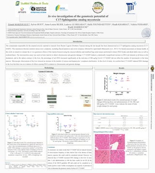
- 1. In vivo investigation of the genotoxic potential of C17-Sphinganine analog mycotoxin Zeineb MARZOUGUI1,2, Sylvie HUET3, Anne-Louise BLIER, Ludovic LE HEGARAT3, Haïfa TOUNSI-KETTITI 4, Riadh KHARRAT1, Valérie FESSARD3, Riadh MARROUCHI1* 1 : Venom and therapeutic biomolecules Laboratory, Institut Pasteur de Tunis, Tunis El Manar University, 13 place Pasteur - BP 74 1002 Tunis-Belvédère 2: National Institute of Agronomy, University of Carthage, Tunis 1082, Tunisia 3: ANSES French Agency for Food, Environmental and Occupational Health and Safety, Fougères Laboratory, Toxicology of Contaminants Unit, 10b rue Claude Bourgelat, Fougères, 35306, France 4:Laboratoire d’Anatomie Pathologique Humaine et Expérimentale, Institut Pasteur de Tunis, Université Tunis El Manar, 13 Place Pasteur, B.P. 74, Tunis-Belvédère, Tunis 1002, Tunisia *Correspondent author: Riadh MARROUCHI (riadh.marrouchi@pasteur.tn), The contaminant responsible for the atypical toxicity reported in mussels from Bizerte Lagoon (Northern Tunisia) during the last decade has been characterized as C17-sphinganine analog mycotoxin (C17- SAMT). This neurotoxin showed common mouse toxic symptoms, including flaccid paralysis and severe dyspnea, followed by rapid death (Marrouchi et al., 2013). For hazard assessment on human health, in this work we aimed to evaluate the in vivo genotoxic effects of this marine biotoxin using the classical alkaline and modified Fpg comet assays performed to detect DNA breaks and alkali-labile sites as well as oxidized bases. The micronucleus assay was used on bone marrow to detect chromosome and genome damage. C17-SAMT induces a statistically insignificant increase in DNA tail intensity at all doses in the duodenum, and in the spleen contrary to the liver, the percentage of tail DNA increased significantly at the mid dose of 300 µg/kg b.w/d. C17-SAMT did not affect the number of micronuclei in the bone marrow. Microscopic observations of the liver showed an increase in the number of mitosis and hepatocytes’ cytoplasm clarification. At this level of study, we confirm that C17-SAMT induced DNA damage in the liver but there was no evidence of effects causing DNA oxidation or chromosome and genome damage. Classic alkaline Comet assay Methodology References Marrouchi, R., Benoit, E., Le Caer, J.-P., Belayouni, N., Belghith, H., Molgó, J., & Kharrat, R. (2013). Toxic C17-Sphinganine Analogue Mycotoxin, Contaminating Tunisian Mussels, Causes Flaccid Paralysis in Rodents. Marine Drugs, 11(12), Art. 12. https://doi.org/10.3390/md11124724 Introduction Comet Assay with and without fpg 5 groups of 5 male mice Groupe 1 Ser.phy Groupe 2 MMS 80 mg/kg Dose 2 at 24h Dose 1 at 0h Dose 3 at 45h Samples at 48 h Groupe 3 C17-SAMT 150 µg/kg b.w Comet Assay without fpg Micronucleus test Liver Duodenum Spleen Bone marrow BM MN assay Groupe 4 C17-SAMT 300 µg/kg b.w Groupe 5 S17-SAMT 600 µg/kg b.w Results Figure 1. Individual results (five animals/group) of weight changes following three oral administrations of 150, 300, and 600 µg/kg of C17-SAMT. Lines indicate the mean of body weight change for each group. Figure 2. Individual results (five animals/group) obtained in the comet assay in the duodenum (a), spleen (b), and liver (c) after 3-day oral administration of different doses of C17- SAMT. DNA damage is expressed as median % of tail DNA intensity (%TI). Lines indicate mean of medians of %TI for each group. * p < 0.05. The positive control MMS induced 15.66 ± 6.48 and 22.73 ± 6.44%TI in the spleen and duodenum, respectively. In liver, 100% of hedgehogs was recorded. Bone marrow micronucleus assay Histopathological Observations Fpg-modified comet assay Figure 3. Individual results (five animals/group) obtained in the modified-comet assay in the spleen after 3-day oral administration of different doses of C17-SAMT. DNA damage is expressed as % Net- Fpg tail DNA intensity. Lines indicate the mean of medians of % TI for each group. The positive control MMS induced 100% of hedgehogs. Table 1. BMMN assay in mice following oral exposure to 150, 300, and 600 µg/kg of C17-SAMT. *** p < 0.001 Conclusion In conclusion, our study reveals that C17-SAMT did not induced increase of micro-nucleus frequency in bone marrow but in the absence of bone marrow exposure this result could be considered relevant. However, we observed that C17-SAMT increased DNA damage in the liver of mice after 3 oral administrations in 45h but these results do not fill all the criteria of the OECD guideline 489 and could not be considered as a clearly positive result. Further investigations are required, in particular in vitro to determine the genotoxic mode of action of C17-SAMT. Figure 5. Examples of histopathological abnormalities observed in the liver from mice treated with three oral doses of C17- SAMT at 150 µg/kg b.w (B–D) compared to the negative control group (A). I: inflammatory infiltrate; M: mitosis; c: cytoplasm clarification. (Magnification ×40). THE INTERNATIONAL BIOTECHNOLOGY DAYS 2022- Hammamet