SlideShare a Scribd company logo
1 of 35
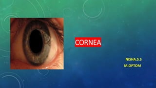
CORNEA
NISHA.S.S
M.OPTOM
ANATOMICAL CONSIDERATIONS
• The cornea is a transparent, avascular, watch glass-like structure. It forms anterior
one sixth of the outer fibrous coat of the eyeball.
DIMENSIONS
• Anterior surface of cornea is elliptical with an average horizontal diameter of 11.75
mm and vertical diameter of 11 mm.
• Posterior surface of cornea is circular with an average diameter of 11.5 mm.
• Thickness of cornea in the centre is about 0.52 mm while at the periphery it is 0.67
mm.
• Radius of curvature: The central 5 mm area of the cornea forms the powerful
refracting surface of the eye. The anterior and posterior radii of curvature of the
central part of cornea are 7.8 mm and 6.5 mm, respectively.
• Refractive power of the anterior surface of cornea is about +48 D and that of its
posterior surface is about -5 D. Thus the net refractive power of cornea is about
+43 D which is three fourth of the total refractive power of the eye (60 dioptres).
• Refractive index of the cornea is 1.37.
HISTOLOGY
• Histologically, originally the cornea was considered to consists of five distinct
layers, from anterior to posterior these are:
 Epithelium
 Bowman's membrane
 corneal stroma (substantia propria)
 Descemet's membrane and
 Endothelium.
• However, recently a new layer between the corneal stroma and Descemet's
membrane named Dua's layer (pre-Descemet's membrane) has been
discovered, and thus now, the cornea is considered to consist of six layers.
Epithelium
• Corneal epithelium is of stratified squamous non-keratinized type and becomes
continuous with epithelium of the bulbar conjunctiva at the limbus.
• It is about 50-90 μm thick, represents 10% of corneal thickness and consists of 5-7
layers of cells.
• The deepest (basal) layer is made up of columnar cells, next mid epithelial 2-3 layers of
wing or umbrella cells and the most superficial two layers are of flattened cells.
• The corneal epithelium sheds at a regular interval and is replaced by growth from its
basal cells.
• It is estimated that the entire epithelium is replaced in a period of 6-8 days.
• Some important features of various layers of epithelium are described below:
 Basal layer
• Basal layer comprises tall columnar polygonal-shaped cells arranged in a palisade like manner on a
basement membrane.
• Basal cells have a width of 12 um and density of approximately 6000 cells/mm².
• It forms the germinal layer of the epithelium and undergoes mitosis to produce daughter cells which
continuously migrate anteriorly into the wing cell layer.
• The basal cell has an oval nucleus and its cytoplasm contains few organelles. Some intermediate
filaments, microtubules, free ribosomes, rough-surfaced endoplasmic reticulum and occasional
Golgi apparatus are seen.
• The basal cells are firmly joined laterally to the other basal cells and anteriorly to the
wing cells by desmosomes and zonula occludens.
• These tight intercellular junctions account for the epithelium's transparency as well as
its resistance to the flow of water, electrolytes and glucose, along with inhibiting
pathological entrance, i.e. its barrier function.
 Limbal stem cells
• The basal cells of the limbal area constitute the so-called limbal stem cells.
• They are the source of new corneal epithelium.
• These slow cycling stem cells divide and give rise to a progeny of daughter cells
(transient amplifying cells) which amplify, proliferate continuously, migrate
centripetally and serve to maintain the corneal epithelium.
• Diffuse damage to the limbal stem cells (e.g. in chemical burns, trachoma) leads
to chronic epithelial surface defects and invasion of conjunctival epithelium on to
the cornea.
 Non-epithelial cells
• Also appear within the corneal epithelial layer especially in the peripheral cornea.
• These cells include wandering histiocytes, macrophages, lymphocyte, and pigmented
melanocyte.
• Antigen presenting Langerhans cells are formed peripherally and move centrally with age or in
response to keratitis.
 Basal lamina
• The basal lamina provides support to the overlying epithelium, limits contact between epithelial
cells and the other cell types in the tissue and acts as a filter allowing only water and small
molecules to pass through.
• Posteriorly, it blends indistinctly into Bowman's membrane.
• Anteriorly, firm adhesions are formed between it and the basal cells by the filaments extending
called as adhesion complex, made up of hemidesmosomes and type VII collagen fibrils.
 Wing cells
• Wing cells form 2-3 layers of the polyhedral shaped cells. Their nuclei are flattened
parallel to the surface and cytoplasmic organelles decrease as compared to the basal
cells. They are attached with the basal cells posteriorly and other wing cells laterally
and anteriorly.
 Flattened cells
• Flattened cells constitute two most superficial cell layers.
• Superficial cells represent the highest level of differentiation and are chronologically the
oldest epithelial cells. These cells are long (45 mm) and thin (4 mm) with flattened nuclei.
• The desmosomal attachment are more numerous in these cells. Further, zonulae
occludentes seen in the lateral cell walls are also found in this layer.
• The anterior cell wall of the most superficial cells has many microvilli (each 0.5 mm in
height) which play an important role in the tear film stability.
• The microvilli contain glycocalyx which is associated with tear film.
Functions of epithelium
• It is extraordinary regular in thickness with smooth wet apical surface for
serving as major refractive surface of the eye.
• It serves as a major surface to respond to wound healing.
• • It helps in providing barrier to fluid loss and pathological entrance to the
organisms.
 Bowman's membrane (layer)
• This layer consists of acellular mass of condensed collagen fibrils. It is about 8-14 um
in thickness and binds the corneal stroma.
• Anteriorly with basement membrane of the epithelium.
• It is not a true elastic membrane but simply a condensed superficial part of the stroma.
• It is composed of randomly packed type I and type V collagen fibres that are enmeshed
in a matrix consisting of proteoglycans and glycoproteins.
• It shows considerable resistance to infection and injury.
• Unlike Descemet's membrane once destroyed, it does not regenerate.
(Bowman's membrane lies just anterior to stroma and is not a true membrane. It is
acellular condensate of the most anterior portion of the stroma. This smooth layer helps
the cornea maintains its shape. When injured, this layer does not regenerate and may
result in a scar)
Function of Bowman membrane
It acts as a smooth base for epithelium uniformity thus helps in refraction.
 Stroma (substantia propria)
• This layer is about 0.5 mm in thickness and constitutes most of the cornea (90% of total
thickness).
• It consists of collagen fibrils (lamellae) and cells embedded in hydrated matrix of
proteoglycans (ground substance).
Corneal lamellae: The lamellae consist of fibrils typical of collagen. The stroma's
collagen types are I, III,V and VI. Among the various subtype, type I predominates.
• Type VII forms the anchoring fibril of the epithelium. The corneal lamellae are arranged
in many layers (200-250). In each layer, they are not only parallel to each other but also
to the corneal plane and become continuous with the scleral lamellae at the limbus. They
vary in disposition according to the area of the cornea.
• They have oblique orientation in the anterior one-third of the stroma. In the posterior, two
thirds of the stroma, the alternating layer of lamellae are at right angles to each other.
• Studies, using X-ray diffraction techniques, have shown that the parallel arrangement of the
central corneal fibrils extends to the periphery where the fibrils adopt a concentric
configuration to form a 'weave' at the limbus.
• This imparts considerable strength to the peripheral cornea and permits it to maintain its
curvature and thus its optical properties.
• Previous studies have shown that the corneal fibrils, running in two preferred orientations in
the central cornea, bend as they approach the peripheral cornea to run circumferentially
and form the peripheral collagen ring.
• The parallel arrangement of lamellae in the cornea allows an easy intralamellar dissection
during superficial keratectomy and lamellar keratoplasty.
• The peculiar arrangement of lamellae has also been implicated in the corneal transparency.
Stromal cells: The cells present among the lamellae are keratocytes, wandering
macrophages, histiocytes and a few lymphocytes.
• Corneal keratocytes about 2.4 million in number constitute 2-4% of the volume of the
stroma in humans.
• The keratocytes are fibroblasts which are found throughout the stroma, between, and
occasionally extending into the lamellae.
• The keratocytes have a flattened cell body, a large eccentric nucleus and long branching
processes which form contact with other cells in the same layer.
• It is believed that these cells produce ground substance and collagen fibrils during
embryogenesis and after injury.
Ground substance of stroma
• Ground substance of cornea consists of hydrated matrix of proteoglycans that
run along and between the collagen fibrils.
• The primary glycosaminoglycans of stroma are keratin sulphate and
chondroitin sulphate in the ratio of 3:1.
• Maximum concentration of keratin sulphate occurs in the centre and that of
chondroitin sulphate in the periphery.
• The glycosaminoglycan components (e.g. keratin sulphate) of the ground
substance are highly charged and account for the swelling property of the
stroma.
• The keratocytes which lie between the corneal lamellae synthesize both the
collagen and proteoglycans.
Functions of stroma
• It acts as a window to the right passage and meshes with surrounding scleral
connective tissue to form a rigid frame for maintaining IOP.
 Pre-Descemet's membrane
• Pre-Descemet's membrane, also known as Dua's layer, has been discovered in 2013 by
Dr. Harminder Dua, an ophthalmologist of Indian origin working in Great Britain.
• Located anterior to Descemet membrane, it is about 15 mm thick acellular structure
which is very strong and impervious to air.
• Dua's layer (DL) primarily composed of collagen type 1. Collagen 4 and 6 are also
present. Collagen 5 is also present.
Descemet's membrane (posterior elastic lamina)
• It is a strong homogenous layer which is separated from the stroma by pre-Descemet's
membrane.
• It represents the basement membrane of the corneal endothelium from which it is
produced. Though elasticity is one of its physical characteristics, it is made up of collagen
and glycoprotein with no elastic fibres visible by electron microscopy.
• Its thickness varies with age, being 3 um at birth and 10-12 um in young adults. It is very
resistant to chemical agents, trauma, infection and pathological processes. Even when
whole of the stroma is sloughed off, the Descemet's membrane can maintain the integrity
of the eyeball for long.
• Further, unlike Bowman's membrane, when destroyed it can regenerate. Normally, it
remains in a state of tension and when torn it curls inwards on itself.
• In the periphery, it appears to end at the anterior limit of the trabecular meshwork as
Schwalbe's line (ring).
• On electron microscopy, Descemet's membrane can be divided into two distinct
regions: An anterior one-third having a vertically banded pattern and the posterior
two-thirds appearing amorphous and granular.
• The posterior surface of the Descemet's membrane, at the periphery, shows rounded
wart-like excrescences called Hassel Henle bodies, which increase with advancing
age.
• Similar central excrescences, known as guttatae, are seen with advancing age in
Fuch's dystrophy.
Endothelium
• Consists of a single layer of flat polygonal (mainly hexagonal) cells, which on slit-
lamp Biomicroscopy appear as a mosaic on Descemet's membrane.
• Cell density of endothelium is around 6000 cells/ mm² at birth. In the human adults,
these cells have hardly any ability to divide.
• The cell count falls by about 26% in the first year and a further 26% is lost over the
next 11 years.
• Therefore, with increasing age, the number of cells is reduced to about 2400-3000
cells/mm² in young adults.
• The defect left by the dying cells is filled by enlargement (polymegathism) of the
remaining cells.
• Hence, these cells vary in diameter from 18 to 20 µ early in life to 40 μ or more in
the aged.
• There is a considerable functional reserve for the endothelium.
• Therefore, corneal decompensation occurs only after more than 75% of the adult age
cells are lost (i.e. when the endothelial cell count becomes less than 500 cells/mm²).
• Endothelial cells are best evaluated by specular microscopy.
• Endothelial cells are attached to the Descemet's membrane by hemidesmosomes and
laterally to each other by tight interdigitating junctional complexes.
• The desmosomal linkages and zonulae occludentes are continuous around the entire
cell, and thus close the intercellular space from the anterior chamber.
• This linkage is calcium-dependent and plays an important role in maintaining the
barrier function of endothelium.
• Endothelium also contains an active pump mechanism and is involved in active secretion and
protein synthesis.
• High metabolic activity and energy production for the above process by the endothelial cells
is evidenced by the presence of abundant mitochondria, free ribosomes, rough- and smooth-
surfaced endoplasmic reticulum and Golgi complexes in the cytoplasm of the cells.
• In the eye, next to photoreceptors, the endothelial cells contain the highest number of
mitochondria.
BLOOD SUPPLY AND NERVE SUPPLY
 Blood supply of the cornea
• The cornea is an avascular structure.
• Small loops derived from the anterior ciliary vessels invade its periphery for about 1
mm and provide nourishment.
• Actually, these loops are not in the cornea but in the subconjunctival tissue which
overlaps the cornea.
• In normal condition, cornea does not contain any blood vessels. Anterior
ciliary artery, a branch ophthalmic artery forms a vascular arcade in the limbal
region and helps in corneal metabolism and wound repair by providing
nourishment. Absence of blood vessel in cornea is one of the contributing
factors for its transparency.
Nerve supply of the cornea
• The cornea has a rich supply of sensory nerve endings derived mainly from the long ciliary
nerves which are branches of the nasociliary nerve (a branch of ophthalmic division of the
trigeminal nerve).
• The long ciliary nerves after arising from the nasociliary nerve enter the eyeball around the
optic nerve along with the short ciliary nerves and run forward in the suprachoroidal space.
• A short distance from the limbus, these nerves pierce the sclera to leave the eyeball, divide
dichotomously and connect with each other and the conjunctival nerves to form a
pericorneal plexus of the nerves.
• About 60 80 myelinated trunks from the pericorneal plexus enter the cornea at various
levels, viz. sclera (the principal regions), episclera and conjunctiva
• After having gone for 1-2 mm in the stroma, the corneal nerves lose their myelin sheath,
branch dichotomously and form a stromal plexus.
• Although, some nerves end in mid-stroma, most pass anteriorly and form a subepithelial
plexus.
• The fibres from here penetrate the pores in Bowman's membrane, lose their Schwann's
sheath, divide into filaments under the basal layer of epithelium which extend between the
cells of all the layers of epithelium, and form intraepithelial plexus.
• The nerves end in the epithelium as fine-beaded filaments.
• Thus the cornea has an extensive innervational density which is highest near the centre
and gradually decreases towards the periphery. However, there are no nerves in the central
posterior part of the cornea, Descemet's membrane and endothelium.
THANKYOU

More Related Content

What's hot

Choroidal melanoma
Choroidal melanoma Choroidal melanoma
Choroidal melanoma SSSIHMS-PG
 
Minimally invasive Glaucoma surgery MIGS
Minimally invasive Glaucoma surgery MIGSMinimally invasive Glaucoma surgery MIGS
Minimally invasive Glaucoma surgery MIGSankita mahapatra
 
Just SMILE (small incision lenticule extraction )
Just SMILE (small incision lenticule extraction )Just SMILE (small incision lenticule extraction )
Just SMILE (small incision lenticule extraction )DiyarAlzubaidy
 
Eyelid anatomy & lacrimal pump physiology
Eyelid anatomy & lacrimal pump physiologyEyelid anatomy & lacrimal pump physiology
Eyelid anatomy & lacrimal pump physiologyPrajakta Matey
 
Circumscribed choroidal hemangioma
Circumscribed choroidal hemangiomaCircumscribed choroidal hemangioma
Circumscribed choroidal hemangiomaJuan Carlos Rivera
 
Vogt koyanagi-harada disease
Vogt koyanagi-harada diseaseVogt koyanagi-harada disease
Vogt koyanagi-harada diseaseBipin Bista
 
Anatomy of cornea & corneal tranparency dr.ysr
Anatomy of cornea &  corneal tranparency dr.ysrAnatomy of cornea &  corneal tranparency dr.ysr
Anatomy of cornea & corneal tranparency dr.ysrDrYajuvendra Rathore
 
Lecture lacrimal gland tumor
Lecture lacrimal gland tumorLecture lacrimal gland tumor
Lecture lacrimal gland tumorPanit Cherdchu
 
Corneal edema after cataract surgery - MALEK AL KOTT
Corneal edema after cataract surgery - MALEK AL KOTTCorneal edema after cataract surgery - MALEK AL KOTT
Corneal edema after cataract surgery - MALEK AL KOTTMalek Al Kott
 
Pseudoexfoliative glaucoma seminar
Pseudoexfoliative glaucoma seminarPseudoexfoliative glaucoma seminar
Pseudoexfoliative glaucoma seminarmithunkishore1
 
Genetics - Ophthalmology
Genetics - OphthalmologyGenetics - Ophthalmology
Genetics - OphthalmologyAhmed Omara
 
Diffuse Lamellar Keratitis (DLK)
Diffuse Lamellar Keratitis (DLK)Diffuse Lamellar Keratitis (DLK)
Diffuse Lamellar Keratitis (DLK)DiyarAlzubaidy
 

What's hot (20)

Phakic iols
Phakic iolsPhakic iols
Phakic iols
 
Choroidal melanoma
Choroidal melanoma Choroidal melanoma
Choroidal melanoma
 
Minimally invasive Glaucoma surgery MIGS
Minimally invasive Glaucoma surgery MIGSMinimally invasive Glaucoma surgery MIGS
Minimally invasive Glaucoma surgery MIGS
 
Limbus
LimbusLimbus
Limbus
 
Just SMILE (small incision lenticule extraction )
Just SMILE (small incision lenticule extraction )Just SMILE (small incision lenticule extraction )
Just SMILE (small incision lenticule extraction )
 
Eyelid anatomy & lacrimal pump physiology
Eyelid anatomy & lacrimal pump physiologyEyelid anatomy & lacrimal pump physiology
Eyelid anatomy & lacrimal pump physiology
 
Circumscribed choroidal hemangioma
Circumscribed choroidal hemangiomaCircumscribed choroidal hemangioma
Circumscribed choroidal hemangioma
 
Cxl
CxlCxl
Cxl
 
Eye lid anatomy
Eye lid anatomyEye lid anatomy
Eye lid anatomy
 
Vogt koyanagi-harada disease
Vogt koyanagi-harada diseaseVogt koyanagi-harada disease
Vogt koyanagi-harada disease
 
Anatomy of cornea & corneal tranparency dr.ysr
Anatomy of cornea &  corneal tranparency dr.ysrAnatomy of cornea &  corneal tranparency dr.ysr
Anatomy of cornea & corneal tranparency dr.ysr
 
Collagen cross linking
Collagen cross linkingCollagen cross linking
Collagen cross linking
 
Lecture lacrimal gland tumor
Lecture lacrimal gland tumorLecture lacrimal gland tumor
Lecture lacrimal gland tumor
 
Corneal edema after cataract surgery - MALEK AL KOTT
Corneal edema after cataract surgery - MALEK AL KOTTCorneal edema after cataract surgery - MALEK AL KOTT
Corneal edema after cataract surgery - MALEK AL KOTT
 
Eyelid reconstraction
Eyelid reconstractionEyelid reconstraction
Eyelid reconstraction
 
FFA AND ICG
FFA AND ICGFFA AND ICG
FFA AND ICG
 
Pseudoexfoliative glaucoma seminar
Pseudoexfoliative glaucoma seminarPseudoexfoliative glaucoma seminar
Pseudoexfoliative glaucoma seminar
 
Genetics - Ophthalmology
Genetics - OphthalmologyGenetics - Ophthalmology
Genetics - Ophthalmology
 
Corneal disorders
Corneal  disordersCorneal  disorders
Corneal disorders
 
Diffuse Lamellar Keratitis (DLK)
Diffuse Lamellar Keratitis (DLK)Diffuse Lamellar Keratitis (DLK)
Diffuse Lamellar Keratitis (DLK)
 

Similar to CORNEA.pptx

Anatomy of Cornea.pptx
Anatomy of Cornea.pptxAnatomy of Cornea.pptx
Anatomy of Cornea.pptxoptometry fans
 
anatomyofthecornea-161101224539-converted.pptx
anatomyofthecornea-161101224539-converted.pptxanatomyofthecornea-161101224539-converted.pptx
anatomyofthecornea-161101224539-converted.pptxArun Kumar A
 
Corneal anatomy and physiology 2
Corneal anatomy and physiology 2Corneal anatomy and physiology 2
Corneal anatomy and physiology 2Om Patel
 
Cornea anatomy simplified
Cornea anatomy simplifiedCornea anatomy simplified
Cornea anatomy simplifiedNayana Gowda
 
CORNEA anat and physio.pptx
CORNEA anat and physio.pptxCORNEA anat and physio.pptx
CORNEA anat and physio.pptxManoAarthi1
 
CORNEA.anatomy of cornea .ophthalmology lecture
CORNEA.anatomy of cornea .ophthalmology lectureCORNEA.anatomy of cornea .ophthalmology lecture
CORNEA.anatomy of cornea .ophthalmology lectureABIDOFFICIALCHANNEL
 
Microscopic anatomy of gingival epithelium
Microscopic anatomy of gingival epitheliumMicroscopic anatomy of gingival epithelium
Microscopic anatomy of gingival epitheliumNishiMahapatra
 
Anatomi fisiologi kornea
Anatomi fisiologi korneaAnatomi fisiologi kornea
Anatomi fisiologi korneahasya arianda
 
Cornea anatomy and physiology PowerPoint presentation
Cornea anatomy and physiology PowerPoint presentationCornea anatomy and physiology PowerPoint presentation
Cornea anatomy and physiology PowerPoint presentationShallyGupta18
 
cornea, conjunctiva and sclera
cornea, conjunctiva and scleracornea, conjunctiva and sclera
cornea, conjunctiva and scleraFawaz Alzweimel
 
Anatomy of cornea and sclera
Anatomy of cornea and scleraAnatomy of cornea and sclera
Anatomy of cornea and scleraHenok Samuel
 
Corneal anatomy and anatomy of limbus !
Corneal anatomy and anatomy of  limbus !Corneal anatomy and anatomy of  limbus !
Corneal anatomy and anatomy of limbus !TrishnaThapa5
 

Similar to CORNEA.pptx (20)

Cornea
CorneaCornea
Cornea
 
Cornea Applied anatomy.pptx
Cornea Applied anatomy.pptxCornea Applied anatomy.pptx
Cornea Applied anatomy.pptx
 
Anatomy of Cornea.pptx
Anatomy of Cornea.pptxAnatomy of Cornea.pptx
Anatomy of Cornea.pptx
 
CORNEA.pptx
CORNEA.pptxCORNEA.pptx
CORNEA.pptx
 
Cornea anatomy
Cornea anatomyCornea anatomy
Cornea anatomy
 
anatomyofthecornea-161101224539-converted.pptx
anatomyofthecornea-161101224539-converted.pptxanatomyofthecornea-161101224539-converted.pptx
anatomyofthecornea-161101224539-converted.pptx
 
Corneal anatomy and physiology 2
Corneal anatomy and physiology 2Corneal anatomy and physiology 2
Corneal anatomy and physiology 2
 
Cornea anatomy simplified
Cornea anatomy simplifiedCornea anatomy simplified
Cornea anatomy simplified
 
CORNEA anat and physio.pptx
CORNEA anat and physio.pptxCORNEA anat and physio.pptx
CORNEA anat and physio.pptx
 
Cornea
Cornea Cornea
Cornea
 
Keratoplasty update 2016
Keratoplasty update 2016Keratoplasty update 2016
Keratoplasty update 2016
 
CORNEA.anatomy of cornea .ophthalmology lecture
CORNEA.anatomy of cornea .ophthalmology lectureCORNEA.anatomy of cornea .ophthalmology lecture
CORNEA.anatomy of cornea .ophthalmology lecture
 
Microscopic anatomy of gingival epithelium
Microscopic anatomy of gingival epitheliumMicroscopic anatomy of gingival epithelium
Microscopic anatomy of gingival epithelium
 
Periodontium 1
Periodontium 1Periodontium 1
Periodontium 1
 
Anatomi fisiologi kornea
Anatomi fisiologi korneaAnatomi fisiologi kornea
Anatomi fisiologi kornea
 
Cornea anatomy and physiology PowerPoint presentation
Cornea anatomy and physiology PowerPoint presentationCornea anatomy and physiology PowerPoint presentation
Cornea anatomy and physiology PowerPoint presentation
 
cornea, conjunctiva and sclera
cornea, conjunctiva and scleracornea, conjunctiva and sclera
cornea, conjunctiva and sclera
 
Anatomy of cornea and sclera
Anatomy of cornea and scleraAnatomy of cornea and sclera
Anatomy of cornea and sclera
 
Histology of eye
Histology of eyeHistology of eye
Histology of eye
 
Corneal anatomy and anatomy of limbus !
Corneal anatomy and anatomy of  limbus !Corneal anatomy and anatomy of  limbus !
Corneal anatomy and anatomy of limbus !
 

Recently uploaded

Call Girls Thane Just Call 9907093804 Top Class Call Girl Service Available
Call Girls Thane Just Call 9907093804 Top Class Call Girl Service AvailableCall Girls Thane Just Call 9907093804 Top Class Call Girl Service Available
Call Girls Thane Just Call 9907093804 Top Class Call Girl Service AvailableDipal Arora
 
Hubli Call Girls 👙 6297143586 👙 Genuine WhatsApp Number for Real Meet
Hubli Call Girls 👙 6297143586 👙 Genuine WhatsApp Number for Real MeetHubli Call Girls 👙 6297143586 👙 Genuine WhatsApp Number for Real Meet
Hubli Call Girls 👙 6297143586 👙 Genuine WhatsApp Number for Real MeetCall Girls Service
 
VIP Call Girl Sector 88 Gurgaon Delhi Just Call Me 9899900591
VIP Call Girl Sector 88 Gurgaon Delhi Just Call Me 9899900591VIP Call Girl Sector 88 Gurgaon Delhi Just Call Me 9899900591
VIP Call Girl Sector 88 Gurgaon Delhi Just Call Me 9899900591adityaroy0215
 
Punjab❤️Call girls in Mohali ☎️7435815124☎️ Call Girl service in Mohali☎️ Moh...
Punjab❤️Call girls in Mohali ☎️7435815124☎️ Call Girl service in Mohali☎️ Moh...Punjab❤️Call girls in Mohali ☎️7435815124☎️ Call Girl service in Mohali☎️ Moh...
Punjab❤️Call girls in Mohali ☎️7435815124☎️ Call Girl service in Mohali☎️ Moh...Sheetaleventcompany
 
❤️♀️@ Jaipur Call Girls ❤️♀️@ Jaispreet Call Girl Services in Jaipur QRYPCF ...
❤️♀️@ Jaipur Call Girls ❤️♀️@ Jaispreet Call Girl Services in Jaipur QRYPCF  ...❤️♀️@ Jaipur Call Girls ❤️♀️@ Jaispreet Call Girl Services in Jaipur QRYPCF  ...
❤️♀️@ Jaipur Call Girls ❤️♀️@ Jaispreet Call Girl Services in Jaipur QRYPCF ...Gfnyt.com
 
Russian Call Girls Kota * 8250192130 Service starts from just ₹9999 ✅
Russian Call Girls Kota * 8250192130 Service starts from just ₹9999 ✅Russian Call Girls Kota * 8250192130 Service starts from just ₹9999 ✅
Russian Call Girls Kota * 8250192130 Service starts from just ₹9999 ✅gragmanisha42
 
Chandigarh Escorts, 😋9988299661 😋50% off at Escort Service in Chandigarh
Chandigarh Escorts, 😋9988299661 😋50% off at Escort Service in ChandigarhChandigarh Escorts, 😋9988299661 😋50% off at Escort Service in Chandigarh
Chandigarh Escorts, 😋9988299661 😋50% off at Escort Service in ChandigarhSheetaleventcompany
 
VIP Call Girls Sector 67 Gurgaon Just Call Me 9711199012
VIP Call Girls Sector 67 Gurgaon Just Call Me 9711199012VIP Call Girls Sector 67 Gurgaon Just Call Me 9711199012
VIP Call Girls Sector 67 Gurgaon Just Call Me 9711199012Call Girls Service Gurgaon
 
Nanded Call Girls 👙 6297143586 👙 Genuine WhatsApp Number for Real Meet
Nanded Call Girls 👙 6297143586 👙 Genuine WhatsApp Number for Real MeetNanded Call Girls 👙 6297143586 👙 Genuine WhatsApp Number for Real Meet
Nanded Call Girls 👙 6297143586 👙 Genuine WhatsApp Number for Real MeetCall Girls Service
 
VIP Call Girl Sector 10 Noida Call Me: 9711199171
VIP Call Girl Sector 10 Noida Call Me: 9711199171VIP Call Girl Sector 10 Noida Call Me: 9711199171
VIP Call Girl Sector 10 Noida Call Me: 9711199171Call Girls Service Gurgaon
 
VIP Call Girls Noida Jhanvi 9711199171 Best VIP Call Girls Near Me
VIP Call Girls Noida Jhanvi 9711199171 Best VIP Call Girls Near MeVIP Call Girls Noida Jhanvi 9711199171 Best VIP Call Girls Near Me
VIP Call Girls Noida Jhanvi 9711199171 Best VIP Call Girls Near Memriyagarg453
 
Bangalore call girl 👯‍♀️@ Simran Independent Call Girls in Bangalore GIUXUZ...
Bangalore call girl  👯‍♀️@ Simran Independent Call Girls in Bangalore  GIUXUZ...Bangalore call girl  👯‍♀️@ Simran Independent Call Girls in Bangalore  GIUXUZ...
Bangalore call girl 👯‍♀️@ Simran Independent Call Girls in Bangalore GIUXUZ...Gfnyt
 
Jodhpur Call Girls 📲 9999965857 Jodhpur best beutiful hot girls full satisfie...
Jodhpur Call Girls 📲 9999965857 Jodhpur best beutiful hot girls full satisfie...Jodhpur Call Girls 📲 9999965857 Jodhpur best beutiful hot girls full satisfie...
Jodhpur Call Girls 📲 9999965857 Jodhpur best beutiful hot girls full satisfie...seemahedar019
 
VIP Call Girl Sector 25 Gurgaon Just Call Me 9899900591
VIP Call Girl Sector 25 Gurgaon Just Call Me 9899900591VIP Call Girl Sector 25 Gurgaon Just Call Me 9899900591
VIP Call Girl Sector 25 Gurgaon Just Call Me 9899900591adityaroy0215
 
Call Girls Service In Goa 💋 9316020077💋 Goa Call Girls By Russian Call Girl...
Call Girls Service In Goa  💋 9316020077💋 Goa Call Girls  By Russian Call Girl...Call Girls Service In Goa  💋 9316020077💋 Goa Call Girls  By Russian Call Girl...
Call Girls Service In Goa 💋 9316020077💋 Goa Call Girls By Russian Call Girl...russian goa call girl and escorts service
 
Call Girl In Zirakpur ❤️♀️@ 9988299661 Zirakpur Call Girls Near Me ❤️♀️@ Sexy...
Call Girl In Zirakpur ❤️♀️@ 9988299661 Zirakpur Call Girls Near Me ❤️♀️@ Sexy...Call Girl In Zirakpur ❤️♀️@ 9988299661 Zirakpur Call Girls Near Me ❤️♀️@ Sexy...
Call Girl In Zirakpur ❤️♀️@ 9988299661 Zirakpur Call Girls Near Me ❤️♀️@ Sexy...Sheetaleventcompany
 
Call Girls Hyderabad Just Call 9907093804 Top Class Call Girl Service Available
Call Girls Hyderabad Just Call 9907093804 Top Class Call Girl Service AvailableCall Girls Hyderabad Just Call 9907093804 Top Class Call Girl Service Available
Call Girls Hyderabad Just Call 9907093804 Top Class Call Girl Service AvailableDipal Arora
 
Udaipur Call Girls 📲 9999965857 Call Girl in Udaipur
Udaipur Call Girls 📲 9999965857 Call Girl in UdaipurUdaipur Call Girls 📲 9999965857 Call Girl in Udaipur
Udaipur Call Girls 📲 9999965857 Call Girl in Udaipurseemahedar019
 
💚😋Chandigarh Escort Service Call Girls, ₹5000 To 25K With AC💚😋
💚😋Chandigarh Escort Service Call Girls, ₹5000 To 25K With AC💚😋💚😋Chandigarh Escort Service Call Girls, ₹5000 To 25K With AC💚😋
💚😋Chandigarh Escort Service Call Girls, ₹5000 To 25K With AC💚😋Sheetaleventcompany
 
💚😋Mumbai Escort Service Call Girls, ₹5000 To 25K With AC💚😋
💚😋Mumbai Escort Service Call Girls, ₹5000 To 25K With AC💚😋💚😋Mumbai Escort Service Call Girls, ₹5000 To 25K With AC💚😋
💚😋Mumbai Escort Service Call Girls, ₹5000 To 25K With AC💚😋Sheetaleventcompany
 

Recently uploaded (20)

Call Girls Thane Just Call 9907093804 Top Class Call Girl Service Available
Call Girls Thane Just Call 9907093804 Top Class Call Girl Service AvailableCall Girls Thane Just Call 9907093804 Top Class Call Girl Service Available
Call Girls Thane Just Call 9907093804 Top Class Call Girl Service Available
 
Hubli Call Girls 👙 6297143586 👙 Genuine WhatsApp Number for Real Meet
Hubli Call Girls 👙 6297143586 👙 Genuine WhatsApp Number for Real MeetHubli Call Girls 👙 6297143586 👙 Genuine WhatsApp Number for Real Meet
Hubli Call Girls 👙 6297143586 👙 Genuine WhatsApp Number for Real Meet
 
VIP Call Girl Sector 88 Gurgaon Delhi Just Call Me 9899900591
VIP Call Girl Sector 88 Gurgaon Delhi Just Call Me 9899900591VIP Call Girl Sector 88 Gurgaon Delhi Just Call Me 9899900591
VIP Call Girl Sector 88 Gurgaon Delhi Just Call Me 9899900591
 
Punjab❤️Call girls in Mohali ☎️7435815124☎️ Call Girl service in Mohali☎️ Moh...
Punjab❤️Call girls in Mohali ☎️7435815124☎️ Call Girl service in Mohali☎️ Moh...Punjab❤️Call girls in Mohali ☎️7435815124☎️ Call Girl service in Mohali☎️ Moh...
Punjab❤️Call girls in Mohali ☎️7435815124☎️ Call Girl service in Mohali☎️ Moh...
 
❤️♀️@ Jaipur Call Girls ❤️♀️@ Jaispreet Call Girl Services in Jaipur QRYPCF ...
❤️♀️@ Jaipur Call Girls ❤️♀️@ Jaispreet Call Girl Services in Jaipur QRYPCF  ...❤️♀️@ Jaipur Call Girls ❤️♀️@ Jaispreet Call Girl Services in Jaipur QRYPCF  ...
❤️♀️@ Jaipur Call Girls ❤️♀️@ Jaispreet Call Girl Services in Jaipur QRYPCF ...
 
Russian Call Girls Kota * 8250192130 Service starts from just ₹9999 ✅
Russian Call Girls Kota * 8250192130 Service starts from just ₹9999 ✅Russian Call Girls Kota * 8250192130 Service starts from just ₹9999 ✅
Russian Call Girls Kota * 8250192130 Service starts from just ₹9999 ✅
 
Chandigarh Escorts, 😋9988299661 😋50% off at Escort Service in Chandigarh
Chandigarh Escorts, 😋9988299661 😋50% off at Escort Service in ChandigarhChandigarh Escorts, 😋9988299661 😋50% off at Escort Service in Chandigarh
Chandigarh Escorts, 😋9988299661 😋50% off at Escort Service in Chandigarh
 
VIP Call Girls Sector 67 Gurgaon Just Call Me 9711199012
VIP Call Girls Sector 67 Gurgaon Just Call Me 9711199012VIP Call Girls Sector 67 Gurgaon Just Call Me 9711199012
VIP Call Girls Sector 67 Gurgaon Just Call Me 9711199012
 
Nanded Call Girls 👙 6297143586 👙 Genuine WhatsApp Number for Real Meet
Nanded Call Girls 👙 6297143586 👙 Genuine WhatsApp Number for Real MeetNanded Call Girls 👙 6297143586 👙 Genuine WhatsApp Number for Real Meet
Nanded Call Girls 👙 6297143586 👙 Genuine WhatsApp Number for Real Meet
 
VIP Call Girl Sector 10 Noida Call Me: 9711199171
VIP Call Girl Sector 10 Noida Call Me: 9711199171VIP Call Girl Sector 10 Noida Call Me: 9711199171
VIP Call Girl Sector 10 Noida Call Me: 9711199171
 
VIP Call Girls Noida Jhanvi 9711199171 Best VIP Call Girls Near Me
VIP Call Girls Noida Jhanvi 9711199171 Best VIP Call Girls Near MeVIP Call Girls Noida Jhanvi 9711199171 Best VIP Call Girls Near Me
VIP Call Girls Noida Jhanvi 9711199171 Best VIP Call Girls Near Me
 
Bangalore call girl 👯‍♀️@ Simran Independent Call Girls in Bangalore GIUXUZ...
Bangalore call girl  👯‍♀️@ Simran Independent Call Girls in Bangalore  GIUXUZ...Bangalore call girl  👯‍♀️@ Simran Independent Call Girls in Bangalore  GIUXUZ...
Bangalore call girl 👯‍♀️@ Simran Independent Call Girls in Bangalore GIUXUZ...
 
Jodhpur Call Girls 📲 9999965857 Jodhpur best beutiful hot girls full satisfie...
Jodhpur Call Girls 📲 9999965857 Jodhpur best beutiful hot girls full satisfie...Jodhpur Call Girls 📲 9999965857 Jodhpur best beutiful hot girls full satisfie...
Jodhpur Call Girls 📲 9999965857 Jodhpur best beutiful hot girls full satisfie...
 
VIP Call Girl Sector 25 Gurgaon Just Call Me 9899900591
VIP Call Girl Sector 25 Gurgaon Just Call Me 9899900591VIP Call Girl Sector 25 Gurgaon Just Call Me 9899900591
VIP Call Girl Sector 25 Gurgaon Just Call Me 9899900591
 
Call Girls Service In Goa 💋 9316020077💋 Goa Call Girls By Russian Call Girl...
Call Girls Service In Goa  💋 9316020077💋 Goa Call Girls  By Russian Call Girl...Call Girls Service In Goa  💋 9316020077💋 Goa Call Girls  By Russian Call Girl...
Call Girls Service In Goa 💋 9316020077💋 Goa Call Girls By Russian Call Girl...
 
Call Girl In Zirakpur ❤️♀️@ 9988299661 Zirakpur Call Girls Near Me ❤️♀️@ Sexy...
Call Girl In Zirakpur ❤️♀️@ 9988299661 Zirakpur Call Girls Near Me ❤️♀️@ Sexy...Call Girl In Zirakpur ❤️♀️@ 9988299661 Zirakpur Call Girls Near Me ❤️♀️@ Sexy...
Call Girl In Zirakpur ❤️♀️@ 9988299661 Zirakpur Call Girls Near Me ❤️♀️@ Sexy...
 
Call Girls Hyderabad Just Call 9907093804 Top Class Call Girl Service Available
Call Girls Hyderabad Just Call 9907093804 Top Class Call Girl Service AvailableCall Girls Hyderabad Just Call 9907093804 Top Class Call Girl Service Available
Call Girls Hyderabad Just Call 9907093804 Top Class Call Girl Service Available
 
Udaipur Call Girls 📲 9999965857 Call Girl in Udaipur
Udaipur Call Girls 📲 9999965857 Call Girl in UdaipurUdaipur Call Girls 📲 9999965857 Call Girl in Udaipur
Udaipur Call Girls 📲 9999965857 Call Girl in Udaipur
 
💚😋Chandigarh Escort Service Call Girls, ₹5000 To 25K With AC💚😋
💚😋Chandigarh Escort Service Call Girls, ₹5000 To 25K With AC💚😋💚😋Chandigarh Escort Service Call Girls, ₹5000 To 25K With AC💚😋
💚😋Chandigarh Escort Service Call Girls, ₹5000 To 25K With AC💚😋
 
💚😋Mumbai Escort Service Call Girls, ₹5000 To 25K With AC💚😋
💚😋Mumbai Escort Service Call Girls, ₹5000 To 25K With AC💚😋💚😋Mumbai Escort Service Call Girls, ₹5000 To 25K With AC💚😋
💚😋Mumbai Escort Service Call Girls, ₹5000 To 25K With AC💚😋
 

CORNEA.pptx

  • 2. ANATOMICAL CONSIDERATIONS • The cornea is a transparent, avascular, watch glass-like structure. It forms anterior one sixth of the outer fibrous coat of the eyeball. DIMENSIONS • Anterior surface of cornea is elliptical with an average horizontal diameter of 11.75 mm and vertical diameter of 11 mm. • Posterior surface of cornea is circular with an average diameter of 11.5 mm. • Thickness of cornea in the centre is about 0.52 mm while at the periphery it is 0.67 mm.
  • 3. • Radius of curvature: The central 5 mm area of the cornea forms the powerful refracting surface of the eye. The anterior and posterior radii of curvature of the central part of cornea are 7.8 mm and 6.5 mm, respectively. • Refractive power of the anterior surface of cornea is about +48 D and that of its posterior surface is about -5 D. Thus the net refractive power of cornea is about +43 D which is three fourth of the total refractive power of the eye (60 dioptres). • Refractive index of the cornea is 1.37.
  • 4. HISTOLOGY • Histologically, originally the cornea was considered to consists of five distinct layers, from anterior to posterior these are:  Epithelium  Bowman's membrane  corneal stroma (substantia propria)  Descemet's membrane and  Endothelium. • However, recently a new layer between the corneal stroma and Descemet's membrane named Dua's layer (pre-Descemet's membrane) has been discovered, and thus now, the cornea is considered to consist of six layers.
  • 5.
  • 6.
  • 7.
  • 8.
  • 9. Epithelium • Corneal epithelium is of stratified squamous non-keratinized type and becomes continuous with epithelium of the bulbar conjunctiva at the limbus. • It is about 50-90 μm thick, represents 10% of corneal thickness and consists of 5-7 layers of cells. • The deepest (basal) layer is made up of columnar cells, next mid epithelial 2-3 layers of wing or umbrella cells and the most superficial two layers are of flattened cells. • The corneal epithelium sheds at a regular interval and is replaced by growth from its basal cells. • It is estimated that the entire epithelium is replaced in a period of 6-8 days.
  • 10. • Some important features of various layers of epithelium are described below:  Basal layer • Basal layer comprises tall columnar polygonal-shaped cells arranged in a palisade like manner on a basement membrane. • Basal cells have a width of 12 um and density of approximately 6000 cells/mm². • It forms the germinal layer of the epithelium and undergoes mitosis to produce daughter cells which continuously migrate anteriorly into the wing cell layer. • The basal cell has an oval nucleus and its cytoplasm contains few organelles. Some intermediate filaments, microtubules, free ribosomes, rough-surfaced endoplasmic reticulum and occasional Golgi apparatus are seen.
  • 11. • The basal cells are firmly joined laterally to the other basal cells and anteriorly to the wing cells by desmosomes and zonula occludens. • These tight intercellular junctions account for the epithelium's transparency as well as its resistance to the flow of water, electrolytes and glucose, along with inhibiting pathological entrance, i.e. its barrier function.
  • 12.  Limbal stem cells • The basal cells of the limbal area constitute the so-called limbal stem cells. • They are the source of new corneal epithelium. • These slow cycling stem cells divide and give rise to a progeny of daughter cells (transient amplifying cells) which amplify, proliferate continuously, migrate centripetally and serve to maintain the corneal epithelium. • Diffuse damage to the limbal stem cells (e.g. in chemical burns, trachoma) leads to chronic epithelial surface defects and invasion of conjunctival epithelium on to the cornea.
  • 13.  Non-epithelial cells • Also appear within the corneal epithelial layer especially in the peripheral cornea. • These cells include wandering histiocytes, macrophages, lymphocyte, and pigmented melanocyte. • Antigen presenting Langerhans cells are formed peripherally and move centrally with age or in response to keratitis.  Basal lamina • The basal lamina provides support to the overlying epithelium, limits contact between epithelial cells and the other cell types in the tissue and acts as a filter allowing only water and small molecules to pass through. • Posteriorly, it blends indistinctly into Bowman's membrane. • Anteriorly, firm adhesions are formed between it and the basal cells by the filaments extending called as adhesion complex, made up of hemidesmosomes and type VII collagen fibrils.
  • 14.  Wing cells • Wing cells form 2-3 layers of the polyhedral shaped cells. Their nuclei are flattened parallel to the surface and cytoplasmic organelles decrease as compared to the basal cells. They are attached with the basal cells posteriorly and other wing cells laterally and anteriorly.
  • 15.  Flattened cells • Flattened cells constitute two most superficial cell layers. • Superficial cells represent the highest level of differentiation and are chronologically the oldest epithelial cells. These cells are long (45 mm) and thin (4 mm) with flattened nuclei. • The desmosomal attachment are more numerous in these cells. Further, zonulae occludentes seen in the lateral cell walls are also found in this layer. • The anterior cell wall of the most superficial cells has many microvilli (each 0.5 mm in height) which play an important role in the tear film stability. • The microvilli contain glycocalyx which is associated with tear film.
  • 16.
  • 17. Functions of epithelium • It is extraordinary regular in thickness with smooth wet apical surface for serving as major refractive surface of the eye. • It serves as a major surface to respond to wound healing. • • It helps in providing barrier to fluid loss and pathological entrance to the organisms.
  • 18.  Bowman's membrane (layer) • This layer consists of acellular mass of condensed collagen fibrils. It is about 8-14 um in thickness and binds the corneal stroma. • Anteriorly with basement membrane of the epithelium. • It is not a true elastic membrane but simply a condensed superficial part of the stroma. • It is composed of randomly packed type I and type V collagen fibres that are enmeshed in a matrix consisting of proteoglycans and glycoproteins. • It shows considerable resistance to infection and injury. • Unlike Descemet's membrane once destroyed, it does not regenerate. (Bowman's membrane lies just anterior to stroma and is not a true membrane. It is acellular condensate of the most anterior portion of the stroma. This smooth layer helps the cornea maintains its shape. When injured, this layer does not regenerate and may result in a scar)
  • 19. Function of Bowman membrane It acts as a smooth base for epithelium uniformity thus helps in refraction.
  • 20.  Stroma (substantia propria) • This layer is about 0.5 mm in thickness and constitutes most of the cornea (90% of total thickness). • It consists of collagen fibrils (lamellae) and cells embedded in hydrated matrix of proteoglycans (ground substance). Corneal lamellae: The lamellae consist of fibrils typical of collagen. The stroma's collagen types are I, III,V and VI. Among the various subtype, type I predominates. • Type VII forms the anchoring fibril of the epithelium. The corneal lamellae are arranged in many layers (200-250). In each layer, they are not only parallel to each other but also to the corneal plane and become continuous with the scleral lamellae at the limbus. They vary in disposition according to the area of the cornea. • They have oblique orientation in the anterior one-third of the stroma. In the posterior, two thirds of the stroma, the alternating layer of lamellae are at right angles to each other.
  • 21. • Studies, using X-ray diffraction techniques, have shown that the parallel arrangement of the central corneal fibrils extends to the periphery where the fibrils adopt a concentric configuration to form a 'weave' at the limbus. • This imparts considerable strength to the peripheral cornea and permits it to maintain its curvature and thus its optical properties. • Previous studies have shown that the corneal fibrils, running in two preferred orientations in the central cornea, bend as they approach the peripheral cornea to run circumferentially and form the peripheral collagen ring. • The parallel arrangement of lamellae in the cornea allows an easy intralamellar dissection during superficial keratectomy and lamellar keratoplasty. • The peculiar arrangement of lamellae has also been implicated in the corneal transparency.
  • 22. Stromal cells: The cells present among the lamellae are keratocytes, wandering macrophages, histiocytes and a few lymphocytes. • Corneal keratocytes about 2.4 million in number constitute 2-4% of the volume of the stroma in humans. • The keratocytes are fibroblasts which are found throughout the stroma, between, and occasionally extending into the lamellae. • The keratocytes have a flattened cell body, a large eccentric nucleus and long branching processes which form contact with other cells in the same layer. • It is believed that these cells produce ground substance and collagen fibrils during embryogenesis and after injury.
  • 23. Ground substance of stroma • Ground substance of cornea consists of hydrated matrix of proteoglycans that run along and between the collagen fibrils. • The primary glycosaminoglycans of stroma are keratin sulphate and chondroitin sulphate in the ratio of 3:1. • Maximum concentration of keratin sulphate occurs in the centre and that of chondroitin sulphate in the periphery. • The glycosaminoglycan components (e.g. keratin sulphate) of the ground substance are highly charged and account for the swelling property of the stroma. • The keratocytes which lie between the corneal lamellae synthesize both the collagen and proteoglycans.
  • 24. Functions of stroma • It acts as a window to the right passage and meshes with surrounding scleral connective tissue to form a rigid frame for maintaining IOP.
  • 25.  Pre-Descemet's membrane • Pre-Descemet's membrane, also known as Dua's layer, has been discovered in 2013 by Dr. Harminder Dua, an ophthalmologist of Indian origin working in Great Britain. • Located anterior to Descemet membrane, it is about 15 mm thick acellular structure which is very strong and impervious to air. • Dua's layer (DL) primarily composed of collagen type 1. Collagen 4 and 6 are also present. Collagen 5 is also present.
  • 26. Descemet's membrane (posterior elastic lamina) • It is a strong homogenous layer which is separated from the stroma by pre-Descemet's membrane. • It represents the basement membrane of the corneal endothelium from which it is produced. Though elasticity is one of its physical characteristics, it is made up of collagen and glycoprotein with no elastic fibres visible by electron microscopy. • Its thickness varies with age, being 3 um at birth and 10-12 um in young adults. It is very resistant to chemical agents, trauma, infection and pathological processes. Even when whole of the stroma is sloughed off, the Descemet's membrane can maintain the integrity of the eyeball for long. • Further, unlike Bowman's membrane, when destroyed it can regenerate. Normally, it remains in a state of tension and when torn it curls inwards on itself. • In the periphery, it appears to end at the anterior limit of the trabecular meshwork as Schwalbe's line (ring).
  • 27. • On electron microscopy, Descemet's membrane can be divided into two distinct regions: An anterior one-third having a vertically banded pattern and the posterior two-thirds appearing amorphous and granular. • The posterior surface of the Descemet's membrane, at the periphery, shows rounded wart-like excrescences called Hassel Henle bodies, which increase with advancing age. • Similar central excrescences, known as guttatae, are seen with advancing age in Fuch's dystrophy.
  • 28. Endothelium • Consists of a single layer of flat polygonal (mainly hexagonal) cells, which on slit- lamp Biomicroscopy appear as a mosaic on Descemet's membrane. • Cell density of endothelium is around 6000 cells/ mm² at birth. In the human adults, these cells have hardly any ability to divide. • The cell count falls by about 26% in the first year and a further 26% is lost over the next 11 years. • Therefore, with increasing age, the number of cells is reduced to about 2400-3000 cells/mm² in young adults. • The defect left by the dying cells is filled by enlargement (polymegathism) of the remaining cells. • Hence, these cells vary in diameter from 18 to 20 µ early in life to 40 μ or more in the aged.
  • 29. • There is a considerable functional reserve for the endothelium. • Therefore, corneal decompensation occurs only after more than 75% of the adult age cells are lost (i.e. when the endothelial cell count becomes less than 500 cells/mm²). • Endothelial cells are best evaluated by specular microscopy. • Endothelial cells are attached to the Descemet's membrane by hemidesmosomes and laterally to each other by tight interdigitating junctional complexes. • The desmosomal linkages and zonulae occludentes are continuous around the entire cell, and thus close the intercellular space from the anterior chamber. • This linkage is calcium-dependent and plays an important role in maintaining the barrier function of endothelium.
  • 30. • Endothelium also contains an active pump mechanism and is involved in active secretion and protein synthesis. • High metabolic activity and energy production for the above process by the endothelial cells is evidenced by the presence of abundant mitochondria, free ribosomes, rough- and smooth- surfaced endoplasmic reticulum and Golgi complexes in the cytoplasm of the cells. • In the eye, next to photoreceptors, the endothelial cells contain the highest number of mitochondria.
  • 31.
  • 32. BLOOD SUPPLY AND NERVE SUPPLY  Blood supply of the cornea • The cornea is an avascular structure. • Small loops derived from the anterior ciliary vessels invade its periphery for about 1 mm and provide nourishment. • Actually, these loops are not in the cornea but in the subconjunctival tissue which overlaps the cornea. • In normal condition, cornea does not contain any blood vessels. Anterior ciliary artery, a branch ophthalmic artery forms a vascular arcade in the limbal region and helps in corneal metabolism and wound repair by providing nourishment. Absence of blood vessel in cornea is one of the contributing factors for its transparency.
  • 33. Nerve supply of the cornea • The cornea has a rich supply of sensory nerve endings derived mainly from the long ciliary nerves which are branches of the nasociliary nerve (a branch of ophthalmic division of the trigeminal nerve). • The long ciliary nerves after arising from the nasociliary nerve enter the eyeball around the optic nerve along with the short ciliary nerves and run forward in the suprachoroidal space. • A short distance from the limbus, these nerves pierce the sclera to leave the eyeball, divide dichotomously and connect with each other and the conjunctival nerves to form a pericorneal plexus of the nerves. • About 60 80 myelinated trunks from the pericorneal plexus enter the cornea at various levels, viz. sclera (the principal regions), episclera and conjunctiva
  • 34. • After having gone for 1-2 mm in the stroma, the corneal nerves lose their myelin sheath, branch dichotomously and form a stromal plexus. • Although, some nerves end in mid-stroma, most pass anteriorly and form a subepithelial plexus. • The fibres from here penetrate the pores in Bowman's membrane, lose their Schwann's sheath, divide into filaments under the basal layer of epithelium which extend between the cells of all the layers of epithelium, and form intraepithelial plexus. • The nerves end in the epithelium as fine-beaded filaments. • Thus the cornea has an extensive innervational density which is highest near the centre and gradually decreases towards the periphery. However, there are no nerves in the central posterior part of the cornea, Descemet's membrane and endothelium.