
Recommended
More Related Content
What's hot
What's hot (20)
Similar to Clinical and biomedical applications of imaging therapy
Similar to Clinical and biomedical applications of imaging therapy (20)
Recently uploaded
Recently uploaded (20)
Clinical and biomedical applications of imaging therapy
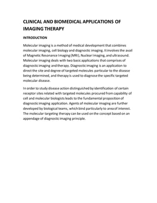
- 1. CLINICAL AND BIOMEDICAL APPLICATIONS OF IMAGING THERAPY INTRODUCTION Molecular imaging is a method of medical development that combines molecular imaging, cell biology and diagnostic imaging. Itinvolves the avail of Magnetic Resonance Imaging (MRI), Nuclear Imaging, and ultrasound. Molecular imaging deals with two basic applications that comprises of diagnostic imaging and therapy. Diagnostic imaging is an application to direct the site and degree of targeted molecules particular to the disease being determined, and therapy is used to diagnosethe specific targeted molecular disease. In order to study disease action distinguished by identification of certain receptor sites related with targeted molecules procured fromcapability of cell and molecular biologists leads to the fundamental proposition of diagnostic imaging application. Agents of molecular imaging are further developed by biological teams, which bind particularly to area of interest. The molecular targeting therapy can be used on the concept based on an appendage of diagnostic imaging principle.
- 2. Fig.1 CLINICAL AND BIOMEDICAL APPLICATIONS OF MRI: MRI is used for softtissueimaging with excellent contrastdetail and anatomic structures like gray and white matter in the brain and small metastatic lesions in the liver whereas x-rays aregood for densestructures such as bone imaging with good resolution. MRI can be used in clinical neurology for segmentation and classification. The measuring volumes of brain structures along with the multiple sclerosis, neurodegeneracy , stroke. Itis used for detection of different cancers like breast, liver, colorectal, prostateand also for soft tissue damage to image cartilages and ligaments. MRI is also used to study cancer growth and brain function. Itis used in cardiology to image fast and to detect the motion of heart. In musculoskeletalsystemit’s application include in assessmentof joint disease, spinal imaging, soft tissuetumors.
- 3. MRI steered the stroketherapy as- clinical situations indicated below as: 1. Spontaneous enhancement 2. Improvementin responseto therapy 3. No unbidden improvementor responseto therapy Only outcome 2 is really effective while outcome 1 and 3 could be worsen by the side effects. Fig.2
- 4. f Fig .3 Even small amount of noise can change the classification.
- 5. Fig.4 The segmentation of brain tissue depends upon evaluating the possibility of each category at individual voxel x and then iteratively updating and transmitting these estimates to the neighbors. Hidden Markov Random Fields are used in this case. Steps involved in this process :
- 6. Fig.5 MULTIPLE SCLEROSIS: Multiple sclerosis are visible as the both spots of plaque, indicating it, in the below figure. As T1-Weighted image gives white grey matter, while white matter appears black.
- 7. Fig.6 MR image of horizontal slice through the brain BREAST CANCER Breast cancer is predominant cancer found in women and it is a second leading cause of death in worldwide. In India every one out of 22 women may have a possibility of carcinoma in breast. It is most common non skin cancer in women as it is a cancer of tissues. Early breast cancer often does not show any symptoms. That’s why regular breast examination and mammograms are important, so cancer that does not show symptoms may be detected earlier. Deform ductal, connective tissues and micro calcifications are largely causes of malignancy. There is high probability of cancer in upper lateral quadrant of breast. X-ray mammography has demonstrated to be precise and easy method of detecting breast cancer, but it is not perform easily. The radiographer and support staff must have exceptional knowledge, skill, and caring.
- 8. In 1992, the US government mandated regulations in the Mammography Quality Standard Act (MQSA), which set standard dose, personnel qualifications, and examination procedures. Most common symptoms of breast cancer are: Breast lump Nipple discharge Change in size, shape or feel of breast or nipple Breast pain or discomfort. Skin ulcers Swelling of the lymph nodes in the armpit Weight loss Risk of breast cancer: In 2010, approximately 2, 60,000 new cases of breast cancer were reported in the United States, and it is growing rapidly. More than 90% of women diagnosed with early stage of disease will survive due to early detection. In 1995, the National Cancer Institute survey the first reduction in breast cancer mortality in 50 years and this trend continues. Age : The older the person, the higher the risk Family history : Mother, sister with breast cancer Genetics : Presence of the BRCA1 or BRCA2 gene Breast architecture : Dense breast tissue and obesity Menstruation : Onset before age 12 years Menopause : Onset after 55 years Hormonal therapy : Prolonged use of estrogen Late age at birth of first child or no children Previous radiation therapy to the chest at an early age Socioeconomics:Risk increases with higher socioeconomic status Education: Risk increases with higher level of education Breast MRI, Breast ultrasound, Breast biopsy, CT scan, PET scan can also use as tests to diagnose breast cancer.
- 9. Refrences: 1. www.imaginis.com/mri-scan/applications-and-clinical-benefits-of-mr- imaging. 2. https://www.ncbi.nlm.nih.gov/pubmed/12817419. 3. https://www.researchgate.net/figure/41089445_fig2_Figure-1-Magnetic- resonance-imaging-MRI-guided-stroke-therapy-Three-clinical (figure 2) 4. https://upload.wikimedia.org/wikipedia/commons/b/b2/MolecularImagingTher apy.jpg (figure 1) 5. https://www.google.co.in/search?q=segmentation+and+measurement+of+brain +tissue+diagram&tbm=isch&source=iu&ictx=1&fir=- iH8HscFu9B76M%253A%252Cp3zBWhNmZDMQcM%252C_&usg=__q6ENF2FVZZ FjAltPFw7sXyZd30k%3D&sa=X&ved=0ahUKEwjan7_fvonYAhUHS48KHZIVA8YQ9 QEILTAA#imgrc=RObIfLNDhfW7LM (figure 5) 6. www.robots.ox.ac.uk/~jmb/lectures/medimanallecture2.pdf. (figure 3,4,6)