
Recommended
More Related Content
Similar to Beckenfragkturen.ppt
Similar to Beckenfragkturen.ppt (20)
Recently uploaded
Recently uploaded (20)
Beckenfragkturen.ppt
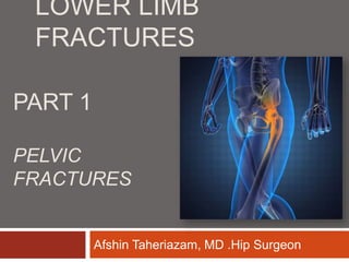
- 1. LOWER LIMB FRACTURES Hip Surgeon . Afshin Taheriazam, MD PART 1 PELVIC FRACTURES
- 3. Objectives: Epidemiology & relevance Anatomical review Classification Systems Examples Management
- 4. Epidemiology ~3% of all fractures in ED (Emergency Department) 50-60% secondary to MVA Motorcycle crashes ~15% Car vs. pedestrian ~15% Falls 10-30% Crush injuries ~5% ( Mortality is about 6 - 50%) : Mortality 6-10% ; Inc’s to ~50% in unstable pt (prothrombin time) – 39% due to bleeding (early). – 30% due to sepsis & multi-organ failure (late). Complications: Hemorrhage, neurological injury, deformity, GU injury, GI injury Mortality quoted tends to be all causes of mortaltiy; less than 1% will die as a direct consequence of their pelvis fracture.
- 5. Anatomy of Pelvis Pelvis = sacrum + 2 inominate bones Inominate bones = ilium, ischium, pubis Strength from ligamentous + muscular supports
- 6. Anatomy of Pelvis Pelvis contains one pair of fused bone Each half contains: ilium, pubis, and ischium Joined together in posterior by sacrum Joined in anterior by symphysis pubis Coalesce at triradiate cartilage. Ilium, ishium and pubis have three separate ossification centers that fuse at sixteen years. Gap in symphysis < 5 mm SI joint 2-4 mm
- 7. Anatomy of Pelvis Ilium Sacrum Male Pelvis Female Pelvis Pubis Ischium Symphysis Pubis
- 8. Anatomy of Pelvis : ligaments Anterior Support: ~40% of strength Symphysis pubis Fibrocartilaginous joint covered by ant & post symphyseal ligaments Pubic rami Posterior Support: ~60% of strength Sacroiliac complex Sacroiliac ligaments Iliolumbar ligaments Pelvic floor Sacrospinous ligament Sacrotuberous ligament Pelvic diaphragm
- 9. Anatomy of Pelvis : ligaments posterior ligaments are stronger than anterior ligaments: Posterior SI Anterior SI Interosseous ligaments Pubic symphysis Sacrotuberous Sacrospinous Sacrospinous resists external rotation Sacrotuberous resists rotational and vertical shearing forces
- 10. ASI ST SS PSI ST
- 11. Posterior Ligaments Ant. SI Joint – resist external rotation Post. SI and Interosseous – posterior stability by tension band (strongest in body) Iliolumbar ligaments augments posterior complex Sacrotuberous (sacrum behind sacro-spinous into ischial tuberosily vertically)Resists shear and flexion of SI joint Sacrospinous – (anterior sacral body to ischial spine horizontally) resists external rotation
- 12. Normal SI Joint Motion with Gait < 6 mm of translation < 6° rotation Intact cadaver resist 5,837 N (1,212 lbs)
- 14. Acetabulum Divided into 3 columns: Anterior superior column (= ilium) Anterior inferior column (= pubis) Posterior Column (= ischium) Ant superior column is the primary weight bearing structure. The ant inferior column is thin and easily fractured. The post column is thick and strong but most commonly fractured.
- 15. Vascular Anatomy Vessels lie closely adherent to posterior pelvic walls Most common cause of bleeding is venous Most commonly injured arteries are superior gluteal and internal pudendal aa. Venous bleeding is most common because of relative deficiency in protective vasospasm, and lack of valves in pelvis with extensive collateral
- 16. Vascular Anatomy Internal iliac artery courses medial to the vein, splits into anterior and posterior branches. Posterior branch is more likely injured (SGA is largest branch). Usual bleeding is from venous plexus.
- 17. Pelvic Stability Stability : ability of pelvic ring to withstand physiologic forces without abnormal deformation. Physiologic load may be sitting, side lying, or standing, as dictated by patient needs , else consider as unstable. Strength of ring: 40% anterior and 60% posterior. Vsphere = 4/3r³.
- 18. Pelvic Ring Stability Posterior ring integrity is important in transferring load from torso to lower extremities
- 19. Common Fractures of Pelvis Pelvic ring fractures Pelvic ring is likely to separate in more than one location Iliac crest fractures Fractures to upper wing of ilium Loss of posterior ring integrity leads to instability Loss of anterior ring integrity may contribute to instability, and may be a marker to posterior ring injury. Young and burgess classification will guide us for stability issues
- 20. Pelvic Fractures Common mechanisms of pelvic injury result from high energy ex. MVC, significant falls, skiing accident Those at risk for pelvic fractures Growing teens (especially those involved in sports) Elderly patients (osteoporosis)
- 21. Classification systems 2 most common are Tile and Young systems Tile Classification system: Advantages Comprehensive Predicts need for operative intervention Disadvantages Does NOT predict morbidity or mortality Young Classification System: Advantages Based on mechanism of injury predicts injury Estimates mortality Disadvantages Excludes more minor injuries
- 22. Tile : Classification System Type A: Stable pelvis post structures intact A1: avulsion injury A2: iliac wing or ant arch A3: Transverse sacrococcygeal
- 23. Tile : Classification System Type B: Partially stable pelvis: incomplete posterior structure disruption B1: open-book injury B2: lateral compression injury B3: contralateral / bucket handle injuries
- 24. Tile : Classification System Type C: Unstable pelvis: complete disruption of posterior structures C1: unilateral C2: bilateral w/ one side Type B, one side Type C C3: bilateral Type C
- 25. Young & Burgess Classification
- 26. Classification System: Young Lateral Compression (50%) – transverse of pubic rami, ipsilateral or contralateral to posterior injury LC I – sacral compression on side of impact LC II – iliac wing on side of impact LC III – LC-I or LC-II on side of impact w/ contralateral APC injury
- 27. Classification System: Young AP Compression (25%) Symphyseal and / or Longitudinal Rami Fractures APC I – slight widening of the pubic symphysis and/or anterior SI joint APC II – disrupted anterior SI joint, sacrotuberous, and sacrospinous ligaments APC III – complete SI joint disruption w/ lateral displacement and disruption of
- 28. Classification System: Young Vertical Shear ( VS) (5%) Symphyseal diastasis or vertical displacement anteriorly and posteriorly Example : Fall from heights Combined Mechanism (CM) combination of injury patterns
- 29. Young: Morbidity & Mortality Fracture Type Severe Bleeding Bladder Rupture Urethral Injury Mortalit y LC - I 0.5% 4% 2% 6% LC – II 36% 7% 0% 6% LC – III 60% 20% 20% 13% APC – I 1% 8% 12% 7% APC – II 28% 11% 23% 7% APC – III 53% 14% 36% 25% VS 75% 15% 25% 25% CM 58% 16% 21% 17%
- 30. Classify the fractures in the next slides
- 31. Tile B1 / Young APC II
- 33. Tile C1/ Young VS
- 35. Tile A1
- 37. Type A1 avulsion
- 39. Tile A2 / Young LC II
- 40. Risks of Pelvic Factures Iliac Crest fracture Typically pelvis still stable Little blood loss Pelvic Ring fracture Internal organ damage Significant blood loss (up to 4 liters) : Fracture and vascular injury can cause the formation of hematoma in the pelvis and retroperitoneum Hypovolemic shock 90% bleeding venous disruption or cancellous bone 10% bleeding an arterial injury Unstable pelvis Risk of death (Mortality of 3.4%-42%)
- 41. Assessment ATLS Approach (Advanced Trauma Life Support) Check Stability : Mechanic Haemodynamic Pelvis specific assessment Check for bruising, deformity, or abrasions Listen/Feel for crepitus Check limb length
- 42. Stability Assessment Check stability of pelvis (DON’T REPEAT) 1) Apply gentle medial pressure with palms by pressing inward on iliac crests 2) With patient supine, apply gentle posterior pressure by pressing downward on iliac crests 3) Apply gentle downward pressure on pubis to check pelvic ring stability 1) Medial pressure 2) Posterior iliac pressure 3) Posterior pubis pressure
- 43. Diagnosis 1. General: abrasion, contusion, hematoma, over bony prominence of pelvis, scrotal, vulvar hematoma. 2. PE ( Physical Exam ) 3. X-ray 4. FAST 5. DPL 6. CT
- 44. IDENTIFY THE HIGH RISK PELVIC DISRUPTION By Physical Exam By Radiography
- 45. Radiographic Evaluation • X-Ray AP view: – Anterior lesions: pubic rami fractures – Symphysis displacement – Sacroiliac joint and sacral fractures – Iliac fractures – L5 transverse process fractures
- 46. Radiographic Signs of Instability Broken ‘Ring’ Symphysis gap > 2.5 cm Sacroiliac displacement of 5 mm in any plane. Avulsion of the 5th lumbar transverse process, the lateral border of the sacrum (sacrotuberous ligament), or the ischial spine (sacrospinous ligament).
- 47. Treatment Treat for life threatening injuries Treat for possible shock Oxygen Intravenous infusion Splinting / Wrap Pain control RAPID TRANSPORT!!!
- 48. Patients with hemorrhagic shock and unstable pelvic fractures have four potential sources of blood loss : (1) fractured bone surfaces (2) pelvic venous plexus (3) pelvic arterial injury (4) extrapelvic sources. The pelvis should be temporarily stabilized or "closed" using an available commercial compression device or sheet to decrease bleeding.
- 49. • In the presence of unstable pelvic ring disruption and a positive abdominal study, stabilization of the pelvis should be undertaken before laparatomy. • If hemodynamic stability is not achieved after placement of the external fixator, arteriography should then be performed.
- 50. Non-Operative Management ( haemodinamically stable ) Lateral impaction type injuries with minimal (< 1.5 cm) displacement Pubic rami fractures with no posterior displacement Minimal gapping of pubic symphysis
- 52. Operative Management Operative indications Pelvic unstable symphysis diastasis > 2.5 cm SI joint displacement > 1 cm sacral fracture with displacement > 1 cm displacement or rotation of hemipelvis open fracture Hemodynamically unstable
- 53. Operative Management Hemodynamically unstable Reduce pelvic volume : promote blood clot as well as reducing blood volume from inside bleeding Technique First aid : pelvic wrap (This is wrapped circumferentially around the pelvis) Next : Ex fix/ C clamp
- 54. • Haemodynamic Status Options for immediate hemorrhage control • Military antishock trousers (MAST): Typically applied in the field. – No impact on survival rate. – Severe complications reported (compartment syndrome, extremity
- 55. Posterior ring structure is important Goal : restoration of anatomy and enough stability to maintain reduction during healing Anterior ring fixation may provide structural protection of posterior fixation Operative Management
- 56. Anterior Fixation of Pelvic
- 57. Posterior Fixation of Pelvic
- 58. • Anterior external fixator: – In the acute phase many advocate external fixation as a temporary device to achieve stabilization of the fracture and a positive effect on haemorrhage.
- 59. External fixation 1. Advantages It helps tamponade bleeding from bone edges . Stabilizing the clots and the bone. Could be done in 20 min. 2. Disadvantages Can’t stop arterial bleeding. Delay the embolization for ongoing arterial hemorrhage. Degrade the quality of CT and Angio.
- 60. Complications • Infection • Thromboembolism • Non-Union • Malunion
- 61. Potentially Damaged Visceral Anatomy Blunt vs. impaled by bony spike Bladder/urethra Rectum Vagina
- 62. Imaging Plain films AP Inlet view / Outlet view Judet view (oblique) AP alone ~90% sensitive; combined w/ inlet / outlet views ~94% sensitive Limited in ability to clearly delineate posterior injuries Pelvic films are NOT necessary in pts with normal physical exam + GCS >13 At least one study shows clinical exam reliable in EtOH Gonzalez et al. J Am Coll Surg. 2002; 194: 121-5 CT scans Evaluates extent of posterior injury better Superior imaging of sacrum and acetabulum More detailed info about associated injuries
- 63. EtOh levels up to 104 mmol/l, most were > 21.7 Prospective study of 2176 consecutive blunt trauma pts of which 4.5% had pelvic #’s AP plevis alone missed more injuries than clinical exam even in intoxicated pts On the other hand, plain films can help to predict bleeding complications and should be done if pelvis is suspected to be busted as the first step in the work up
- 64. Inlet & Outlet Views Inlet view X-ray beam at 60o to plate directed towards feet Used to look for vertical & horizontal fracture displacement, and SI widening Outlet view Beam aimed 30o towards head Used to look at sacral fractures & SI disruption
- 65. Imaging What you really want to know is if there has been damage to the posterior structures Clues on X-rays: L5 transverse process avulsion (iliolumbar ligament) Ischial spine avulsion (sacrospinous ligament) Unable to clearly make out sacral foramina Assymmetry of sacral foramina Significant displacement of anterior arch fracture Sacral avulsion (sacrotuberous ligament) Ist 2 always denote mechanical instability
- 67. 6 lines of the pelvis: 1. Iliopubic (arcuate) line – disruption indicates ant column injury 2. Ilioischial line which defines the posterior column 3. Teardrop or Roentgenographic U formed by roof of acetabaulum and ilioischial spine defines quadrangular plate – disruption means intraplevic penetration 4. Roof of acetabulum 5. Post rim of acetabulum 6. Ant rim of acetabulum 7. Shenton’s line = medial femoral shaft obturator foramen: disruption in hip dislocation or femoral neck #’s
- 69. Pelvic Fracture Complications Early complications Delayed complications
- 70. Hemorrhage The most dangerous & life threatening condition ( hypovolemic shock ) Sources : Retroperitoneal (Bone-Small & Large vessels ) Multiple trauma (Chest-Abdomen- Long bone fractures)
- 71. • Evaluating Pelvic Hemorrhage (EPH) Study – 724 consecutive pelvic fractures at Harborview • 62 % male • Average age = 34 • Mechanism – Motor vehicle crash 57% – Car versus pedestrian 21% – Fall (>3.3 meters) 11% – Crush 5%
- 72. • Hemodynamic shock in Emergency Dept. – Blood pressure<90 27% – Pulse>130 30% – Transfuse in ED 29% • Blood requirement – Any 80% – 6 or more units 41% – Range (0 to 171 units) • Death 13%
- 73. Sign & Symptom Back pain Abdominal pain Swelling & Echymosis (Flank – Buttock – Inguinal – Perineum ) Hypotension & Shock
- 74. X-ray : Soft tissue shadow displacement (Int.obturator, Iliopsoas, Gluteal Fat pad Bladder , Uterus) CT scan : Hematoma Angigraphy :
- 75. Fracture Type APC (anterior posterior compression) & VS (vertical shear) ( high risk) Artery & Vein Inj. Iliac – Iliolumbar – Sup.Gluteal – Internal Pudental. LC (rare) Fx site – Visceral Inj. Stable Fx (very rare)
- 76. Treatment Transfusion Pelvic belt Antishock garment Reduction & Fixation Angiographic embolization
- 77. Thromboembolism Pelvic bone trauma & Immobilization Ipsilateral or contralateral Calf – Thigh – Pelvic veins Proximal thrombosis has Greatest risk of embolism
- 78. Increased risk of DVT Older age Spinal cord Inj. Lower extremity Inj. History of DVT
- 79. Rate MR Venography 35% Thrombosis Contrast Venography 29% Dopler Sonography 9% Pulmonary Embolism 2 – 12% Fatal Pulmonary Embo. 0.5 – 10%
- 80. Prophylaxy Routin prophylaxis is mandatory Method is controversial Drug : Aspirin – Warfarin Low dose Heparin Low M.W.Heparin Mechanical devices : Compresion stocking Foot pump Compresion device thigh & leg Vena cava filter
- 81. Fat Embolism Gasterointestinal Inj. Open fracture Deep pelvic infection Retroperitoneal absces Peritonitis High mortality rate
- 82. Gasterointestinal Inj. Wound in perineum Blood in rectum More proximal Injury (Contrast CTscan) Direct Inj. (Bone fragment) Indirect Inj. (Ext.Rot. Streching)
- 83. Summary Pelvic fracture High morbidity and mortality Multiple trauma Team work (ATLS Approach) Check stability (Mechanic and Haemodynamic) Early immobilization Pelvic Wrap Diagnostic tools Definitive treatment