
Recommended
Recommended
More Related Content
Similar to motor system.pptx
Similar to motor system.pptx (20)
Recently uploaded
Recently uploaded (20)
motor system.pptx
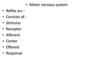
- 1. • Motor nervous system • Reflex arc : • Consists of : • Stimulus • Receptor • Afferent • Center • Efferent • Response
- 3. • White matter Grey matter posterior horn sensory neurons anterior horn motor neurons
- 4. • Spinal reflexes: Monosynaptic reflexes Polysynaptic reflexes -Reflex having only one synapse -No interneurones -The strech reflex is the only mono synaptic reflex -Reflex containing more than one synapse ---may reach 100 interneurones -Ex –Flexor withdrawal reflex
- 5. • Stretch reflex: • Def: Reflex contraction of the muscle in response to stretch. • Components of the stretch reflex: Stimulus –stretch of the muscle extrafusal fibers Receptor – Muscle spindle Afferent -thick myelinated fibers (primary,secondary) Center – spinal cord (monosynaptic) Efferent – thick myelinated A alpha fibers Response – muscle contraction
- 6. • Structure of the muscle spindle: • 1-mehanoreceptors • 2-monitor change in muscle length • OR rate of change of muscle length • 3-spindle shape • 4-encapsulated & ends of capsule are attached to tendons on both sides of muscle • 5-distributed through out the muscle length • 6-each muscle has many spindles • 7-each spindle contain 2-10 intrafusal muscle fibers
- 8. • Intrafusal muscle fibers: • -smaller than extrafusal fibers • -less striated than ordinary fibers • -Parallel with muscle fibers • -intrafusal muscle fibers are 2 types • -end of chain fibers are attached to side of bag fibers Nuclear bag fibers Nuclear chain fibers - Thicker than nuclear chain - Longer than nuclear chain - 1-3 nuclei in each spindle -thinner -shorter -3-9 nuclei in each spindle
- 9. • -the central portion of the intrafusal fibers is • *the receptor area • *stretcheable • *not contractile • *contain no Actin or Myosin • -the peripheral part of intrafusal fibers is contractile (contain actin & myosin)
- 10. • Innervation of muscle spindle: • Afferent (sensory) ----*primary ending • *circle central part of nuclear bag and chain • *secondary ending • *circle nuclear chain only • Efferent (motor) • *dynamic gamma efferent---to peripheral contractile part of nuclear bag *static gamma efferent ----to peripheral contractile part of nuclear chain
- 13. • Properties of stretch reflex: • -monosynaptic • -fastest reflex in body • -restricted to stretched muscles • -show receprocal innervation • -more in antigravirty muscles • -does not show fatigue
- 14. • Components (types) 0f stretch reflex: • Static stretch reflex • Dynamic stretch reflex • Static stretch reflex Dynamic stretch reflex *It is the base of MUSCLE TONE as the tone in the antigravity muscles *its stimulus is SUSTAINED STRETCH of the muscle *the receptor is muscle spindle(NUCLEAR CHAIN TYPE) *afferent ---primary & seondary endings *response is CONTINUOUS CONTRACTION OF THE MUSCLE *it is the base of TENDON JERK *its stimulus is SUDDEN STRETCH OF MUSCLE *the receptor is muscle spindle (NUCLEAR BAG TYPE) *afferent ---primary ending *response is SUDDEN CONTRACTION FOLLOWED BY RELAXATION
- 15. • Supraspinal regulation of stretch reflex: • *the stretch reflex is a spinal reflex which is controlled by supra spinal centers • The supra spinal centers regulating the stretch reflex are: Facilitatory supra spinal centers Inhibitory supra spinal centers 1-facilitatory reticular formation 2-motor area 4 3-neocerebellum 4-vestibular nuclei NB: *facilitatory reticular formation has intrensic activity *send impulses through facilitatory reticular formation then to gamma motor neurons 1-inhibitory reticular formation 2-area 4 S (cortical suppressor area) 3-paleocerebellum 4-red nucleus 5-basal ganglia NB:*inhibitory reticular formation has no intrensic activity *send impulses through inhibitory reticular formation
- 16. • Function of stretch reflex: • 1-static stretch reflex is the base of MUSCLE TONE • 2-role of stretch reflex in controlling voluntary movement. • 3-clinical importance of stretch reflex
- 17. • Static stretch reflex is the base of muscle tone: • Skeletal muscle tone: • Def: • reflex • subtetenic contraction of skeletal muscle fibers • alternating • • lead to muscle tension • it occurs especially in antigravity muscles
- 18. • Mechanism: • During rest the muscle is continuously stretched as length of the muscle is shorter than the distance between the origin and insertion • (Intrensic activity) of higher centers form continuous impulses from them through gamma fibers-----keep muscle spindle stretched
- 19. • NB: • The muscle tone acquire NO FATIGUE. • Causes: • 1-alternating activity of different motor neurons --- alternating contraction of group of muscle fibers and the other group relax and rest • 2-the reflex contraction is SUBTETANIC
- 20. • Function of muscle tone: • 1-keep body position against gravity • 2-keep viscera in position • 3-maintain body temperature----muscle contract -- -release energy----increase metabolic rate • 4-help venous return and lymph drain
- 21. • 2-role of stretch reflex in controlling voluntary movements. • 1-Damping function of stretch reflex: • the brain discharges the impulses irregularly • to motor neurones • this leads to JERKY OSCILLATORY movements • The stretch reflex prevents oscillation of movements & make them smooth • due to • Simultaneous discharge to gamma motor neurones & alpha motor neurones • this leads to • Contraction of extra & intra fusal fibers at same time • This make the contraction smooth
- 22. • 2-servoassist function during muscle contraction: • lift weight • cortex send signals to • Alpha & Gamma motor neurons • (coactivation between alpha & gamma) +++ Extra fusal fibers +++ Intra fusal fibers At same time (the stimulation of the gamma fibers reflexly stimulate the alpha fibers thus potentiate it)
- 23. • Decerebrate rigidity: • make transverse section of spinal cord • between SUPERIOR COLLICULI & INFERIOR • COLLICULI • This will remove the INHIBITORY EFFECT OF • SUPRA SPINAL INHIBITORY CENTER & RED • NUCLEUS • & • leave the facilitatory effect of facilitatory • reticular formation & vestibular nucleus •
- 24. • This will lead to • marked increase in the tone of antigravity muscles • Animal will have---1-extended neck • 2-extended back • 3-extended limbs • 4-elevated tail. • NB: • The same condition occur in humans if there is lesion in the INTERNAL CAPSULE ----person will have increased tone in antigravity muscles
- 25. • Clinical importance of stretch reflex: • Tendon jerk (Deep reflexes) • (Dynamic stretch reflex) • Def: sudden strike of tendon of muscle • lead to • sudden stretch of the muscle • which lead to • reflex contraction of muscle followed by relaxation
- 26. • NB: the tendon jerk is facilitated by: • 1- putting muscle in stretched position • 2- increase gamma efferent discharge by • *clinching teeth • * hook fingers of hands together & pull • hands apart •
- 27. • 1-Biceps jerk: • Stimulus ---sudden tap on tendon of biceps • Receptor---muscle spindle (nuclear bag) • Afferent----primary ending • Center -----C5,C6 • Efferent ----myelinated • Response---contraction of biceps (flex elbow) then • relax
- 28. • 2-Triceps jerk: • Stimulus ---sudden tap on tendon of triceps • Receptor---muscle spindle (nuclear bag) • Afferent----primary ending • Center -----C6,C7 • Efferent ----myelinated • Response---contraction of triceps (extend elbow) • then relax •
- 29. • 3-Knee jerk: • Stimulus ---sudden tap on tendon of Quadriceps • Receptor---muscle spindle (nuclear bag) • Afferent----primary ending • Center -----L2,L3,L4 • Efferent ----myelinated • Response---contraction of Quadriceps (extend • knee) then relax •
- 30. • 4-Ankle jerk: • Stimulus ---sudden tap on tendon of Achilles • Receptor---muscle spindle (nuclear bag) • Afferent----primary ending • Center -----S1,S2 • Efferent ----myelinated • Response---contraction of gastrocnemius muscle • (planter flexion of ankle)
- 31. • Inverse stretch reflex: • Def: marked stretch of the muscle ----lead to reflex relaxation of the muscle • Stimulus----increase tension in the muscle • (as in marked stretch OR strong contraction) • Receptor---GOLGI TENDON ORGAN • *receptor • *in the tendon • *parallel with muscle fibers • *stimulated by high tension in muscle
- 32. • Afferent: myelinated • Center: spinal cord (BI SYNAPTIC) (2 interneurons • Efferent: alpha motor neuron fiber inhibitory to • muscle fiber • Response: relaxation of muscle (decrease muscle • tone to prevent tear of muscle fibers)
- 33. • Clinical application of inverse stretch reflex: • Clasp knife rigidity: • Def: resistance then sudden release • felx upper limb • (extensors are stretched) • stimulate muscle spindle • increase muscle tone (Resistance) • stimulate golgi tendon receptor • inhibit Alpha motor neuron • relaxation of muscle (Release)
- 34. • Polysynaptic reflexes: • 1-flexor withdrawal reflex: • Def: flexion & withdrawal of limb away of injurious stimulus. • Stimulus: painful stimulus on limb • Receptor: F.N.E. (free nerve ending) • Afferent: A delta fibers , C fibers • Center : spinal cord (polysynaptic) • Efferent: Alpha motor neurone • Response: flexion & withdraw of limb away of pain stimulus
- 35. • Polysynaptic reflexes: • 1-flexor withdrawal reflex: • Def: flexion & withdrawal of limb away of injurious stimulus. • Stimulus: painful stimulus on limb • Receptor: F.N.E. (free nerve ending) • Afferent: A delta fibers , C fibers • Center : spinal cord (polysynaptic) • Efferent: Alpha motor neurone • Response: flexion & withdraw of limb away of pain stimulus
- 36. • 4-pattern of contraction depend on site of stimulation • 5-tetanus (there will be continuous ontraction) • Types of tetanus are: • Reflex tetanus Motor tetanus *Repeated Stimulation of AFFERENT nerve *conduction of impulse along AFFERENT INTERNEURONES EFFERENT MUSCLE *continuous contraction of muscle *Repeated stimulation of MOTOR EFFERENT nerve of the muscle *impulse move to muscle directly
- 37. • Characters of reflex tetanus , & motor tetanus Reflex tetanus Motor tetanus *Longer latent period *Tension rises gradually (due to increase number of activated motor cells) (Recrutement) The tension decreases gradually (due to fatigue in the synapse) After removal of stimulus the tension persists for short period (due to after discharge) *Short latent period *Tension rise rapidly maintained as the stimulus is applied drop rapidly after removal of stimulus
- 38. • 2- Crossed extensor reflex: • Def: application of injurious stimulus to a limb • this leads to • Reflex flexion of Reflex extension of • Ipsilateral limb contralateral limb to • support body weight • *properties of C.E.R.: • 1-the latent period in C.E.R. LONGER than F.W.R. • 2-after discharge in C.E.R. LONGER than F.W.R. • 3-it has receprocal innervation
- 39. • 3-Reflexes of posture & locomotion: Positive supporting reaction Stepping movement Deep pressure on sole of foot lead to Reflex contraction of flexors & extensors (make limb rigid column) (no receprocal innervation) Support body weight Rhythmic stepping of limb & Receprocal stepping of the other limb (walking)
- 40. • 4-superficial skin reflexes: • 1-abdominal reflexes • 2-cremasteric reflex • 3-planter reflex • 5-Autonomic reflexes: • 1-micturation reflex • 2-defecation reflex • 3-sweating
- 41. • Complete transection of spinal cord: • Due to 1-accidents • 2-penetrating wounds • The effects of complete transverse section of spinal cord are • Motor orders • sensory information
- 42. • Effect of complete T.S. of spinal cord: Permanent loss of all sensations below level of lesion No neurolemmal sheath Permanat loss of voluntary movements below level of lesion No neurolemmal sheath *If the lesion in 1-upper cervical region Respiratory failure & death 2-lower cervical region Quadriplegia , respiration is diaphragmatic 3-mid thoracic region paraplegia Reflexes The reflexes pass by 3 stages which are 1-Spinal shock immediately after T.S. & last 2-6 weeks 2-Recovery of reflexes *Early recovery --- immediately follow spinal shock *late recovery----after 6 month 3-Failure of reflexes If infection occur
- 43. • A: spinal shock • Def: complete loss of all reflexes below level of lesion • Reflexes lost are: • 1-stretch reflex: • Its loss leads to-----Atonia , Areflexia • 3-Blood pressure: • there will be loss of the vasomotor tone • Due to interruption of fibers connecting vasomotor center with sympathetic • this leads to -----V.D. ----decrease B.P • The higher the lesion the lower is the BP
- 44. • 4-Loss of Autonomic reflexes: • Loss of -----*defecation reflex • *micturition reflex • (retension with over flow) • Accumulate urine in bladder till pressure in bladder overcomes tone of sphincter---drippling • * no Sweating reflex ---skin is dry • * no erection
- 46. he • Duration of spinal shock: • *The duration of the spinal shock depends on THE DEGREE OF DEVELOPMENT OF THE BRAIN (ENCEPHALIZATION) • *lower animals as frogs----1-2 hours • dogs-----days • • * in humans ------------------2-6 weeks •
- 47. • Cause of spinal shock: • Sudden withdraw of the supra spinal facilitatory impulses form higher centers • this leads to • hyperpolarization of the neurons in spinal cord & they do not respond so there will be no reflexes
- 48. • Recovery of reflexes:(due to denervation hypersenstivity) • I- Early recovery of reflexes: • Immediately after the end of spinal shock (after 2- 6 weeks). • The reflexes which return early are: • 1- stretch reflex: • *first reflex to return • *the tone in the muscles are weak • *the tone appear in the flexor muscles at first--- lead to ------ paraplegia in flexion.
- 49. • planter reflex: (NEW REFLEX APPEAR NOT PRESENT AT PAST) • Stimulus: ----scratch skin of foot • Response:----dorsiflexion of big toe
- 50. • 3- Deep reflexes: • First knee jerk appear----weak • Later Ankle jerk ----------- weak • 4- Mass reflex: (NEW ABNORMAL REFLEX APPEAR NOT PRESENT AT PAST) • Stimulus: scratching the skin of the • *abdominal wall • *lower limb • lead to • Response: *flexion and withdraw lower limbs • *evacuate bladder , & retum • *sweating • *increase Blood pressure
- 51. • 5-Autonomic reflexes: • *the patient will be shifted to the automatic bladder & automatic rectum • (reflex micturition , reflex defecation) • *there will be increase in the BP • *there will be erection & ejaculation on manipulating the glans but not the complete act
- 52. • 6- the skin will be warm , ulcers heal , good color due to increased BP • Late recovery: • *after 6 months • *marked reflex activity appear • *the tone in the extensor muscles become greater (paraplegia in extension) • *Mass reflex disappear • *FWR is accompanied by CER • *NEW REFLEXES APPEAR • 1-positive supporting reaction • 2-stepping reflex
- 53. • NB: if the patient had : • *severe urinary tract infection • *severe bed sores • The patient will go to stage of FAILURE OF REFLEXES where reflexes disappear and patient die • Care of patient with complete T.S. of spinal cord: • The aim of the care is to pass the patient from stage of SPINAL SHOCK to RECOVERY OF REFLEXES • 1-catheterization • 2-rectal enema • 3-frequent mobilization • 4-antibiotics
- 54. Role of brain stem in controlling voluntary function: brain stem Mid brain pons medulla diencephalon 1-thalamus 2-subthalamus 3-hypothalamus NB: *brain stem is formed of dense network called RETICULAR FORMATION *The reticular formation include nuclei as CVC, RC,sleep centers
- 55. • The reticular nuclei are Pontine reticular nuclei (facilitatory reticular formation) medullary reticular formation (inhibitory reticular formation) *Discharge spontaneously *Send the excitatory impulses to 1-UP -Ascending reticular activating system (RAS) -This stimulate cortex -----lead to alertness 2-DOWN -stimulate STRECH REFLEX----maintain tone in antigravity muscles *Have NO intrensic activity *Activated by signals from -BASAL GANGLIA _RED NUCLEUS _PALEOCEREBELLUM *They send signals to -inhibit stretch reflex
- 56. • RAS : • Def: it is the ascending branches of the FACILITATORY RETICULAR FORMATION • *Fibers of the RAS go up to all area of the cerebral cortex • Function of the RAS: • 1-it is responsible for ALERT,& CONSCIOUSNESS • 2-If depressed -----sleep & its damage lead to coma
- 57. • Factors affecting activity of RAS: Factors increase activity of RAS Factors decrease activity of RAS 1-sensory signals *Auditory signals more effective than visual signals *Pain signals 2-Signals from cerebral cortex *From temporal lobe, Frontal lobe as in Emotions *Performing Voluntary movements this help to resist sleep 3-Drugs---*sympathomymetics (adrenaline) (caffeine) (amphetamine) 1-Decrease sensory signals 2-Decrease signals from cerebral cortex 3-Damage of RAS as by tumors 4-General anaesthesia as by *drugs *alcohol 5- stimulation of sleep centers
- 58. • Electroencephalogram EEG: • Def: recording electric activity of BRAIN through INTACT SKULL • The normal waves are: • ALPHA, BETA ,DELTA, THETA, LAMDA Alpha waves Beta waves *low frequency *high amplitude *recored when 1-person relaxed 2-closed eyes *high frequency *low amplitude *recored when 1-alert 2-eyes open 3-anxiety
- 59. • Sleep : • Def: state of unconsciousness from which person is easily aroused. • The sleep occur in 2 alternate types which are: Slow wave sleep(non REM) non Rapid Eye Movement Rapid eye movement (REM) *first to occur *represent 80% of sleep *last for 90 min. *No-------- rapid eye movement -dreams -swallowing -teeth grinding -erection , ejaculation *decrease –HR -BP -Respiratory rate -Basal metabolic rate -muscle tone *Occur later after the 4th stage of non REM sleep. *represent 20% of sleep *Last 20 min. *it is accompanied by: -dreams -rapid eye movement -increase HR BP Respiratory rate -teeth grinding -swallowing -erection , ejaculation
- 60. Non REM sleep REM sleep *EEG show 4 stages: Stage I ----waves are ---low amplitude (BETA) high frequency Stage II ----waves are – spindle shape Stage III ---waves are –high amplitude (ALPHA) low frequency Stage IV ---waves are –very large *easy to be aroused -decrease muscle tone. *EEG show -small -rapid -irregular -Beta waves -like alert *not easy to be aroused
- 61. • Mechanism of sleep: • The sleep is due to inhibition of the RAS by: Passive mechanism of sleep Active mechanism of sleep *passive inhibition of the RAS by 1-fatigue after long time wakefulness 2-eleminating the visual, auditory exciting stimuli *active inhibition of the RAS by: subcortical centers release chemical transmitters 1- serotonin 2-prostaglandin 3-acetylcholine 4-noradrenaline inhibit RAS
- 62. • Equilibrium is maintained by afferent fibers from:
- 63. 1-Pressure receptors in foot (detect support) 2-Body proprioceptors (detect position of parts of body Related to each other) 3-neck proprioceptors (detect position of head related EQUILIBRIUM To body) 4-visual system (position in space) 5-vestibular system (detect position of head in space)
- 64. • The vestibular system & its role in maintenance of equilibrium: • Vestibular apparatus(filled Endolymph) Membranous labyrinth bony labyrinth *inside body of labyrinth * formed of Non Auditory labyrinth Auditory labyrinth Semicircular Utricle & Saccule canals
- 65. Semicircular canals 3 in each vestibular apparatus Superior posterior vertical Horizontal (Anterior vertical) NB: the semicircular canals contain ENLARGEMENT AT ONE OF ITS ENDS _____AMPULLA NB: inside the ampulla there present the RECEPTOR (CRISTA AMPULLARIS) *mechanoreceptor *found in the ampulla of the semicircular canals *formed of hair cells in gelatinous CUPOLA *detect ANGULAR ACCELERATION
- 66. • Mechanism of stimulation of semicircular canals: During rotation to right At start of rotation During rotation At end of rotation *By ENERTIA the ENDOLYMPH rotate to left this lead both CRISTAE bend to left Left crista Right crista Bend bend Away from towards UTRICLE UTRICLE inhibited stimulated *the unbalanced discharge cause SENSE OF ROTATION TO RIGHT (you feel to rotate to right) *constant speed within 30 seconds Both crista regain normal resting position sense of rotation disappear *By MOMENTUM the ENDOLYMPH continue to rotate to right after stop rotation this lead both CRISTA bend to right Left crista Right crista Bend bend towards from away from UTRICLE UTRICLE Stimulated inhibited *person feel sense of rotation to left despite stop of rotation (VERTIGO)
- 67. • Effect of stimulation of semicircular canals: Nystagmus Vertigo Postural reflexes (change in muscle tone) Autonomic changes *Def: oscillatory eye movement *it has 2 components - FAST component—in same direction of rotation - SLOW - Component –in - Opposite direction of rotation *causes of nystugmus: 1-look from widow (optokinetic) 2-Menier disease (enlarge SCCs) 3-Neocerebellar lesion Def: false sense of rotation ----leads to loss of balance *causes of vertigo: 1-Alcohol toxicity 2-streptomycin drug 3-Motion sickness 4-Menier disease Stimulation of SCCs During angular acceleration Change muscle tone To maintain balance *stimulate SCCs 1-decrease HR 2-decrease BP 3-increase RR 4-nausea 5-vomiting 6-sweating 7-pallor
- 68. • The UTRICLE & SACCULE: • Receptors (MACULAE)(OTOLITH ORGAN) • mechanoreceptors • in each UTRICLE & SACCULE • Macula of utricle Macula of saccule • Lie horizontal lie vertical
- 69. • Function of otolith organ: Macula detect change in position of head Macula detect linear acceleration *Detect tilt of head to front & back or side to side *Macula of UTRICLE detect orientation of head in space when person is upright *Macula of saccule detect orientation of head in space when person is lying *Macula detect orientation of head in space during SWIMMING when -No vision -No proprioception *Macula in utricle -----respond to horizontal acceleration *Macula in saccule ----respond to vertical acceleration
- 71. • Cortical control of motor action: • The motor cortex is divided into: Primary motor cortex, (area 4) , (pyramidal area) Premotor cortex 1-in the precentral gyrus 2-contain large neurones 3-contain highly excitable neurones 4-neurones are giant cells (called BETZ cells) 5-body is represented as: -crossed -inverted -depend on motor value (large area for hands , lips , less for back 6-function: *stimulate stretch reflex *initiate & control fine movement 7-if lesion occur in motor area 4 lead to: *paralysis of opposite side of body *hypotonia , hyporeflexia *loss of superficial reflexes *+ve babiniski sign 1-less excitable 2-the body is represented *crossed *inverted 3-function: *control voluntary movement need group of muscles to act together to form a task *inhibit stretch reflex *inhibit grasp reflex *contain specific areas: 1-word formation area----memory of words 2-Head rotation area 3-voluntary eye movement 4-hand skills
- 72. • Brocas area: (word formation area) (speech area) • Function: • -coordinated patern of vocalization • -proper movement of tongue—form speech • Damage: • -
- 73. • Upper & lower motor neurons: pyramidal tract(UMN) pyramidal tract (UMN) LMN LMN • Brain (cortex cells) Alpha motor cells (spinal cord) muscle Brain (cortex cells) Alpha motor cells (spinal cord) muscle
- 74. • Function of pyramidal system: • 1-complex skilled movement • 2-facilitate Stretch reflex • Lesion in pyramidal pathway results in • 1-paralysis of opposite side of body • 2-hypotonia • 3-hyporeflexia
- 75. • The extra pyramidal system: • Def: all parts of brain and brain stem concerned by motor control other than pyramidal system • 1-premotor area • 2-basal ganglia • 3-reticular formation • 4- red nucleus • 5- vestibular nucleus • Function of extra pyramidal system: • -control gross movements involving group of muscles
- 76. UMNL LMNL 1-Causes: *hemorrhage OR thrombosis lead to lesion in the posterior limb internal capsule . 2-Effects: *paralysis: -wide spread paralysis. -contralateral hemiplegia -permanent loss of voluntary movement -increase muscle tone (spastic paralysis) *reflexes: A-(stretch reflex) increased tone in paralyzed muscles -in antigravity muscles -clasp knife spasticity -due to cut inhibitory pathways B-exaggerated tendon jerk C-superficial reflexes absent D-+ve babiniski sign 1-causes: -Damage of LMN ---poliomyelitis DM B12 deficiency myasthenia gravis 2-Effects: *paralysis: -on same side -localized paralysis -decrease muscle tone (flaccid paralysis) -Recovery ----nerves have NEUROLEMMA *Reflexes: -Atonia ---absent stretch reflex -Absent deep reflexes -Absent superficial reflexes *Muscle: -marked atrophy -abnormal response to electric stimulus (reaction of degeneration) -prolonged Chronaxie
- 77. UMNL LMNL *muscle -minimal wasting of paralyzed muscles -normal response to electric stimuli (no reaction of degeneration). *gait: -circumduction gait *contralateral hemi anaesthesia -cut sensory radiation -some recovery for pain , temperature , touch *hemianopia -lesion for optic radiation *slight affection of hearing -bilateral representation of hearing
- 78. • Basal ganglia: • -nuclei • -deep in cortex • -interconnected • -lateral to thalamus • -have high O2 consumption • -have high copper content • -5 on each side • -3 of them are large--*Caudate • *Putamen Corpus striatum • *Globus pallidus • -2 of them are related functionally • *Subthalamus • *Substantia nigra
- 79. • Nervous connection of basal ganglia: • (2) cerebral cortex • basal ganglia • (1)(interconnection between them) • (3) Brain stem
- 80. • Neurotransmitters in basal ganglia: • -there are EXCITATORY & INHIBITORY transmitters • -the normal function of BG depends on balance between them • -the EXCITATORY transmitters are: • 1-Acetyl choline • 2-Noradrenaline • -the INHIBITORY transmitters are: • 1-Dopamine • 2-GABA
- 81. • Function of BG: • 1-the BG in BIRDS,& FISH do all voluntary movement • 2-BG inhibit muscle tone • 3-BG have a role in controlling voluntary movement: (1)*Caudate------Convert the thoughts into plans to • form complex goal • *if the caudate is damaged • -no thoughts are converted to plans • -person can not write, or draw • -no timing , no scaling of movement
- 82. (2)*the Putamen ----help to form SUBCONSCIOUS • LEARNED MOVEMENT • -store familiar automatic • movement • - as in driving, walking, writing • *damage of Putamen—Apraxia (inability to do • familiar movement with no • paralysis. (3)*BG help planning & programming of movement (4)*BG help to take posture (position) taken by body to do certain movement
- 83. • Lesion of BG: • Involuntary movements • 1-Chorea----rapid • involuntary • jerky • dancing movement • due to lesion in caudate , putamen • 2-Athetosis---continuous • slow • snake like movement • due to lesion in globus pallidus
- 84. • 3-Hemiballismus: involuntary • intense • violent movement • due to lesion in subthalamus
- 85. • Parkinsonism (paralysis agitans) • -disease • -due to lesion in substantia nigra • -there is loss of Dopamine inhibitory transmitter • Causes of parkinsonism: • Idiopathic phenothiazine tranquilizers • -old age
- 86. • Manifestation: • 1-Rigidity: • (lead pipe) (Cog wheel) type of rigidity : • -there is resistance all through bending of limb • -occur in both antigravity , & progravity muscles • -more in flexors (person has flexed position)
- 87. • 2-hyperkinesia: (static tremors) • -rhythmic • -involuntary • -alternating contraction • -of antagonist muscles • -occur at distal joints • -with rate of 4-8/sec • -occur at rest • -disappear on voluntary movement • -have the form of up & down movement of mandible , or pill rolling of hands
- 88. • 3-Akinesia: • -difficult to initiate voluntary movement • -decrease associative movement • -mask face • -monotonous speech • -bent forward---flexors tone are stronger • -shuffling gait----short steps • no swinging arms • Treatment: • 1-L-dopa----change in CNS to dopamine • 2-Anticholinergic drugs---inhibit acetylcholine
- 89. • Cerebellum: • It is divided functionally into Vestibulocerebellum (flocculonodular lobe) (Archicerebellum) Spinocerebellum (intermediate zone) (paleocerebellum) Cerebrocerebellum (lateral zone of hemisphere) (Neocerebellum) -concerned with equilibrium -concerned with coordination -concerned with planning and programming of movement
- 90. • Function of cerebellum: • 1-control posture and equilibrium • 2-effect on muscle tone • 3-control voluntary movement • *servocomparter function • *prevent over shoot (damping function) • *timing of movement
- 91. • 1-control posture and equilibrium: • During rapid movement • Vestibular apparatus send impulses to vestibulocerebellum (archicerebellum) • To maintain equilibrium through changing muscle tone
- 92. • 2-effect of cerebellum on muscle tone: • Neocerebellum is facilitatory to SR---increase muscle tone • Paleocerebellum is inhibitory to SR---inhibit muscle tone
- 93. • 3-control voluntary movement: • A* Servocomparter function: • The spinocerebellum compares between the intended plan (intention) of motor cortex • & • The performance of the muscles • & • It sends corrective signals to motor cortex
- 94. • B-cerebellum prevent over shoot (damping function) • *The cerebellum sends signals to stop movement at the intended point &prevent over shoot • C-cerebellum function in timing of movement • *Cerebrocerebellum forms the timing for the start & termination of each movement • this helps the ability • To progress smooth from one movement to the next (this is needed in complex movements ) writing, running
- 95. • Abnormalities of cerebellum: • -neocerebellar syndrome • -lesion in deep cerebellar nuclei • *Manifestation: • -on the same side of lesion • -hypotonia---due to loss of facilitatory effect of cerebellum on muscle tone • -Asthenia---muscle weakness ----due to difficult to maintain muscle contraction • -Ataxia ---incoordination of voluntary movement in absence of UMNL , or LMNL
- 96. • Manifestation of ataxia: • -dysmetria----movement over shoot the intended point • -decomposition of movement • -disdiadokinesia----inability to do rapid alternating opposite movements • -dysarthria-----(stacato-speech) • Difficult to form correct speech----as the person can not progress from one movement to another • -eye ball tremors • -kinetic tremors (intention tremors )---due to absence of damping function
- 97. • -Rebound phenomenon: • Person can not stop the motor act at the intended point • -Staggering gait: • Patient walks on wide base