2. 2 | International Journal of Neuropsychopharmacology, 2015
et al., 2012; Rais et al., 2012; Vita et al., 2012), but greater tissue
loss over time has been observed in patients with established
schizophrenia compared with healthy subjects, suggesting that
there are also progressive brain changes throughout the dis-
ease (Andreasen et al., 2011; Olabi et al., 2011). These studies all
demonstrate the structural heterogeneity between subjects and
regions and over time, however, which may be related to some
extent to the diversity of symptoms patients experience and
their individual responses to antipsychotic medication.
Treatment-resistant schizophrenia is defined as either a poor
or no symptomatic response to multiple (at least two) antipsy-
chotic trials lasting an adequate duration (at least six weeks)
and at a therapeutic dose (American Psychiatric Association,
2004). Approximately 30% of people with schizophrenia do not
respond adequately to first-line antipsychotics and are consid-
ered treatment-resistant (American Psychiatric Association,
2004). Clozapine is the gold-standard antipsychotic for peo-
ple with treatment-resistant schizophrenia due to its superior
efficacy, although some people prescribed clozapine for treat-
ment-resistant schizophrenia still remain symptomatic and
are considered ultra-treatment-resistant (Mouaffak et al., 2006).
Recently, Farooq et al. (2013) suggested using treatment response
to classify subtypes of schizophrenia, which has several advan-
tages over current classification systems and could establish a
way to distinguish variations of the illness that may better repre-
sent differences in the underlying pathophysiology. Determining
factors associated with treatment response may help to identify
mechanisms responsible for treatment resistance and could aid
in the early identification of people for whom clozapine or even
combined antipsychotic treatment may be appropriate.
In first-episode schizophrenia, it has been shown that people
who respond poorly to antipsychotic medication have greater
structural brain abnormalities than those who respond well
to treatment over periods of 12–18 weeks (Bodnar et al., 2010;
Szeszko et al., 2012; Palaniyappan et al., 2013). Whilst it is valu-
able to investigate the treatment response and outcomes in
first-episode patients, it is unclear whether patients who do
not respond well to initial antipsychotic treatment will be treat-
ment-resistant using the current criteria and what the potential
impact of clozapine is on these patients. In addition, the struc-
tural abnormalities observed in first-episode patients may be
acute and related to the short-term treatment response.
It is therefore important to investigate whether there are dif-
ferences in brain morphology associated with a lack of response
to first-line antipsychotics and treatment resistance in schizo-
phrenia also. A region-of-interest analysis has shown that treat-
ment-resistant patients had significantly less GM in frontal and
occipital lobes and significantly more WM in the frontal, parietal,
and occipital regions compared with controls (C), whilst no sig-
nificant differences between non-treatment-resistant patients
and controls were observed (Molina et al., 2008). Conversely, this
same study found that treatment-resistant patients who had
commenced clozapine showed significant increases in fron-
tal, parietal, and occipital GM over 6 months compared with
controls, and a more marked decrease in frontal, parietal, and
occipital WM, suggesting that clozapine may alter patterns of
tissue loss. However, treatment-resistant and non-treatment-
resistant patients were not directly comparable, as the former
group had higher mean Positive and Negative Syndrome Scale
(PANSS) scores at baseline and, in addition, only began clozap-
ine at study entry, which may mean the findings were due to
an acute response to medication. A second study using voxel-
based morphometry (VBM) to directly compare treatment-
resistant and non-treatment-resistant patients found smaller
volumes of the basal ganglia, precentral, and right medial
occipital brain regions in treatment-resistant compared with
non-treatment-resistant patients (Molina et al., 2010). However,
none of the treatment-resistant patients were taking clozapine.
More recently, a large study demonstrated reduced left dorsolat-
eral prefrontal cortex in treatment-resistant patients compared
with non-treatment-resistant patients (Zugman et al., 2013).
However, the treatment-resistant patients were taking a variety
of antipsychotics, including typical antipsychotics.
In summary, further studies investigating comparable groups
of treatment-resistant and non-treatment-resistant patients and
the role of clozapine are required to ascertain whether there are
underlying differences in the pathophysiology of these patients.
The specific aim of this study was to use 3 Tesla MRI to examine
the brain volumes and patterns of GM loss in three distinct groups
of patients with established schizophrenia who were matched
for disease duration and psychopathology: (1) first-line atypical
antipsychotic responders; (2) treatment-resistant but responding
well to clozapine; and (3) ultra-treatment-resistant (clozapine-
resistant).This is the first study to include a group of patients with
ultra-treatment-resistant schizophrenia. We hypothesised that,
despite clozapine therapy, treatment-resistant patients would
show greater tissue loss compared with first-line antipsychotic
responders, and that ultra-treatment-resistant patients would
show greater tissue loss than both first-line responders and treat-
ment-resistant (clozapine-responsive) patients. We also aimed to
investigate the association between regional GM loss and psycho-
pathology by regression with the PANSS and antipsychotic doses.
Methods
Participants
Fifty-two people diagnosed with schizophrenia according to
the Diagnostic and Statistical Manual of Mental Disorders cri-
teria (American Psychiatric Association, 2000) were recruited
from inpatient and outpatient mental health services for a
study investigating treatment-resistant schizophrenia. Twenty
control subjects without histories of a psychiatric illness were
directly recruited from the same geographic location through
staff involved in the study. Selection of subjects for this study
was based on a medication history that matched criteria for the
treatment groups we were investigating (see below). All partici-
pants were aged between 18 and 45 years, and exclusion crite-
ria included a history of any other Axis I disorder, history of a
head injury (loss of consciousness greater than three minutes),
other significant physical disorders that were uncontrolled and
may have impacted brain structure or functioning (eg, hyperten-
sion), active substance dependence, and contraindications for
MRI acquisition. All participants with schizophrenia were taking
atypical antipsychotics and were clinically stable for at least six
weeks at the time of the investigation to minimize the impact
on data of acute relapse and/or large doses of medication. At the
time of recruitment, average duration of antipsychotic treatment
at the current dose was 378 days.The study was approved by the
Northern X Regional Ethics Committee (Health and Disability
Ethics Committee, New Zealand), and written informed consent
was obtained from all participants after description of the study.
Medication History and Clinical Assessment
Patients were assigned to one of three groups based on their treat-
ment history and response to antipsychotic medication, which
identified them as a first-line antipsychotic responder (FLR, atypical
byguestonDecember3,2015http://ijnp.oxfordjournals.org/Downloadedfrom
3. Anderson et al. | 3
non-clozapine antipsychotic), treatment-resistant (TR, responding
to clozapine), or ultra-treatment-resistant (UTR, clozapine-resist-
ant; Table 1). UTR participants were put on alternative or additional
antipsychotics because their symptoms did not respond to clozapine
alone, and was not due to clozapine-induced side effects that led to
a decrease in clozapine dose and the subsequent need for additional
antipsychotics.Treatment resistance was defined as a lack of signifi-
cant symptom improvement following at least two trials of differ-
ent antipsychotic agents at therapeutic doses for a minimum of 6
weeks each (American Psychiatric Association, 2004). Each patient’s
antipsychotic dose at the time of assessment was converted to chlor-
promazine equivalents (CPZEs) using formulae with power transfor-
mation (Andreasen et al., 2010), except for amisulpride, which in the
absence of a power formula was calculated using expert consensus
regarding antipsychotic dosing (Gardner et al., 2010). Duration of ill-
ness was calculated as the interval between first contact with psy-
chiatric services and study assessment. Psychotic symptoms were
evaluated at the time of MRI using the PANSS (Kay et al., 1987). Only
four patients had been ill for less than three years (three FLR and one
TR), indicating that most patients were chronically ill. The Alcohol,
Smoking and Substance Involvement Screening Test (ASSIST; WHO
ASSIST Working Group, 2002) was used to assess past and present
substance use; all participants provided a urine sample to test for
current recreational drug use.
Magnetic Resonance Imaging
All participants underwent MRI in a Siemens 3T Skyra scanner to
obtain a T1-weighted MPRAGE. A 32-channel head coil was used
for all acquisitions except five, in which a 20-channel head coil
was used (2 FLR, 1 TR, 2 UTR). This was due to some participants
having a larger head size, which led to discomfort when using
the slightly smaller 32-channel coil. Acquisition parameters
were: repetition time = 1900 ms; echo time = 2.39 ms; inversion
time = 900 ms; flip angle 9°; one repetition; parallel imaging
(GRAPPA) factor of 2; field-of-view 230 x 230 mm; matrix = 256
x 256; resulting in 0.9 x 0.9 x 0.8 mm voxels. Three-dimensional
gradient distortion correction was applied to images to correct
for non-linear changes in the magnetic field that could lead to
image warping. All subjects had good GM/WM contrast and no
or minimal artefact.
MRI data was analyzed with FSL version 5.0.2 (http://fsl.fmrib.
ox.ac.uk/fsl). Whole brain, GM, and WM tissue volumes, normal-
ized for subject head sizes, were estimated with SIENAX (Smith
et al., 2002). Voxel-wise differences in GM volume between
groups were investigated using FSL-VBM (Douaud et al., 2007).
Structural images were brain-extracted and the GM was seg-
mented before being registered to MNI-152 standard space using
non-linear registration.The resulting images were averaged and
flipped along the x-axis to create a left-right symmetric, study-
specific GM template. All native GM images were subsequently
non-linearly registered to this study-specific template and
modulated to correct for local expansion (or contraction) due
to the non-linear component of the spatial transformation. The
modulated GM images were then smoothed with an isotropic
Gaussian kernel with a sigma of 3 mm (~7 mm FWHM).
Statistical Analyses
Analyses of demographic and clinical variables and brain volumes
were performed using SPSS version 20.0 (SPSS Inc.). Age, sex, and
disease duration were matched between groups at the design
stage. Group differences in demographic and clinical data were
analyzed using a one-way between-subjects ANOVA with Tukey
post hoc tests for continuous variables, and Fisher’s ExactTest for
categorical variables. Linear regression models were employed
to compare differences in brain volumes between groups, using
a group indicator and including age and sex as covariates. VBM
Table 1. Demographic Data of Different Schizophrenia Treatment Groups and Controls.
Controls
(n = 20)
First-line
antipsychotic
responders (n = 18)
Treatment-
resistant
(n = 19) Ultra-treatment-resistant (n = 15)
Age (years) 33.3 (8.4) 32.2 (7.9) 33.3 (8.0) 34.3 (7.1) F(3, 68) = 0.187, p = 0.905
Sex (M:F) 17:3 14:4 14:5 13:2 p = 0.766 (FET)
Education (years) 13.9 (2.0) 12.3 (2.8) 11.1 (2.6) 12.1 (2.0) F(3, 68) = 4.498, p = 0.006
(C > TR)
Illness duration (years) - 10.0 (7.9) 13.0 (6.9) 11.4 (4.7) F(2, 49) = 0.899, p = 0.414
Duration of prodromal
phase (months)*
- 12.5 (14.7) 10.4 (17.8) 23.4 (26.0) F(2, 49) = 2.060, p = 0.138
Pre-morbid IQ (z-score) -0.52 (0.97) -1.04 (1.13) -0.79 (0.99) -1.46 (1.14) F(3, 68) = 2.428, p = 0.073
PANSS - 60 (11) 57 (12) 62 (11) F(2, 49) = 0.788, p = 0.460
PANSS positive - 13 (5) 11 (4) 13 (5) F(2, 49) = 1.106, p = 0.339
PANSS negative - 17 (6) 18 (7) 20 (7) F(2, 49) = 1.093, p = 0.343
PANSS general - 29 (6) 28 (6) 29 (4) F(2,49) = 0.210, p = 0.811
ASSIST score 26.4 (17.3) 50.2 (21.4) 36.4 (21.1) 45.7 (23.4) F(3,68) = 4.853, p = 0.004
Current
antipsychotics used
- Olanzapine (n = 8) Clozapine
(n = 19)
Clozapine + Aripiprazole (n = 4)
Risperidone (n = 6) Clozapine + Amisulpride (n = 4)
Aripiprazole (n = 3) Clozapine + Risperidone (n = 3)
Amisulpride (n = 1) Clozapine + Quetiapine (n = 2)
Aripiprazole + Quetiapine (n = 2)
Current antipsychotic
medication dosage†
- 421.6 (191.6) 459.9 (221.9) 847.4 (342.7) F(2,49)=13.785, p < 0.001
(FLR & TR < UTR)
All data given as mean (SD) except sex and antipsychotics used. Bolded p values are statistically significant at p < 0.05.
ASSIST, Alcohol, Smoking and Substance Involvement Screening Test; C, control; FET, Fisher’s Exact Test; FLR, first-line antipsychotic responder; PANSS, Positive and
Negative Syndrome Scale; TR, treatment-resistant; UTR, ultra-treatment-resistant.
*Time prior to first psychiatric contact based on treating physician’s notes and self-report. †Chlorpromazine mg equivalents.
byguestonDecember3,2015http://ijnp.oxfordjournals.org/Downloadedfrom
4. 4 | International Journal of Neuropsychopharmacology, 2015
analyses to investigate voxel-wise differences in the GM den-
sity between groups were performed using permutation-based
non-parametric testing (5000 permutations) implemented using
Randomise (FSL version 5.0.2). Threshold-free cluster enhance-
ment was used, and family-wise error (FWE)-corrected values (to
correct for multiple comparisons across space) of p < 0.05 were
considered significant. Age and gender were used as covariates
in VBM analyses. To obtain the anatomical localization of signifi-
cant cluster peaks, the Harvard-Oxford cortical structural atlas
was used (Desikan et al., 2006), and only clusters consisting of
more than 20 voxels are reported. In addition, we performed
separate regression analyses using Randomise with: (1) the
antipsychotic dosage (CPZE) within the patient group, to explore
whether current medication dose is related to structural abnor-
malities; and (2) PANSS scores, to explore whether psychopathol-
ogy is related to structural abnormalities.
Results
Mean age, sex distribution, illness duration, PANSS scores, and
pre-morbid IQs did not differ significantly between our subject
groups (Table 1). A significant difference in years of education
was found, with the treatment-resistant group having fewer
years of education than control subjects. In addition, a signifi-
cant difference in total ASSIST scores was shown, with the FLR
and UTR groups having higher scores than control subjects.
When looking at the different classes of recreational drugs, use
of tobacco, cannabis, inhalants, and hallucinogens were found
to be significantly different between groups (tobacco C < FLR and
C < UTR; cannabis C < FLR; inhalants C < TR; hallucinogens C <
UTR). There were no significant differences between groups in
scores for alcohol, cocaine, amphetamines, sedatives, or opiods.
Whole Brain Differences
The mean brain volumes in controls and the three treatment
groups are presented in Table 2, adjusted for age and sex.
Significantly smaller whole brain and WM volumes were seen
in all patient groups compared with controls, and GM volumes
were significantly smaller in TR and UTR treatment groups com-
pared with both controls and FLR. Only UTR patients showed
significantly larger ventricular CSF volumes compared with con-
trols and the FLR group.
VBM GM Differences Between Schizophrenia
Treatment Groups and Controls
VBM analyses revealed an extensive bilateral pattern of
decreased GM volume in UTR patients compared with controls,
with the two largest clusters bilaterally including the superior
and middle temporal gyri, Heschl’s gyrus, central and parietal
operculum, post-central gyrus, and insula, but with clusters also
seen in the left cerebellum, bilateral ventromedial prefrontal
cortex, right lateral occipital cortex, and bilateral anterior cin-
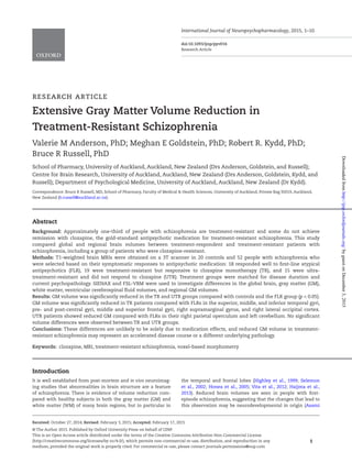
gulate gyrus (Table 3 and Figure 1). A less widespread pattern
of reduced GM was seen in TR patients compared with controls,
with significant clusters observed in the right central operculum
(MNI peak coordinates x = 64, y = -8, z = 10; 136 voxels; p = 0.019)
and right inferior temporal gyrus (MNI peak coordinates x = 56,
y = -36, z = -20; 106 voxels; p = 0.033). In FLR, increased GM volume
was observed compared with control subjects in the right lateral
occipital cortex (MNI peak coordinates x = 42, y = -72, z = -14; 30
voxels; p = 0.037). Reverse contrasts showed no regions where TR
or UTR had significantly more GM volume than controls, or FLR
had significantly less GM volume than controls.
VBM GM Differences Between Schizophrenia
Treatment Groups
Both TR and UTR patient groups showed areas of significantly
less GM compared with the FLR group. Areas showing signifi-
cantly less GM in TR patients compared with FLRs included the
superior, middle, and inferior temporal gyri, pre- and post-cen-
tral gyri, middle and superior frontal gyri, supramarginal gyrus,
and lateral occipital cortex (Table 4, Figure 2). Two significant
clusters were observed when comparing UTR patients with FLRs,
in the right parietal operculum (MNI peak coordinates x = 50,
y = -26, z = 18; 594 voxels; p = 0.008) and the left cerebellum (MNI
Table 2. Normalized Volumes and Significant Group Differences.
Controls (n = 20)
Antipsychotic
responders (n = 18)
Treatment-
resistant (n = 19)
Ultra-treatment-
resistant (n = 15) Mean difference (cm3
) (95% CI, p-value)
Whole brain
volume (cm3
)
1603.0 (58.5) 1560.8 (66.0) 1530.1 (68.9) 1501.7 (55.7) C > FLR 42.2 (8.6–75.8, p = 0.015)
C > TR 72.9 (39.8–106.1, p < 0.001)
C > UTR 101.4 (66.2–136.5, p < 0.001)
FLR > UTR 59.1 (22.9–95.5, p = 0.002)
Gray matter
volume (cm3
)
830.3 (40.8) 817.3 (46.6) 781.5 (38.5) 764.1 (27.5) C > TR 48.8 (30.7–67.0, p < 0.001)
C > UTR 66.1 (46.9–85.4, p < 0.001)
FLR > TR 35.9 (17.3–54.4, p < 0.001)
FLR > UTR 53.2 (33.3–73.1, p < 0.001)
White matter
volume (cm3
)
772.8 (32.7) 743.5 (37.7) 748.7 (42.7) 737.5 (38.1) C > FLR 29.3 (5.0–53.5, p = 0.019)
C > TR 24.1 (0.1–48.1, p = 0.049)
C > UTR 35.2 (9.8–60.7, p = 0.007)
Peripheral cortex
gray matter
volume (cm3
)
676.7 (35.5) 664.9 (40.8) 628.5 (35.1) 620.2 (23.8) C > TR 48.2 (32.7–63.8, p < 0.001)
C > UTR 56.6 (40.1–73.0, p < 0.001)
FLR > TR 36.4 (20.5–52.3, p < 0.001)
FLR > UTR 44.7 (27.7–61.7, p < 0.001)
Ventricular CSF
volume (cm3
)
29.3 (10.2) 30.4 (9.7) 32.0 (12.7) 38.6 (13.0) C < UTR -9.4 (-16.0 to -2.7, p = 0.007)
FLR < UTR -8.3 (-15.2 to -1.4, p = 0.019)
Data are given as mean (SD). Volumes adjusted for age and sex.
C, control; CSF, ; FLR, first-line antipsychotic responder; TR, treatment-resistant; UTR, ultra-treatment-resistant.
byguestonDecember3,2015http://ijnp.oxfordjournals.org/Downloadedfrom
5. Anderson et al. | 5
peak coordinates x = -42, y = -40, z = -44; 145 voxels; p = 0.016).
Direct comparison of the two treatment-resistant patient groups
showed no significant differences. Reverse contrasts showed no
regions where TR or UTR schizophrenia groups had significantly
greater GM volume than the FLR group.
Correlation of GM with Antipsychotic Dosage and
PANSS Scores
There was no association between GM volumes and either
antipsychotic dose or PANSS scores (total, positive, negative, or
general) in patients.
Discussion
This study investigated structural brain differences between
people with schizophrenia whose symptoms responded to first-
line conventional antipsychotic medication and those who are
treatment-resistant (but clozapine-responsive) and ultra-treat-
ment-resistant (clozapine-resistant). Our main finding is that TR
and UTR schizophrenia patients show a widespread GM volume
reduction, including areas of the temporal, parietal, frontal, and
occipital lobes, compared with those people with schizophrenia
who respond to first-line atypical antipsychotic medication. Our
study is unique in several key areas compared with previous MRI
studies investigating treatment response and treatment-resist-
ance: (1) we defined our schizophrenia treatment groups accord-
ing to current guidelines on treatment resistance, and included
a separate group of patients who were treatment-resistant to
clozapine; (2) all patients who were treatment-resistant were
receiving clozapine (or had received clozapine in the past if
ultra-treatment-resistant); (3) schizophrenia treatment groups
were matched for disease duration and psychopathology; and
(4) we used advanced imaging techniques, including 3T MRI and
unbiased VBM analytical methods.
Reduced whole brain, WM, and GM volumes in patients
with schizophrenia relative to control subjects were observed,
although the reduction in GM volume in first-line antipsychotic
responders did not reach statistical significance. VBM corrobo-
rated our findings of GM volume reduction in TR and UTR schiz-
ophrenia treatment groups compared with controls, whereby
a similar pattern of regional GM tissue loss as in other studies
of schizophrenia was seen, focused on the superior and middle
temporal gyri but also including medial frontal areas and the
anterior cingulate cortex (Honea et al., 2005; Meisenzahl et al.,
2008; Palaniyappan et al., 2010; Tanskanen et al., 2010; Colibazzi
et al., 2013; Zierhut et al., 2013; Liu et al., 2014; Ohtani et al.,
2014). Our results are comparable with those of Molina et al.
(2008), who found that TR patients had significantly less GM
in the frontal and occipital regions relative to controls, whilst
Figure 1. Reduced gray matter in ultra-treatment-resistant patients compared with controls, overlaid on the MNI-152 template brain.
Table 3. Local Peaks of Significant Clusters Showing Reduced Gray Matter in the Ultra-Treatment-Resistant Group Compared with the Controls.
Cortical region Side Cluster size
MNI peak
coordinates (mm)
Local maximum p
value*
Superior temporal gyrus Right 3896 48 -22 -6 0.002
Superior temporal gyrus (planum polare) Left 3121 -42 -6 -18 <0.001
Cerebellum Left 815 -32 -60 -48 0.010
Ventromedial prefrontal cortex Bilateral 363 6 32 -16 0.021
Cerebellum Left 217 -32 -50 -24 0.027
Lateral occipital cortex Right 178 12 -58 66 0.018
Anterior cingulate gyrus Bilateral 55 0 42 14 0.039
*With family-wise error correction for multiple comparisons across space. p < 0.05 and >20 voxels.
byguestonDecember3,2015http://ijnp.oxfordjournals.org/Downloadedfrom
6. 6 | International Journal of Neuropsychopharmacology, 2015
there were no significant differences between non-treatment-
resistant patients and controls. The more extensive pattern of
GM loss seen in TR patients in the current study may be due
to the fact that VBM rather than region-of-interest analysis
was used, or that our patient groups were matched for PANSS
scores. The most extensive region of GM loss in UTR patients
was seen in the superior temporal gyrus (STG), a region that has
been implicated in studies of first-episode schizophrenia (Kasai
et al., 2003; Asami et al., 2012), suggesting that this is a relatively
stable deficit but that extensive tissue loss in this area may be
associated with more severe symptomatology and treatment
resistance. A meta-analysis of longitudinal studies of schizo-
phrenia found a significantly higher volumetric decrease over
time in the left and right STG, left Heschl’s gyrus, and left pla-
num temporal (Vita et al., 2012). Similarly, a review investigating
the trajectory of brain structural impairments in schizophrenia
found the STG was already impaired at the onset of symptoms,
which worsened during the acute disease phase followed by sta-
bilization and subsequent age-related progression (Chiapponi
et al., 2013). The STG is critical for auditory processing, and has
therefore been proposed as the region responsible for symptoms
such as auditory hallucinations and thought disorder in schizo-
phrenia (Modinos et al., 2013; Zierhut et al., 2013). We also found
two regions of reduced GM in the cerebellum in the UTR group
compared with controls, a structure that has been implicated
in first-episode and schizophrenia patients (Rasser et al., 2010;
Tanskanen et al., 2010). Recent evidence suggests that the cer-
ebellum plays a role in cognition (Andreasen and Pierson, 2008),
and deficits in a variety of cognitive domains are known symp-
toms of schizophrenia which do not necessarily improve with
the administration of antipsychotics. Cognitive dysfunction
plays a critical role in the pathogenesis and prognosis of schizo-
phrenia, and reduced volume in the cerebellum in TR schizo-
phrenia may reflect a poorer prognosis in these patients.
The group of FLRs showed no significant regions of volume
reduction compared with controls, and actually showed a region
of increased volume in the right lateral occipital cortex.The fact
that we did not find extensive regions of GM tissue loss in this
group of patients, despite a mean disease duration of 10 years,
may reflect responsiveness to medication. Studies showing tis-
sue loss in schizophrenia are likely to have included patients
that exhibited a wide range of responses to their antipsychotic
medication that could have masked minimal volume changes
in some subjects. Antipsychotic drugs might also act to reverse
pathology in the dopaminergic pathway during the early treat-
ment phase of schizophrenia (Wang et al., 2004; Kippin et al.,
2005), and this may be more pronounced in early treatment
responders compared with those who are treatment resistant.
Figure 2. Reduced gray matter in treatment-resistant patients compared with first-line antipsychotic responders, overlaid on the MNI-152 template brain.
Table 4. Local Peaks of Significant Clusters Showing Reduced Gray Matter in the Treatment-Resistant Patients Compared with the First-Line
Antipsychotic Responders.
Cortical region Side
Cluster
size
MNI peak
coordinates (mm)
Local maxi-
mum p value*
Inferior temporal gyrus Right 1685 58 -56 -24 0.001
Post-central gyrus Bilateral 1193 -2 -46 72 0.006
Middle frontal gyrus Left 797 -36 2 48 0.007
Anterior supramarginal gyrus Right 656 66 -24 26 0.010
Superior frontal gyrus Bilateral 625 0 14 62 0.006
Superior temporal gyrus (temporal pole) Right 175 60 10 -10 0.036
Lateral occipital cortex Right 74 14 -70 50 0.028
Supplementary motor cortex Left 35 -12 2 38 0.039
*With family-wise error correction for multiple comparisons across space. p < 0.05 and >20 voxels.
byguestonDecember3,2015http://ijnp.oxfordjournals.org/Downloadedfrom
7. Anderson et al. | 7
Direct comparison showed that TR and UTR groups had
significantly less GM both globally and regionally compared
with the group of FLRs. Regional GM differences were similar
in location to those shown between TR/UTR and control sub-
jects, including the inferior and superior temporal gyri, mid-
dle frontal gyrus, lateral occipital cortex, and cerebellum. This
finding agrees with that of Zugman et al. (2013) who found a
significant reduction in the left dorsolateral prefrontal cortex
in TR schizophrenia in comparison to non-treatment-resistant
schizophrenia.There is some prior evidence to support the view
that GM reduction is associated with poor treatment response
and poor outcomes in schizophrenia. Some studies have found
significantly greater loss of frontal, parietal, and occipital GM
in treatment-resistant patients compared with non-treatment-
resistant patients, and that the best predictor of response to clo-
zapine treatment was temporal and dorsolateral prefrontal GM
volume (Molina et al., 2003, 2008, 2010). Moreover, patients with
poor outcomes (based on Global Assessment of Functioning) had
significantly greater decreases in cortical thickness within the
left middle temporal cortex, superior temporal cortex, Heschl’s
gyrus, and anterior cingulate over a 5-year period (van Haren
et al., 2011). Regions where we found reductions in the TR and
UTR groups compared with the FLRs have also been implicated
in studies investigating the response to antipsychotics in those
with first-episode schizophrenia. Over a 16-week period, non-
responders to olanzapine or risperidone were found to have
significant cortical thinning in occipital and prefrontal regions
compared with responders (Szeszko et al., 2012). Likewise, a
12-week follow-up of first-episode patients showed that non-
responders to antipsychotics showed hypogyria in the insula,
superior frontal, middle frontal, inferior and superior temporal
cortices, and temporal pole (Palaniyappan et al., 2013). Whilst
we do not propose that GM volume loss is directly related to
treatment resistance, and our study cannot determine whether
the differences we observed indirectly contribute to or are a con-
sequence of treatment resistance, these studies in first-episode
patients suggest there may be a neurobiological underpinning
in people with treatment-resistant schizophrenia. One could
also propose that people with treatment-resistant schizophre-
nia may have experienced a greater number of relapses and/
or relapse duration and that these may have had a toxic effect
on the brain (Andreasen et al., 2013; Hyza et al., 2014). However,
it has been shown that brain volume change is not associated
with duration of untreated illness (Boonstra et al., 2011). It is still
undetermined whether there is an independent pathophysi-
ological process taking place in treatment-resistant schizophre-
nia indicating a distinct group of patients, or whether treatment
resistance represents an accelerated form of the same underly-
ing disease process.
We found no evidence that the two treatment-resistant
groups (clozapine-responsive vs clozapine-resistant) differed
from each other. These results are somewhat unexpected, as
ultra-treatment-resistant (clozapine-resistant) schizophre-
nia may be considered a more severe form of schizophrenia
than treatment-resistant (clozapine-responsive) schizophre-
nia. Previous studies investigating the response to clozap-
ine in treatment-resistant patients showed that there was
significantly greater prefrontal sulcal prominence in those
that did not respond to clozapine compared with those who
did (Friedman et al., 1991; Konicki et al., 2001). In addition,
Honer et al. (1995) found that an increased size of the poste-
rior frontal and lateral temporal sulci was related to a poor
response to clozapine. However, these studies used computed
tomography to assess specific brain measurements, rather
than the unbiased whole-brain MRI method of analysis we
employed, which may reveal brain changes more sensitively.
More recently, two MRI studies found that whilst there were
no differences in caudate volume cross-sectionally (Scheepers
et al., 2001a), there was a significant decrease in volume of
the left caudate over 1 year in responders to clozapine rela-
tive to non-responders (Scheepers et al., 2001b). Patients were
taking typical antipsychotics prior to initiation of clozapine,
and therefore it was postulated that clozapine had a “correct-
ing” effect on the increase in caudate volume that has been
observed in patients after treatment with typical antipsy-
chotics. The patients in our study had been taking atypical
antipsychotic treatment for some time, and it is therefore
unlikely that we would have observed this restoring effect on
the caudate in either treatment group. The absence of volume
differences between TR and UTR groups in our investigation
indicates that non-response to clozapine is not related to GM
volume. It must also be considered that if the trajectory of vol-
ume changes in TR and UTR groups are different, this would
not be apparent due to the cross-sectional nature of the study.
However, investigation of a cohort of patients from the same
treatment-resistant study using diffusion tensor imaging
found that the UTR group did not show any significant deficits
in fractional anisotropy compared with the TR group; instead,
significantly higher fractional anisotropy (better WM integrity)
was shown in the right superior longitudinal fasciculus in the
UTR group compared with the TR group (unpublished data).
Although current antipsychotic doses were significantly
higher in the UTR group compared with both the TR and FLR
groups, we found no significant association between regional
GM volume and antipsychotic dose in patients. However it is
possible that both past and current exposure to antipsychotic
medication may have had an effect on brain tissue volume.
A longitudinal study of 211 people with first-episode schizo-
phrenia found that antipsychotic treatment was significantly
related to a decrease in GM and WM volumes after correcting
for the effects of follow-up duration, illness severity, and sub-
stance misuse (Ho et al., 2011), whilst a meta-analysis found
that longitudinal GM volume decreases in patients with schizo-
phrenia were associated with higher cumulative exposure to
antipsychotics over time (Fusar-Poli et al., 2013). Conversely, in
105 patients with schizophrenia no association was observed
between cumulative antipsychotic exposure and brain volume
change over 5 years (Collin et al., 2012). Moreover, in people with
chronic schizophrenia who have had little or no exposure to
antipsychotic medication similar regional brain volume reduc-
tions as those seen in our study have been reported (Liu et al.,
2014), and first-episode patients who are medication-naive
show structural brain changes that suggest the differences we
observed are unlikely to be solely due to medication effects (Lui
et al., 2009; Radua et al., 2012). There was also no significant dif-
ference in GM between our TR and UTR groups, despite it being
likely that UTR schizophrenia patients had higher cumulative
antipsychotic exposure, suggesting that our findings are related
to treatment resistance rather than the effects of antipsychotic
medication exclusively. Studies have also suggested that clo-
zapine may in fact preserve or reverse GM volume and cortical
thickness, which would strengthen our finding that greater GM
loss is in fact related to treatment resistance rather than medi-
cation effects (Molina et al., 2005, 2008; van Haren et al., 2011).
Additionally, no association was observed between regional GM
volume and PANSS scores in our patients, contrary to some pre-
vious studies (Palaniyappan et al., 2010; Zierhut et al., 2013).This
also suggests that the differences in brain morphology observed
byguestonDecember3,2015http://ijnp.oxfordjournals.org/Downloadedfrom
8. 8 | International Journal of Neuropsychopharmacology, 2015
in the current study are related to treatment resistance rather
than psychopathology per se.
Whilst we have already mentioned the unique and advanta-
geous features of this study, which potentially make it a valu-
able addition to the current literature on treatment-resistant
schizophrenia, there are some limitations. This was a cross-
sectional study, so we could not determine the trajectory of
tissue loss between our treatment groups or draw any conclu-
sions regarding causal relationships between brain volume
loss, treatment resistance, and antipsychotic medication. In
addition, as with most studies investigating patients with
established schizophrenia, it is likely that patients differed
in the amount of medication they had used before inclusion
in the study, and we were unable to reliably calculate life-
time chlorpromazine equivalents based on patients’ medical
charts. Similarly, it was difficult to assess medication adher-
ence in our population, which had a mean disease duration
of 10 years. Whilst we attempted to minimize these effects by
only including clinically stable patients taking atypical antip-
sychotics at the time of investigation (and therefore not receiv-
ing large doses of medication) it was not possible to assess
whether GM volumes were related to the long-term effects of
antipsychotic medication. Due to the unique way that we dif-
ferentiated our patients with schizophrenia, we had relatively
small sample sizes within each group. However, this was a pre-
liminary study of treatment resistance and our results indicate
that larger studies should be performed. Although we had to
use a 20-channel head coil rather than the 32-channel head
coil for five of our participants, these subjects were distrib-
uted across the three patient groups and analyses run whilst
excluding these participants showed no changes in our main
findings. A recent study has also shown VBM results were not
significantly affected when MPRAGE images were acquired
using a 12-channel versus a 32-channel head-coil (Streitburger
et al., 2014). Finally, our patients with schizophrenia showed
some differences in recreational drug history compared with
control subjects and six of our participants tested positive for
tetrahydrocannabinol (three FLR, one TR, and two UTR) at the
time of assessment, which could have affected our results.
However, these six subjects were distributed across the three
patient groups and analyses were run again excluding these
participants with no major changes in our findings. Moreover,
there were no significant differences in ASSIST scores between
patient groups, suggesting that our main finding of differences
in GM volume between treatment-resistant and non-treat-
ment-resistant patients remains valid.
In conclusion, this study has shown that there is decreased
GM volume in people with treatment-resistant and ultra-treat-
ment-resistant schizophrenia relative to those who respond to
first-line atypical antipsychotic medication. The discovery of
factors associated with treatment resistance will aid in under-
standing the underlying causes of treatment resistance and
may assist in identifying and treating people with treatment-
resistant schizophrenia more effectively; regional GM volume
loss may be one such factor. Large-scale prospective longitudi-
nal studies following people from the first episode to treatment
response or resistance are required to confirm our findings and
the true relationship between brain tissue loss, medication, and
treatment resistance.
Acknowledgments
This work was supported by The University of Auckland Faculty
Research and Development Fund, with support from the New
Zealand Schizophrenia Research Group and the Oakley Mental
Health Research Foundation.
Dr Anderson was supported by an Edith C. Coan Research
Fellowship from The Auckland Medical Research Foundation. Dr
Goldstein was supported by a University of Auckland Doctoral
Scholarship and a New Zealand Federation of Graduate Women
Fellowship. We would like to thank all the participants who
took part in this study and their support persons and staff at
The Mason Clinic, The Cottage Community Health Centre,
and Te Rawhiti Community Mental Health Centre. We would
also like to acknowledge the Centre for Advanced MRI at the
University of Auckland for performing the MRI scanning, and Dr
Frederick Sundram for providing suggestions on analyses and
the manuscript.
Statement of Interest
None
References
American Psychiatric Association (2000) Diagnostic and statisti-
cal manual of mental disorders. 4th ed., text rev. Washington,
DC: American Psychiatric Association.
Lehman AF, Lieberman JA, Dixon LB, McGlashan TH, Miller AL,
Perkins DO, Kreyenbuhl J, American Psychiatric Association,
Steering Committee on Practice Guidelines (2004) Practice
guideline for the treatment of patients with schizophrenia,
second edition. Am J Psych 161:1–56.
Andreasen NC, Pierson R (2008) The role of the cerebellum in
schizophrenia. Biol Psychiatry 64:81–88.
Andreasen NC, Pressler M, Nopoulos P, Miller D, Ho BC (2010)
Antipsychotic dose equivalents and dose-years: a standard-
ized method for comparing exposure to different drugs. Biol
Psychiatry 67:255–262.
Andreasen NC, Nopoulos P, Magnotta V, Pierson R, Ziebell S, Ho
BC (2011) Progressive brain change in schizophrenia: a pro-
spective longitudinal study of first-episode schizophrenia.
Biol Psychiatry 70:672–679.
Andreasen NC, Liu D, Ziebell S, Vora A, Ho BC (2013) Relapse dura-
tion,treatment intensity,and brain tissue loss in schizophrenia:
a prospective longitudinal MRI study. Am J Psych 170:609–615.
Asami T, Bouix S, Whitford TJ, Shenton ME, Salisbury DF, McCa-
rley RW (2012) Longitudinal loss of gray matter volume in
patients with first-episode schizophrenia: DARTEL auto-
mated analysis and ROI validation. Neuroimage 59:986–996.
Bodnar M, Malla AK, Czechowska Y, Benoit A, Fathalli F, Joober R,
Pruessner M, Pruessner J, Lepage M (2010) Neural markers of
remission in first-episode schizophrenia: a volumetric neuro-
imaging study of the hippocampus and amygdala. Schizophr
Res 122:72–80.
Boonstra G, Cahn W, Schnack HG, Hulshoff Pol HE, Minderhoud
TC, Kahn RS, van Haren NE (2011) Duration of untreated ill-
ness in schizophrenia is not associated with 5-year brain vol-
ume change. Schizophr Res 132:84–90.
Chiapponi C, Piras F, Fagioli S, Piras F, Caltagirone C, Spalletta G
(2013) Age-related brain trajectories in schizophrenia: a system-
atic review of structural MRI studies. Psychiatry Res 214:83–93.
Colibazzi T, Wexler BE, Bansal R, Hao X, Liu J, Sanchez-Pena J,
Corcoran C, Lieberman JA, Peterson BS (2013) Anatomical
abnormalities in gray and white matter of the cortical surface
in persons with schizophrenia. PLOS ONE 8:e55783.
Collin G, Derks EM, van Haren NE, Schnack HG, Hulshoff Pol HE,
Kahn RS, Cahn W (2012) Symptom dimensions are associated
byguestonDecember3,2015http://ijnp.oxfordjournals.org/Downloadedfrom
9. Anderson et al. | 9
with progressive brain volume changes in schizophrenia.
Schizophr Res 138:171–176.
Desikan RS, Segonne F, Fischl B, Quinn BT, Dickerson BC, Blacker
D, Buckner RL, Dale AM, Maguire RP, Hyman BT, Albert MS,
Killiany RJ (2006) An automated labeling system for subdivid-
ing the human cerebral cortex on MRI scans into gyral based
regions of interest. Neuroimage 31:968–980.
Douaud G, Smith S, Jenkinson M, Behrens T, Johansen-Berg H,
Vickers J, James S, Voets N, Watkins K, Matthews PM, James A
(2007) Anatomically related grey and white matter abnormal-
ities in adolescent-onset schizophrenia. Brain 130:2375–2386.
Farooq S, Agid O, Foussias G, Remington G (2013) Using treat-
ment response to subtype schizophrenia: proposal for a new
paradigm in classification. Schizophr Bull 39:1169–1172.
Friedman L, Knutson L, Shurell M, Meltzer HY (1991) Prefrontal
sulcal prominence is inversely related to response to clozap-
ine in schizophrenia. Biol Psychiatry 29:865–877.
Fusar-Poli P, Smieskova R, Kempton MJ, Ho BC, Andreasen NC,
Borgwardt S (2013) Progressive brain changes in schizophrenia
related to antipsychotic treatment? A meta-analysis of longi-
tudinal MRI studies. Neurosci Biobehav Rev 37:1680–1691.
Gardner DM, Murphy AL, O’Donnell H, Centorrino F, Baldessarini
RJ (2010) International consensus study of antipsychotic dos-
ing. Am J Psych 167:686–693.
Haijma SV, Van Haren N, Cahn W, Koolschijn PC, Hulshoff Pol
HE, Kahn RS (2013) Brain volumes in schizophrenia: a meta-
analysis in over 18 000 subjects. Schizophr Bull 39:1129–
1138.
Highley JR, McDonald B, Walker MA, Esiri MM, Crow TJ (1999)
Schizophrenia and temporal lobe asymmetry. A post-mor-
tem stereological study of tissue volume. Br J Psychiatry
175:127–134.
Ho BC, Andreasen NC, Ziebell S, Pierson R, Magnotta V (2011)
Long-term antipsychotic treatment and brain volumes: a
longitudinal study of first-episode schizophrenia. Arch Gen
Psychiatry 68:128–137.
Honea R, Crow TJ, Passingham D, Mackay CE (2005) Regional
deficits in brain volume in schizophrenia: a meta-analysis of
voxel-based morphometry studies.Am J Psych 162:2233–2245.
Honer WG, Smith GN, Lapointe JS, MacEwan GW, Kopala L,
Altman S (1995) Regional cortical anatomy and clozapine
response in refractory schizophrenia. Neuropsychopharma-
cology 13:85–87.
Hyza M, Huttlova J, Kerkovsky M, Kasparek T (2014) Psychosis
effect on hippocampal reduction in schizophrenia. Prog Neu-
ropsychopharmacol Biol Psychiatry 48:186–192.
Kasai K, Shenton ME, Salisbury DF, Hirayasu Y, Lee CU, Ciszewski
AA,Yurgelun-Todd D, Kikinis R, Jolesz FA, McCarley RW (2003)
Progressive decrease of left superior temporal gyrus gray
matter volume in patients with first-episode schizophrenia.
Am J Psych 160:156–164.
Kay SR, Fiszbein A, Opler LA (1987) The positive and negative
syndrome scale (PANSS) for schizophrenia. Schizophr Bull
13:261–276.
Kippin TE, Kapur S, van der Kooy D (2005) Dopamine specifically
inhibits forebrain neural stem cell proliferation, suggesting a
novel effect of antipsychotic drugs. J Neurosci 25:5815–5823.
Konicki PE, Kwon KY, Steele V, White J, Fuller M, Jurjus GJ, Jaskiw
GE (2001) Prefrontal cortical sulcal widening associated with
poor treatment response to clozapine. Schizophr Res 48:173–
176.
Liu X, Lai Y, Wang X, Hao C, Chen L, Zhou Z, Yu X, Hong N (2014)
A combined DTI and structural MRI study in medicated-naive
chronic schizophrenia. Magn Reson Imaging 32:1–8.
Lui S, Deng W, Huang X, Jiang L, Ma X, Chen H, Zhang T, Li X, Li
D, Zou L, Tang H, Zhou XJ, Mechelli A, Collier DA, Sweeney JA,
Li T, Gong Q (2009) Association of cerebral deficits with clini-
cal symptoms in antipsychotic-naive first-episode schizo-
phrenia: an optimized voxel-based morphometry and resting
state functional connectivity study. Am J Psych 166:196–205.
Meisenzahl EM, Koutsouleris N, Bottlender R, Scheuerecker J,
Jager M, Teipel SJ, Holzinger S, Frodl T, Preuss U, Schmitt G,
Burgermeister B, Reiser M, Born C, Moller HJ (2008) Structural
brain alterations at different stages of schizophrenia: a voxel-
based morphometric study. Schizophr Res 104:44–60.
Modinos G, Costafreda SG, van Tol MJ, McGuire PK, Aleman A,
Allen P (2013) Neuroanatomy of auditory verbal hallucina-
tions in schizophrenia: a quantitative meta-analysis of voxel-
based morphometry studies. Cortex 49:1046–1055.
Molina V, Reig S, Sarramea F, Sanz J, Francisco Artaloytia J, Luque
R, Aragues M, Pascau J, Benito C, Palomo T, Desco M (2003)
Anatomical and functional brain variables associated with
clozapine response in treatment-resistant schizophrenia.
Psychiatry Res 124:153–161.
Molina V, Reig S, Sanz J, Palomo T, Benito C, Sanchez J, Sar-
ramea F, Pascau J, Desco M (2005) Increase in gray matter and
decrease in white matter volumes in the cortex during treat-
ment with atypical neuroleptics in schizophrenia. Schizophr
Res 80:61–71.
Molina V, Reig S, Sanz J, Palomo T, Benito C, Sarramea F, Pascau
J, Sanchez J, Martin-Loeches M, Munoz F, Desco M (2008) Dif-
ferential clinical, structural and P300 parameters in schizo-
phrenia patients resistant to conventional neuroleptics. Prog
Neuropsychopharmacol Biol Psychiatry 32:257–266.
Molina V, Hernandez JA, Sanz J, Paniagua JC, Hernandez AI,
Martin C, Matias J, Calama J, Bote B (2010) Subcortical and
cortical gray matter differences between Kraepelinian and
non-Kraepelinian schizophrenia patients identified using
voxel-based morphometry. Psychiatry Res 184:16–22.
Mouaffak F, Tranulis C, Gourevitch R, Poirier MF, Douki S, Olie JP,
Loo H, Gourion D (2006) Augmentation strategies of clozapine
with antipsychotics in the treatment of ultraresistant schizo-
phrenia. Clin Neuropharmacol 29:28–33.
Ohtani T, Levitt JJ, Nestor PG, Kawashima T, Asami T, Shenton ME,
Niznikiewicz M, McCarley RW (2014) Prefrontal cortex volume
deficit in schizophrenia: A new look using 3T MRI with man-
ual parcellation. Schizophr Res 152:184–190.
Olabi B, Ellison-Wright I, McIntosh AM, Wood SJ, Bullmore E,
Lawrie SM (2011) Are there progressive brain changes in
schizophrenia? A meta-analysis of structural magnetic reso-
nance imaging studies. Biol Psychiatry 70:88–96.
Palaniyappan L, Mallikarjun P, Joseph V, White TP, Liddle PF
(2011) Reality distortion is related to the structure of the sali-
ence network in schizophrenia. Psychol Med 41:1701–1708.
Palaniyappan L, Marques TR, Taylor H, Handley R, Mondelli V,
Bonaccorso S, Giordano A, McQueen G, DiForti M, Simmons
A, David AS, Pariante CM, Murray RM, Dazzan P (2013) Corti-
cal folding defects as markers of poor treatment response in
first-episode psychosis. JAMA Psychiatry 70:1031–1040.
Radua J, Borgwardt S, Crescini A, Mataix-Cols D, Meyer-Linden-
berg A, McGuire PK, Fusar-Poli P (2012) Multimodal meta-
analysis of structural and functional brain changes in first
episode psychosis and the effects of antipsychotic medica-
tion. Neurosci Biobehav Rev 36:2325–2333.
Rais M, Cahn W, Schnack HG, Hulshoff Pol HE, Kahn RS, van
Haren NE (2012) Brain volume reductions in medication-
naive patients with schizophrenia in relation to intelligence
quotient. Psychol Med 42:1847–1856.
byguestonDecember3,2015http://ijnp.oxfordjournals.org/Downloadedfrom
10. 10 | International Journal of Neuropsychopharmacology, 2015
Rasser PE, Schall U, Peck G, Cohen M, Johnston P, Khoo K, Carr VJ,
Ward PB, Thompson PM (2010) Cerebellar grey matter deficits
in first-episode schizophrenia mapped using cortical pattern
matching. Neuroimage 53:1175–1180.
Scheepers FE, de Wied CC, Hulshoff Pol HE, van de Flier W, van
der Linden JA, Kahn RS (2001a) The effect of clozapine on
caudate nucleus volume in schizophrenic patients previously
treated with typical antipsychotics. Neuropsychopharmacol-
ogy 24:47–54.
Scheepers FE, Gispen de Wied CC, Hulshoff Pol HE, Kahn RS
(2001b) Effect of clozapine on caudate nucleus volume in rela-
tion to symptoms of schizophrenia. Am J Psych 158:644–646.
Selemon LD, Kleinman JE, Herman MM, Goldman-Rakic PS (2002)
Smaller frontal gray matter volume in postmortem schizo-
phrenic brains. Am J Psych 159:1983–1991.
Smith SM, Zhang Y, Jenkinson M, Chen J, Matthews PM, Federico
A, De Stefano N (2002) Accurate, robust, and automated longi-
tudinal and cross-sectional brain change analysis. Neuroim-
age 17:479–489.
Streitburger DP, Pampel A, Krueger G, Lepsien J, Schroeter ML,
Mueller K, Moller HE (2014) Impact of image acquisition on
voxel-based-morphometry investigations of age-related
structural brain changes. Neuroimage 87:170–182.
Szeszko PR, Narr KL, Phillips OR, McCormack J, Sevy S, Gunduz-
Bruce H, Kane JM, Bilder RM, Robinson DG (2012) Magnetic
resonance imaging predictors of treatment response in first-
episode schizophrenia. Schizophr Bull 38:569–578.
Tanskanen P, Ridler K, Murray GK, Haapea M, Veijola JM, Jaaskel-
ainen E, Miettunen J, Jones PB, Bullmore ET, Isohanni MK
(2010) Morphometric brain abnormalities in schizophrenia
in a population-based sample: relationship to duration of ill-
ness. Schizophr Bull 36:766–777.
van Haren NE, Schnack HG, Cahn W, van den Heuvel MP, Lep-
age C, Collins L, Evans AC, Hulshoff Pol HE, Kahn RS (2011)
Changes in cortical thickness during the course of illness in
schizophrenia. Arch Gen Psychiatry 68:871–880.
Vita A, De Peri L, Deste G, Sacchetti E (2012) Progressive loss of
cortical gray matter in schizophrenia: a meta-analysis and
meta-regression of longitudinal MRI studies. Transl Psychia-
try 2:e190.
Wang HD, Dunnavant FD, Jarman T, Deutch AY (2004) Effects of
antipsychotic drugs on neurogenesis in the forebrain of the
adult rat. Neuropsychopharmacology 29:1230–1238.
WHO ASSIST Working Group (2002) The alcohol, smoking and
substance involvement screening test (ASSIST): develop-
ment, reliability and feasibility. Addiction 97:1183–1194.
Zierhut KC, Schulte-Kemna A, Kaufmann J, Steiner J, Bogerts B,
Schiltz K (2013) Distinct structural alterations independently
contributing to working memory deficits and symptomatol-
ogy in paranoid schizophrenia. Cortex 49:1063–1072.
Zugman A, Gadelha A, Assuncao I, Sato J, Ota VK, Rocha DL, Mari
JJ, Belangero SI, Bressan RA, Brietzke E, Jackowski AP (2013)
Reduced dorso-lateral prefrontal cortex in treatment resist-
ant schizophrenia. Schizophr Res 148:81–86.
byguestonDecember3,2015http://ijnp.oxfordjournals.org/Downloadedfrom