1. MARK BOTIRIUS
Page 1 of 8
Describe the steps in CTAB DNA extraction and explain what is
happening in each step
1. Establishthe bigpicture.
The firststepwhenperformingaCTAB DNA extractionistoconsiderthose factorsthat may
affecthowyouproceedwiththe extraction. These factorsinclude suchthingsaswhattype of tissue
isrepresentedinthe sample,the size of the organism’sgenome,how freshthe sample is,how much
tissue there istoworkwith,and evensuchthingsashow manycentrifuge tubeswill be neededand
labeling. Forexample,isthe sample fromaplant,animal,orfungus? Each of these organismshave
differentcell wall characteristicsthatmayalterhow those stepsdesignedtorelease the DNA into
the extractionbufferare executed. Plantsandfungi have cell wallscomposedof cellulose andchitin
respectively,inadditiontomembranesthatmustbe brokendownto allow accesstothe DNA,
whereasmostanimalsonlyhave membranes. Consequently,if the sampleisfroma plant,itwould
needtobe groundup,while amuscle sample mayonlyneedtohave itscell membranesdisrupted
by a detergent,suchasCTAB. Anotherexampleiswhetherornotthe sample isfreshor frozen,and
howoldthe sample is. Freshsamples,of course,containthe highestqualityof DNA,whereasolder
DNA is subjecttodegradationthatvarieswithage andthe conditionsinwhichthe DNA was
preserved. Applyingthissteptothe extractionwe didinclassrevealsthatthe organismwasa plant
(Vicia faba) andthe sample wasfresh. Also,we hadplentyof material toworkwith. Withthese
factors inmind,itwas determinedthatwe hadto freeze the tissue solidindryice andthengrindit
ina coffee grinder. Why? Because of the tissue type factor. Planttissue meansthe cellshave cell
wallsmade of cellulose,freezingmakesthembrittle (andhelpspreserve the DNA),andgrinding
breaksthemapart. If we hadbeenworkingwithanimal cells,forexample,we mayhave beenable
to skipthese steps,andwentstraighttothe detergentstepsince itispossiblethatthe onlything
neededistodisruptthe membranes. Havingestablishedthat Vicia faba ismymodel organismfor
thisdiscussionandthatall of myprerequisitepreparationshave beencompleted(suchaslabelingall
tubesandsuch),it istime for the nextstep. (Rogers,2016)
2. Gain accessto the DNA
Great. I have plentyof Vicia faba tissue,butitisall sequesteredbehindprotective cellwalls
and membranes. Ineedtogainaccess to thatDNA (Ican’t extractsomethingthatI have noaccess
to) while atthe same time,protectingitfromdamage anddegradation. How thisisdone,asI have
alreadypointedout,canvary dependingonseveral circumstancesthatIhave alreadydescribed. In
thiscase,I have planttissue,sofor thisstepthe tissue isfrozeninliquidnitrogen(ordryice,aswas
done inlab),andeverythingthatcomesintocontactwiththe tissue isfrozenaswell. AsI have
alreadymentioned,thismakesthe cell wallsbrittle andhelpspreservethe DNA. The frozentissue is
thengroundup (eithermanuallyinamortar and pestle,orbyusinga small coffee grinder) tobreak
the alreadybrittle wallsandmembranesapartandexpose the DNA. Of course,the dry ice isallowed
to sublime before goingfurther. (Rogers,2016)
3. Breakapart the membranes,protectthe DNA andkeepitinsolution.
Okay. I have succeededingettingpastthe cell walls. However,muchof the DNA may still
be protectedbehind nuclearandcellularmembranes. Therefore,Imusttake stepstobreakapart
2. MARK BOTIRIUS
Page 2 of 8
those membranesandthenprotectthe DNA withoutdelay. Toaccomplishthis,immediatelyafter
grindingthe DNA indry ice and allowingthe ice tosublime,anequal volume of hot (65℃) 2X CTAB
bufferisaddedtothe tissue. Cell membranesconsistof aphospholipidbilayer. Because CTABisa
detergent,itsolubilizesthe phospholipidsandcausesthe membranestodisintegrate,exposingthe
DNA. Unfortunately,the DNA isnowexposedtoanythingthatcandamage or degrade it,such as
nucleases. Fortunately,CTABbufferalsocontainsEDTA,whichchelatesthe cofactorsneededby
these nucleases(Mg2+
) andtherebystoppingthemfromdegradingthe DNA. Trisisalsopresentto
maintaina stable pH(around8.0 to furtherprotectthe DNA from degradation). Lastly,itis
necessarytokeepthe DNA insolution. If itwere to precipitate outatthistime,itwouldsimplybe a
part of all of the other junksolidspresentinthe sample andit wouldbe impossible toisolate.
Therefore,CTABbufferalsocontains1.4MNaCl. This createsa sodiumsaltof DNA whichkeepsitin
solution. Inorderto ensure thatthe CTAB bufferhassufficienttime tosolubilize the membranesthe
solutionisplaced inawater bath(65℃) forone to five minutes. (Rogers,2016)
4. Extract proteins
At thisstepinthe procedure,Ihave an aqueoussolutionthatalsohasa lot of junksolidsthat
were neverinsolution(suchasplantfibers) andsome moleculesthathave precipitatedoutby
interactingwithCTAB(CTAB,beingacationicmolecule,willcomplex withsome proteinsand
polysaccharidesthathave anegative charge somehow associatedwiththe molecule,takingthem
out of solution). Regrettably,however,cellsnaturallycontainawhole hostof watersoluble proteins
because,let’sface it,muchof the cell isan aqueousenvironment. Whichmeansthat,althoughI
have a lot of unwantedstuff thatisnot insolutionandtherefore canbe removed,Istill have alotof
unwantedproteinsinsolutionalongwithmyDNA,andIneedtoseparate those proteinsfrommy
DNA. In otherwords,I needtoextractthe proteins,andleave the DNA saltinsolution. Buthow can
thisbe done? I do thisby addinganotherphase tomy solution.
So far,my solutionhasconsistedof onlyone phase,anaqueousphase,representedbythe
CTAB buffersolution. Mostsaltsare happyinaqueousphases,andrightnow,myDNA existsasa
sodiumsaltof DNA. To extractthe aqueousproteins,Ineedtoaddanotherphase,anorganic phase.
Aqueousphasesare polar(waterisa polarmolecule),andmostorganicphasesare non-polar(orless
polarthan water). Thisdifference inpolarityisthe basisbehindseparatingtwo differentsubstances
ina solution,orextractingone substance fromanother. Since the twosolventshave different
polarities,theyalsohave differentsolubility,andwill separateinsolution.Forexample,suppose I
have a solutionof benzoicacid,m-nitroaniline,andnaphthaleneinanon-polarorganicphase such
as ether. Ether,isnon-polar. To separate outthe benzoicacid,Iadd a NaOH aqueousphase. The
polar,aqueousphase andthe non-polarorganicphase formtwolayers. Whentheyare forcedto
mix,however,the benzoicacidinthe organicphase (ether) reactswiththe NaOHinthe aqueous
phase,formingasalt (benzoate-
+Na+
) whichishighlysoluble inthe aqueousphase(notunlike our
DNA salt) and insolubleinthe organicphase. Thiscausesthe benzoicacidtotransferto the aqueous
phase as benzoate (the conjugate base of benzoicacid) fromthe organic(ether) phase. Toget the
benzoicacidback,simplysiphonoff the aqueousphase (remember,the twophasesdon’tmix,but
formtwo layers) andaddHCl to convertthe benzoate backto benzoicacid. Benzoicacidhasjust
beenextractedfromthe ether. The ethersolutionnow primarilycontainsonlym-nitroanilineand
naphthalene. (Chung,2015)
The principlesbehindextractingthe proteinsfromourCTAB solutionare verysimilar.
Insteadof extractingoutof an organic phase intoan aqueousphase,however,we are extractingout
3. MARK BOTIRIUS
Page 3 of 8
of an aqueousphase (CTAB) intoanorganic
one (chloroform). Inaddition,inorderto
getbenzoicacidto change its solubility
frometherto waterwe usedNaOH to
change it to benzoate (itsconjugate base).
Similarly,the proteinsinourCTABsolution
alsochange. However,thistime acid-base
chemistryisn’tused,the propertythat
proteinscanchange conformationiswhatis
used. Whenthe chloroform(organicphase)
ismixedwiththe CTAB(aqueousphase) to
forman emulsion,the chloroformcauses
the proteinstodenature thusadoptinga
radicallydifferentconformation. They
don’tjustadopt anyoldconformation,
however, they adopta non-polarone,
thereby changing theirsolubility frompolar
(aqueous/CTAB) to a non-polar
(organic/chloroform)causing themto move
fromthe CTABto the chloroformsolution. (Oswald,2016) Now thatthe proteinsare inthe
chloroform,the aqueous/CTABphase containsmostlyjustthe DNA salt. The CTAB/chloroform
solutioniscentrifugedto separate the twophases(chloroformisalittle heavier) andthe phase
containingthe DNA (aqueous,ortop) issiphonedoff.Of course,nochemical reactionis100%,and
so to maximize the removal of proteins,the chloroformextractionisperformedtwice. (Rogers,
2016)
5. Precipitate the DNA
Awesome,nowIhave removedall of the plantdebris,andalsoremovedmostof the soluble
proteins. Myaqueoussolution,however, mostlikelystill containsimpurities. Forexample,although
chloroformismuchlesspolarthan water,itnonethelesshasadefinite dipolemomentthatis
apparentwhenlookingatthe molecularstructure. Chlorine ismore
electronegativethancarbon,and therefore the electronsspendmore
time at the chlorine atomsthantheydo at the hydrogenatom. This
allowsthe moleculetoparticipate insome hydrogenbonding,although
not nearlyasmuch as water. Which meansthatchloroformisslightly
soluble inwater,andnomatterhow muchthe solutioniscentrifuged
to remove it,there isprobablysome dissolvedinsolutionregardless. A
goodway to getrid of the impuritiesstillleftinsolution,isto
precipitate the DNA outof solution,anddiscardthe solutionalongwith
itsimpurities,leavingonlythe DNA behind. Todothis,CTAB
precipitationbufferisaddedtothe aqueousphase. Thisbufferisthe same as the CTAB buffer,only
the CTAB precipitationbufferhasnoNaCl. As a result,the DNA formsa saltwiththe CTAB molecule
itself insteadof the Na+
. Recall thatCTAB iscationic,and therefore inthe absence of the sodium
cation,it will formasalt withthe DNA molecule aswell.However,the solubilityof ionicsubstances
(suchas salts) exhibitgreatvariability. Forexample,36grams of NaCl can be dissolvedin100 mL of
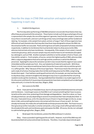
Figure 1. Polar proteins that reside inthe aqueous portion of
the cytoplasm denature to become non-polar. Thiscauses
them to transfer from an aqueous phase to an organic phase,
where theycanbe removedfromsolution. This figure is from
BitesizeBio.com
Figure 2. Chloroform, the
picture is fromWikipedia
4. MARK BOTIRIUS
Page 4 of 8
water,while only0.002 grams of calciumphosphate canbe dissolved. Tosee whythe CTABsalt of
DNA is muchlesssoluble thanthe sodiumsalt,one needonlylookatthe sodium andCTAB ions.
Obviously,the CTABcationhasa relativelyhuge nonpolar“tail”16 carbons long. There isno doubt,
thiswouldaffectthe solubilityof the saltitformswithDNA. Afterthe precipitationbufferisadded
and the CTAB saltof DNA precipitatesout,DNA ispelletedinacentrifuge andthe supernatantis
discarded(alongwithanyremainingimpurities,suchasproteins,orevenchloroform). (Rogers,
2016)
6 Convertback to a sodiumsaltof DNA
It lookslike Ishouldbe done. Ihave a relativelypure pelletof DNA. There isonlyone
problem,however. CTABis,afterall,acationic detergent,andassuch, isn’tacceptable inalotof
biological procedures. Therefore,inthisform, the DNA isprettymuchuseless. Tobe useful,it
needstobe convertedbackto a sodiumsaltof DNA. Thisis nota problem;the DNA pelletissimply
dissolvedinahighsodiumsaltbuffercalled“highsaltTE”. Thisbuffernotonlycontains1M of salt,it
alsohas Tris (tomaintainpH),andEDTA (to protectthe DNA fromnucleases). Inthe highsodium
solution,the CTABionisreplacedwithsodium, andthe DNA goesbackinsolution. (Rogers,2016)
7. Re-pelletthe sodiumsaltof DNA
Once again,we have our sodiumsaltof DNA,howeveritisinsolution. We cannot
add CTAB to precipitate ourDNA,sowe change our solventinstead. Lasttime we usedCTABto
make our molecule more non-polarinahighlypolarsolvent. Thistime,we change oursolventtobe
lesspolarwhenourmolecule ishighlypolar. This,alongwithlowertemperature,givesusthe same
result. OurDNA precipitatesoutof solutionandcanbe pelleted. The lesspolarsolventusedis
100% coldethanol. The DNA pelletiscentrifuged,andagainthe supernatantisdiscarded. Finally,
our DNA pelletis“washed”incold80% ethanol andcentrifuged. Finally,it isrehydratedin0.1XTE
whenitis to be usedinmolecularbiologymethods. (Rogers,2016)
Define and explain protein trafficking, as it exists in eukaryotic cells.
Be sure to address the movement of all of the cell compartments that
are the destinations of various proteins.
Proteinsserve awide varietyof functions. Theyserve enzymatic,structural,movement,
defense,storage,andregulatoryfunctionsjusttoname a few. Eventhoughtheyserve many
differentfunctions throughoutthe organism, theyare all synthesizedbythe ribosome(non-
ribosomal peptidesnotwithstanding). Because theirfunctionsare sovaried,the locationswhere
theyserve these functionsare equallyvaried,andsothere mustbe some wayto direct a particular
Figure 3. The difference between a sodium
ion andCTAB ionclearlyillustrate whythe
CTAB salt is less soluble.VS Na+
5. MARK BOTIRIUS
Page 5 of 8
proteinfromitssite of synthesis(the ribosome) toitssite of function. Forexample,amembrane
proteinmustbe directedfromthe ribosome tothe membrane,while anenzymaticprotein
synthesizedatthe ribosome mustbe directedto,forthe sake of example,the cytoplasm(Iamnot
sayinghere,thatall enzymeswindupinthe cytoplasm). Therefore,proteintraffickingreferstothe
transportof proteinsfromwhere theyare synthesizedtoa particulardestinationwhere theyare
needed. This canbe an extremelycomplexquestionbecause,forexample,inhumansthere are well
over100,000 proteinseachwithitsownfunctionanddestination. Luckily,however,almostall of
those proteinscanbe placedinone of twocategories;those proteinsthatmove fromthe ribosome
to the lumenof the endoplasmicreticulum,andthose thatmove fromthe ribosome tothe cytosol.
Proteinsthattravel throughthe endoplasmicreticulumeitherstayinthe endomembrane system,
become partof the membranesthemselves,orare secretedfromthe cell. Onthe otherhand,those
proteinsthattravel intothe cytosol usuallyremaininthe cell andeitherstayinthe cytosol or they
travel to variouscellulardestinationssuchasthe organelles,orthe cytoskeletontoname a few.
(Rogers,2016)
Proteins that travel to the lumen of the endoplasmic reticulum
Althoughitistrue that the rough endoplasmic
reticulumisa place where the ribosomesare attachedtothe
cytosolicsurface of the endoplasmicreticulum,itshouldbe
notedthat these ribosomesare notpermanentlyattached.
In theory,all ribosomesare actuallyfree. The attachmentof
the ribosome isactuallypartof proteintrafficking. Itis
where proteintraffickingbeginsforthose proteinsthatmust
travel intothe lumenof the endoplasmicreticulum. In
proteinsynthesis,aminoacidsare addedtothe carboxyl end
of the growingpeptide chain,whichmeansthatthe amino
endof the proteinfirstemergesfromthe ribosome. Those
aminoacidsthat are destinedforthe ERlumencontaina
signal sequence closetothe aminoendthatis recognizedby
a signal recognitionproteininthe ERmembrane thatbinds
the sequence therebystoppingtranslation. Whenthe SRP
comesintocontact withan SRP receptor,itbindstothe
receptorandin so doingattachesthe ribosome tothe ER
cytosolicmembrane. SRPreceptorsare locatednextto
transmembrane proteinscalledtranslocons,andthe bound
ribosome,now attachedtothe ER, continuestranslationas
the growingpeptide isfedintothe transloconandthrough
the ER membrane intothe lumen. Forthisreason,this
processiscalledco-translationaltranslocation. If,onthe
otherhand,the final destinationof the proteinhappensto
be inside the ER membrane itself,itwill containa
hydrophobicsequence thatcausesthe translocontoeject
the growingpeptide laterallyandintothe ERmembrane
where itbecomesanintegral membrane protein. (Iwasa,
2016)
Figure 5. Whenthetranslocon (theblue
transmembraneprotein) encounters a
hydrophobic domainofthegrowing peptide,it
expels thenascent protein to the sideandinto the
ER membrane. This figureis from
www.zoology.ubc.ca
Figure 4. Those proteins that are
traffickedintothe ER lumen travel
through the Golgi complex andhave one
of three fates. Theyeither become an
integralmember protein, stayin the ER
lumen, or are packaged intoa secretory
vesicle and leave the cell. This figure is
from the classlecture.
6. MARK BOTIRIUS
Page 6 of 8
The traffickingof nascentproteinsthatwindupwithinthe ERlumenisdone byvesicular
transport.The destinationof these proteinsisdeterminedbyvarioussequences. Forexample,those
proteinsdestinedtostaywithinthe ERlumencontaina shortretrieval sequence of lys-asp-glu-leu
(KDEL incanonical nomenclature)thatisrecognizedbyareceptorina COPIcoatedvesicle that
ensuresthe proteinwillstayinthe ER where itbelongs. Those proteinswithoutKDELsequences
continue onto the Golgi complex inCOPIIcoatedvesicles. Inthe Golgi complex,proteinsare
identifiedbycertainsortingsignalsandprocessedfortraffickingaccordingly. Forexample,those
proteinsdestinedforthe lysozymecontainphosphorylatedmannose residuesthatare recognizedby
receptorsinmembranesthatformclathryncoatedvesiclesthatbudoff fromthe Golgi complex and
travel to the lysosome. Mostproteinswithoutthese signalresiduesendupinasecretoryvesicle or
theyhave signal sequencesintheircytoplasmic membranedomainsthatdestine themtobecome
integral cellularmembrane proteins. (Iwasa,2016)
That prettymuch sumsup the traffickingassociatedwiththose proteinsthattravel intothe
lumenof the ER. However,there isone lastthingthatI thinkI shouldaddressthatdoesnotinvolve
proteinsdirectly,butnonethelessplaysarole intheirtransport. AsI have previouslystated,
proteinsthatendup inthe ER lumenare traffickedintransportvesiclesthatmove fromthe ERto
the Golgi complex andbeyond. These vesiclesalsomove the opposite direction,asinthe case of
COPIvesicles. Althoughthesevesiclesaren’tproteinsthemselves,theyhave protein“cargo”. Sothe
questionis,howdothese proteintransportingvesiclesmove? (Iwasa,2016)
Most likely,theymove alongmicrotubule “tracks”thatexistwithinthe cell. Microtubules
are cytoskeletalelementsconstructedof α andβ tubulinsubunitsthatgive the microtubulepolarity.
Microtubulesare oftensynthesizedsothattheir“plus”endpointsawayfromthe centerof the cell
whichmeanstheir“minus”endspointtowardsthe center. Thisisimportantbecause there are two
motor proteinsassociatedwithmicrotubulesthatuse ATPto move cell elementssuchasvesicles.
Kinesinisthe motorproteinresponsible formovingvesiclestowardsthe plusendof the microtubule
(towardsthe outside of the cell) anddyneinisresponsible formovingvesiclestowardsthe minus
end(towardsthe inside of the cell). Thismeans,thatCOPIIandclathryncoated vesiclesare most
likelypoweredbykinesin,while COPIcoatedvesiclesare poweredbydynein. The associationof a
particularvesicle withaparticulardirectionismediatedbyvesicularproteins(suchas“Rabs”and
“SNARE’s”) thatsetthe vesicularspecificity. (Iwasa,2016)
Proteins that move from the ribosome to the cytosol
Those proteinsthatdo not have a signal sequence
recognizedbyanER membrane receptorare releasedbythe
ribosome intothe cytosol andtheyhave differentsequencesat
theiraminoendsthatare recognizedbyreceptorslocatedon
the organellestheyare destinedfor. Generallyspeaking,those
target organellesare the peroxisome,the mitochondrion,the
chloroplast,andthe nucleus. (Rogers,2016) (Iwasa,2016)
Those proteinsintendedforthe peroxisomehave a
peroxisomal targetingsignalsequence thatisrecognizedbya
membrane receptoronthe peroxisome. Whenthe protein
encountersthe peroxisomereceptor(alsocalledan
Figure 6. Those proteins that go from
the ribosome directlyinto the cytosol
are destinedfor cellular organelles.
This figure is from the classlecture.
7. MARK BOTIRIUS
Page 7 of 8
importomer) itisshuttleddirectlyintothe peroxisome initscompleted,foldedtertiary
configuration.Howthisisdone isnotcompletelyunderstood. Withregardstothe mitochondrion,
the nascentproteinscontainaremovable sequence (calledapresequence) andtheyassociate with
molecularchaperones(Hsp70andHsp90) that helpthemmaintainarelativelyunfolded
configurationsothattheycan make it throughthe mitochondrial membrane. Whentheyencounter
a mitochondrial receptor(calledaTOMcomplex) theyare translocatedacrossthe membrane.
Figure 7. This figure is fromclass illustrating mitochondrial proteintrafficking
Once inside the intermembranespace,those proteinsdestinedtobecome innermembrane proteins
enterthe TIM22 complex,whereasthose destinedforthe mitochondrial matrixpassthroughthe
TIM23 complex. Once inside the matrix,theirpresequence issplicedoff. Chloroplastprotein
traffickingisverysimilar. Italsoreliesontranslocationprocessesandpeptidesignal sequences,with
the maindifference havingtodowiththe fact that the chloroplasthassix subcompartmentstothe
fourpossessedbymitochondria. Finally,proteinsdestinedforthe nucleusare similar tothose
destinedforthe othercellularorganellesinthattheyalsocontainasequence thatservesasan
“address”directingthemtotheirdestination. (Iwasa,2016)
8. MARK BOTIRIUS
Page 8 of 8
References
Chung.(2015, 9 25). Organic ChemistryClassLecture onExtractionLab.BowlingGreen,Ohio,United
Statesof America.
Iwasa,J. a. (2016). Karp'sCell and MolecularBiology (8thed.).Hoboken:Wiley.
Oswald,N.(2016, July16). RetrievedfromBitesizeBio: http://bitesizebio.com/384/the-basics-how-
phenol-extraction-works
Rogers,S. (2016, October).Classlecture.BowlingGreen,Ohio.