Stability of Tubular DNA Origami in Protein Crystallization Buffers
•
0 likes•107 views

1) The document studied the stability of tubular DNA origami structures in different protein crystallization buffers using three methods: abrupt buffer exchange, gradient buffer exchange, and gradual overnight dialysis. 2) Only the abrupt and gradient buffer exchange methods were successful in maintaining the DNA origami structure, and only for the catalase crystallization buffer. 3) Further experiments examined the impact of individual components in crystallization buffers - salts, buffers, precipitants, and pH - on the stability of the DNA origami structures. The results provide a strategy for screening suitable buffer conditions for using DNA nanostructures to promote protein crystallization.

Recommended
More Related Content
What's hot
Similar to Stability of Tubular DNA Origami in Protein Crystallization Buffers
Similar to Stability of Tubular DNA Origami in Protein Crystallization Buffers (20)
Stability of Tubular DNA Origami in Protein Crystallization Buffers
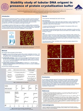
- 1. Dianming Wang, ‡a Ziran Da,‡b Bohan Zhang,a Mark Antonin Isbell,a,b Yuanchen Dong,a Xu Zhou, a Huajie Liu,c Jerry Yong Yew Heng*b and Zhongqiang Yang*a a Key Laboratory of Organic Optoelectronics & Molecular Engineering of the Ministry of Education, Department of Chemistry, Tsinghua University, Beijing 100084, P. R. China. b Surfaces and Particle Engineering Laboratory (SPEL), Department of Chemical Engineering, Imperial College London, South Kensington Campus, London SW7 2AZ, U.K. c Laboratory of Physical Biology, Shanghai Institute of Applied Physics, Chinese Academy of Sciences, Shanghai 201800, China. ‡ The authors contributed equally to this work. Stability study of tubular DNA origami in presence of protein crystallization buffer Acknowledgments This work was supported by the Royal Academy of Engineering – Research Exchange China and India (RECI) programme (Reference: 1314RECI047), National Natural Science Foundation of China (21474059, 21174077, 21421064), and the National Basic Research Program of China (2013CB932803). Methods We employed buffer exchanges by three methods 1) one-off: an abrupt buffer exchange by adding 400 µL protein crystallisation into 40 µL, 1 nM DNA origami solution, removing the buffer by a 10,000 molecular weight cut-off, centrifuging at 3,000 g for 5 min. The exchange was repeated 3 times. 2) gradient buffer exchange: in order to minimise the damage to the DNA origami structures, we used five kinds of gradient buffers with crystallisation/DNA origami buffer (v/v) ratios at 1:7, 1:3, 1:2, 3:1, up to pure crystallisation buffer respectively, with the removal of the DNA buffer identical to the one-off method 3) gradual buffer exchange by overnight dialysis: we added 40 µL, 1 nM DNA origami solution to a dialysis bag (Mw cut-off 12,000), placing it into 500 mL of protein crystallisation and stirred it overnight. Only the first two methods were successful. Introduction Research into the 3D structures of proteins is a huge topic in the field of molecular biology but many challenges remain for scientists1. In the early 1980s, Professor Seeman proposed the idea of utilising DNA as a scaffold to facilitate protein crystallisation for the determination of their 3D structures2 . Nowadays, the construction of nanostructures with nearly any arbitrary geometry from 2D3 to 3D4 can be realised by DNA origami techniques5. To investigate the potential of DNA origami as a scaffold to crystallise proteins, we tested the stability of DNA origami with a tubular structure6 in protein crystallisation buffer. The advantages of using this tubular structure are that the preparation is quick and straightforward, additionally, the porous structure may favour the protein crystallisation7 in future studies. Results Three systematic methodologies were used in this study. First and Second The shot gun approach refers to DNA origami either assembled in protein crystallisation buffer, or assembled in DNA assembly buffer first and followed by buffer exchange. Table 1 lists crystallisation buffers for four model proteins. 1) It turned out that these solutions were not compatible in the assembly of DNA, and no complete origami was observed 2) Exchanging the original DNA assembly buffer with protein crystallisation via either one-off or gradient buffer exchange, the DNA origami structure was retained only for catalase. The results are shown in Figure 2. Third The common variations of the four components in protein crystallisation buffer conditions are summarized in Table 1. The impact of salts, buffering agents, precipitants (alcohol, polymer and salt at high concentration) and pH were examined individually. In each experiment, the DNA assembly buffer was employed as the baseline recipe, apart from the factor of interest, all other aspects were maintained the same. The results are given in Table 1 with either a tick or cross if the conditions allowed for the tubular structure to remain. Figure 1. (left) Scheme for the design of the DNA tubes from rectangles. (Right) AFM image of tubular DNA origami in DNA assembly buffer. The structures are 100 nm in length, 22 nm in diameter and 2 nm in wall thickness. References 1. A. McPherson, 1999; A. McPherson, Methods, 2004, 34, 254-265; D. C. I. Y. P. C. W. Tian-xi Liu, Chinese J. Polym. Sci., 2014, 32, 115- 122. 2. N. C. Seeman, J. Theor. Biol., 1982, 99, 237-247; N. C. Seeman, Annu. Rev. Bioph. Biom., 1998, 27, 225-248; N. C. Seeman, Trends. Biotechnol., 1999, 17, 437-443; N. C. Seeman, Nature, 2003, 421, 427-431. 3. P. W. K. Rothemund, Nature, 2006, 440, 297-302. 4. S. M. Douglas, H. Dietz, T. Liedl, B. Högberg, F. Graf and W. M. Shih, Nature, 2009, 459, 414-418; D. Han, S. Pal, J. Nangreave, Z. Deng, Y. Liu and H. Yan, Science, 2011, 332, 342-346. 5. J. Nangreave, D. Han, Y. Liu and H. Yan, Curr. Opin. Chem. Biol., 2010, 14, 608-615; C. E. Castro, F. Kilchherr, D.-N. Kim, E. L. Shiao, T. Wauer, P. Wortmann, M. Bathe and H. Dietz, Nat. Methods, 2011, 8, 221-229; T. Torring, N. V. Voigt, J. Nangreave, H. Yan and K. V. Gothelf, Chem. Soc. Rev., 2011, 40, 5636-5646; C. Fan, DNA Nanotechnology: From Structure to Function, Springer Science & Business Media, 2013. 6. Y. Fu, D. Zeng, J. Chao, Y. Jin, Z. Zhang, H. Liu, D. Li, H. Ma, Q. Huang and K. V. Gothelf, J. Am. Chem. Soc., 2012, 135, 696-702. 7. N. E. Chayen, E. Saridakis and R. P. Sear, Proc. Natl. Acad. Sci. U. S. A., 2006, 103, 597-601; U. V. Shah, D. R. Williams and J. Y. Y. Heng, Cryst. Growth Des., 2012, 12, 1362-1369; U. V. Shah, M. C. Allenby, D. R. Williams and J. Y. Y. Heng, Cryst. Growth Des., 2012, 12, 1772-1777; J. V. Parambil, S. K. Poornachary, R. B. H. Tan and J. Y. Y. Heng, CrystEngComm, 2014, 16, 4927-4930; U. V. Shah, C. Amberg, Y. Diao, Z. Yang and J. Y. Y. Heng, Curr. Opin. Chem. Eng., 2015, 8, 69-75. Figure 2. AFM images of DNA origami after one-off buffer exchange by the protein crystallisation buffer of (a) lysozyme, (b) thaumatin, (c) human serum albumin, and (d) catalase. Table 2. AFM image of tubular DNA origami in DNA assembly buffer. The structures are 100 nm in length, 22 nm in diameter and 2 nm in wall thickness Table 1. Crystallisation buffer for four model proteins Conclusions Our work offers a systematic approach for studying tubular DNA origami stability in various protein crystallisation buffers. Based on the findings on catalase, we found that assembled structures were stable following buffer exchanges. Systematic studies demonstrated that individual factors in protein crystallisation buffers at certain ranges can still result in the successful assembly of DNA origami. This is a fundamental study that provides a strategy for screening suitable buffer conditions for DNA nanostructures, potentially to promote crystallisation of target proteins.