Chandrapur Call girls 8617370543 Provides all area service COD available
An exophytic lesion of the vagina cytological findings
1. EDUCATIONAL CASE REPORT
An exophytic lesion of the vagina – cytological findings
M. Pajtler*, M. Milojkovic´
and M. Mrcˇelaà
*Department of Clinical Cytology, Department of Gynecology and àDepartment of Pathology,
University Hospital ÔOsijekÕ, Osijek, Croatia
Accepted for publication 11 March 2003
Introduction
Primary melanoma of the vagina is rare, with less
than 250 reported cases to date.1
This tumour
constitutes less than 3% of all vaginal malignancies1,2
and less than 1% of all melanomas in women.3,4
The
majority of patients in the literature are isolated case
reports.5–7
By comparison, 93% of melanomas are
cutaneous, 5.2% are ocular, and 0.4% are oronasal.8
Its clinical behaviour in the vagina is notoriously
more aggressive than that of cutaneous and vulvar
melanoma, with 5-year survival rate ranging from
13% to 19%.3,4
The most appropriate treatment for vaginal melan-
oma has been the subject of some debate. Some
authors9,10
have found no difference in overall 5-year
survival between conservative surgery, radical sur-
gery, radiation, or chemotherapy. Others 11–13
have
recently argued at least better 2-year survival with
radical surgery (total colpectomy or pelvic exentera-
tion). These and other studies have been hampered by
too few cases over many years, precluding any
prospective controlled trials.
These tumours appear to originate from melano-
cytes that are present in the vaginal mucosa of
approximately 3% of women.14,15
They occur more
commonly in the lower third of the vagina,9,16
more
often on the anterior surface, and commonly produce
symptoms of bleeding (79%) or discharge (24%).16
Diagnosis has been made in the absence of symptoms
in only a few patients.
Case of primary malignant melanoma of the vagina
with special reference to the cytological findings has
been presented.
Case report
A 74-year-old woman who sought gynaecological
help due to prolapse of the anterior vaginal wall has
been presented. Gynaecological examination detected
a lesion consisting of few pink pale exophytic nodules
0.5 cm in diameter, among which black-pigmented
areas at mucosa level could be seen. A direct smear
was taken and stained by the Papanicolaou method.
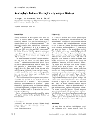
The cytological smear had a clean background and
showed, in addition to the predominantly interme-
diate squamous cells, numerous malignant cells,
either isolated or in small sheets. The cells had
polygonal, oval, fusiform or irregular shapes, with
marked anisocytosis. The cytoplasm was wispy and
cyanophilic, relatively clear with indistinct borders.
The nuclei were very large and located centrally,
although some were eccentric. They were oval,
elongated or lobulated in shape, with a moderately
granular, uneven chromatin pattern, prominent nuc-
lear membranes and prominent nucleoli. Giant multi-
nucleated cells were also found (Figure 1–2). The
cytological diagnosis was squamous cell carcinoma.
The patient had a colposcopically guided biopsy of the
vaginal lesion which showed tumour tissue consisting
of clusters of atypical oval, rounded and polygonal
cells, many of which contained brown pigment
granules of melanin. The nuclei were large, round to
oval, with prominent nucleoli and abnormal mitoses.
The surface consisted of patchily thinned squamous
epithelium, infiltrated with tumour tissue. The histo-
logical findings were those of a malignant melanoma
(Figure 4). Owing to the site of the tumour and the
patient’s age, no oncological therapy was undertaken
and the patient did not come for any follow-up.
Discussion
The smear from the polypoid vaginal lesion identi-
fied malignant cells, which differed from the
Correspondence:
Marija Pajtler, MD, Department of Clinical Cytology, Clinical
Hospital Osijek, 31000 Osijek, J. Huttlera 4, Croatia.
Tel.: 031-511 510;
E-mail: pajtler.marija@kbo.hr
Cytopathology 2003, 14, 150–152 ª 2003 Blackwell Publishing Ltd150
2. common cytological findings in Papanicolaou-stained
cervicovaginal smears.
They were misdiagnosed for several reasons, the
first being lack of experience, as primary malignant
melanoma of vagina occurs very rarely, and the
cytological findings have been described only infre-
quently.17–22
Secondly, most of the malignant cells
did not contain pigment to indicate the tumour type.
On review after the histological diagnosis, brown-
green pigment granules were noted in the cytoplasm
of only one cluster of malignant cell, as well as in one
benign squamous cell, difficult to detect under low
power magnification (Figure 3). This finding,
together with the clinical description perfectly
matched the histology, in which most of the malig-
nant cells were without pigment (Figure 4). The
differential diagnosis was thought to include adeno-
carcinoma because of the nuclear characteristics and
prominent nucleoli, but the distribution of cells and
absence of three-dimensional structures suggested
otherwise. In spite of being atypical, the findings
matched the description of poorly differentiated
squamous carcinoma. Identical cytomorphological
characteristics from other locations, especially in
smears stained by May–Grunwald–Giemsa would be
suspected of malignant melanoma, even without
pigment. However, the gynaecologist failed to men-
tion the prominent black-pigmented areas within the
lesion, which might have helped to diagnose the
exact type of malignant lesion.
Figure 1. Vaginal smear (Papanicolaou, ·400). Figure 2. Vaginal smear (Papanicolaou, ·1000).
Figure 3. Vaginal smear (Papanicolaou, ·1000). Figure 4. Histology of the lesion (H&E, ·400).
Exophytic lesion of the vagina 151
Cytopathology 2003, 14, 150–152 ª 2003 Blackwell Publishing Ltd
3. In order to accurately diagnose cytologically in the
case of rare lesions, the gynaecologist should describe
the clinical findings fully, and the cytologist should
analyse all morphological details thoroughly, bearing
in mind the question ÔWhat other diagnosis might also
be considered?Õ
References
1 Piura B, Rabinovich A, Yanai-Inbar I. Primary malignant
melanoma of the vagina: case report and review of
literature. Eur J Gynaecol Oncol 2002; 23: 195–8.
2 Chung AF, Casey MJ, Flannery JT, Worded JM, Lewis JL,
Jr. Malignant melanoma of the vagina – report of 19
cases. Obstet Gynecol 1980; 55: 720–7.
3 Weinstock MA. Malignant melanoma of the vulva and
vagina in the United States: patterns of incidence and
population-based estimates of survival. Am J Obstet Gynecol
1994; 171: 1225–30.
4 Ragnarsson-Olding B, Johansson H, Rutquist LE, Ring-
borg U. Malignant melanoma of the vulva and vagina.
Cancer 1993; 71: 1893–7.
5 Buchanan DJ, Schlaerth J, Kurosaki T. Primary vaginal
melanoma: thirteen-year disease-free survival after wide
local excision and review of recent literature. Am J Obstet
Gynecol 1998; 178: 1174–84.
6 Borazjani G, Prem KA, Okagaki T, Twiggs LB, Adcock LL.
Primary malignant melanoma of the vagina: a clinico-
pathological analysis of 10 cases. Gynecol Oncol 1990; 37:
264–7.
7 Kimura C, Furuya K. A case of pedunculated malignant
melanoma of the vaginal mucosa. J Dermatol 2001; 28:
564–8.
8 Chiu N, Weinstock MA. Melanoma of oronasal mucosa:
population based analysis of occurrence and mortality.
Arch Otolaryngol Head Neck Surg 1996; 122: 985–8.
9 Morrow CP, DiSaia PJ. Malignant melanoma of the
female genitalia: a clinical analysis. Obstet Gynecol Surv
1976; 31: 233–71.
10 Levitan Z, Gordon AN, Kaplan AL, Kaufman RH. Primary
malignant melanoma of the vagina: report of four cases
and review of the literature. Gynecol Oncol 1989; 33: 85–
90.
11 Van Nortstrand KM, Lucci JA, Schell M, Berman ML,
Manetta A, DiSaia PJ. Primary vaginal melanoma:
improved survival with radical pelvic surgery. Gynecol
Oncol 1994; 55: 234–7.
12 Geisler JP, Look KY, Moore DA, Sutton GP. Pelvic
exeneteration for malignant melanoma of the vagina or
urethra with over 3mm of invasion. Gynecol Oncol 1995;
59: 338–41.
13 Brand E, Fu YS, Lagasse LD, Berek JS. Vulvovaginal
melanoma: report of seven cases and literature review.
Gynecol Oncol 1989; 33: 54–60.
14 Nigogosyan G, DeLaPava S, Pickren JW. Melanoblasts in
vaginal mucosa. Cancer 1964; 17: 1912–3.
15 Lotem M, Anteby S, Peretz T, Ingber A, Avinoach I, Prus
D. Mucosal melanoma of the female genital tract is a
multifocal disorder. Gynecol Oncol 2003; 88: 45–50.
16 Reid GC, Schmidt RW, Roberts JA, Hopkins MP, Barret
RJ, Morley GW. Primary melanoma of the vagina: a
clinicopathological analysis. Obstet Gynecol 1989; 74:
190–9.
17 Ehrmann RL, Younge PA, Lerch VL. The exfoliative
cytology and histogenesis of an early primary malignant
melanoma of the vagina. Acta Cytol 1962; 6: 245–54.
18 Aschitaka Y, Taki I, Yanagida T. The cytologic findings of
malignant melanoma of the vaginal wall. J Jap Soc Clin
Cytol 1963; 4: 27.
19 Masubuchi K, Fujii J, Jyuzoji S, Kimura M, Yamazaki M.
Cytologic studies in malignant melanoma of the vagina.
J Jap Soc Clin Cytol 1969; 8: 66–9.
20 Masubuchi S, Jr, Nagai I, Hirata M, Kubo H, Masubuchi
K. Cytologic studies of malignant melanoma of the
vagina. Acta Cytol 1975; 19: 527–32.
21 Sagebiel RW, Gates EA, Hill LC. Cytologic detection of
recurrent vaginal melanoma. Acta Cytol 1978; 22: 353–7.
22 Genton CY, Kunz J, Schreiner WE. Primary malignant
melanoma of the vagina and cervix uteri. Report of a case
with ultrastructural study. Wirchows Arch (Pathol Anat)
1981; 393: 245–50.
M. Pajtler et al.152
Cytopathology 2003, 14, 150–152 ª 2003 Blackwell Publishing Ltd