
Recommended
More Related Content
What's hot
What's hot (18)
Similar to Anticancer Mechanisms of Cucurbitacins Review
Similar to Anticancer Mechanisms of Cucurbitacins Review (20)
More from LucyPi1
More from LucyPi1 (20)
Recently uploaded
Recently uploaded (20)
Anticancer Mechanisms of Cucurbitacins Review
- 1. REVIEW 68 TMR | March 2019 | vol. 4 | no. 2 | Submit a manuscript: https://www.tmrjournals.com/tmr doi: 10.12032/TMR20190225102 Traditional Chinese Medicine Advances in research on the anticancer mechanism of the natural compound cucurbitacin from Cucurbitaceae plants: a review Jing Liang1 , Dan Chen1 * 1 Department of Pharmacology, School of Basic Medical Sciences, Tianjin Medical University, Tianjin, China. *Corresponding to: Dan Chen, Department of Pharmacology, School of Basic Medical Sciences, Tianjin Medical University, No. 22, Qixiangtai Road, Heping District, Tianjin, China. E-mail: ilvcd@163.com. Highlights This review summarizes the advances in research on cucurbitacins B, D, E, and I in inducing tumor cell apoptosis, cytoskeletal destruction, cell cycle arrest, and autophagy and in regulating various cancer-related signaling pathways. Traditionality Cucurbitacins are present in some traditional Chinese herbs (TCH) such as Gualou (Fructus Trichosanthis), Tianhuafen (Radix Trichosanthis), and Tianguadi (Pedicellus Melo). An ancient book named Shennong Bencao Jing (1602 A.D., Donghan Dynasty of China) has reported that TCH Tianguadi (Pedicellus Melo) and the dried fruit stalk of Cucumis melo L. have been used for treating jaundice because components present in these herbs induce vomiting and aid in expelling phlegm.
- 2. REVIEW 69 Submit a manuscript: https://www.tmrjournals.com/tmr TMR | March 2019 | vol. 4 | no. 2 | doi: 10.12032/TMR20190225102 Abstract Cucurbitacins are highly oxidized tetracyclic triterpenoids that are widely found in plants belonging to Cucurbitaceae family and exert various pharmacological effects. Many cucurbitacin derivatives are available, of which cucurbitacins B, D, E, and I are important members of the cucurbitacin family and exert anticancer effects against various cancers. This review summarizes the advances in research on cucurbitacins B, D, E, and I in inducing tumor cell apoptosis, cytoskeletal destruction, cell cycle arrest, and autophagy and in regulating various cancer-related signaling pathways. In addition, this review summarizes the latest research on the synergistic effects of the combination of cucurbitacins and clinically approved chemotherapeutic drugs. The findings summarized in this review suggest that cucurbitacins are multi-targeting and multi-functional anticancer drugs and that their complex anticancer mechanisms should be examined in future studies. Because of their proven benefits, cucurbitacins have the potential to be used as anticancer drugs in the clinical setting. Keywords: Natural compound, Cucurbitacins, Anticancer Abbreviations: TCH, Traditional Chinese herbs; STAT3, Signal transducer and activator of transcription 3; JAK, Janus kinase; Bcl-2, B-cell lymphoma 2; ROS, Reactive oxygen species; MAPK, Mitogen-activated protein kinase; ERK, Extracellular signal-regulated kinase; MAPK, Mitogen-activated protein kinase; NF-κB, Nuclear factor kappa-light-chain-enhancer of activated B cells; PI3K, Phosphoinositide 3-kinase; AKT, Protein kinase B; mTOR, Mammalian target of rapamycin; LC3, Light chain 3; p-STAT3, Phosphorylated STAT3; NF2, Neurofibromatosis type 2. Competing interests: The authors declare that there is no conflict of interests regarding the publication of this paper. Citation: Jing Liang, Dan Chen. Advances in research on the anticancer mechanism of the natural compound cucurbitacin from Cucurbitaceae plants: a review. Traditional Medicine Research 2019, 4(2): 68-81. Executive Editor: Cui-Hong Zhu. Submitted: 20 September 2018, Accepted: 16 November 2018, Online: 17 December 2018.
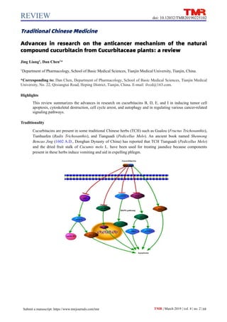
- 3. REVIEW 70 TMR | March 2019 | vol. 4 | no. 2 | Submit a manuscript: https://www.tmrjournals.com/tmr doi: 10.12032/TMR20190225102 Background The aggressiveness of various cancers and progressive tumor resistance and side effects of currently available chemotherapeutic drugs are the main concerns associated with cancer treatments used worldwide. Natural compounds extracted from traditional Chinese herbs (TCH) exert obvious anticancer effects against various cancers and thus are new potential anticancer agents. Increasing number of studies have shown that cucurbitacins, which are highly unsaturated tetracyclic triterpenoids produced mainly by plants belonging to Cucurbitaceae family [1], can be administered in combination with various chemotherapeutic drugs to reduce tumor resistance and enhance the efficacy of these drugs [2-4]. Although most cucurbitacins have a similar four-ring scaffold structure, they show a diversity in substituents (Figure 1). Several types of cucurbitacins are available, of which cucurbitacins B and E are the main cucurbitacin types [5]. Other cucurbitacin types are produced through enzymatic reactions under certain environmental conditions [6]. Cucurbitacins are present in some TCH such as Gualou (Fructus Trichosanthis), Tianhuafen (Radix Trichosanthis), and Tianguadi (Pedicellus Melo). An ancient book named Shennong Bencao Jing (1602 A.D., Donghan Dynasty of China) has reported that TCH Tianguadi (Pedicellus Melo) and the dried fruit stalk of Cucumis melo L. have been used for treating jaundice because components present in these herbs induce vomiting and aid in expelling phlegm. In the early 1970s, cucurbitacins B and E were isolated from Tianguadi (Pedicellus Melo) [7]. Some researchers have shown that cucurbitacins have hepatoprotective, anti-inflammatory, antidiabetic, and anticancer properties [8-10]. Cucurbitacin capsule (approval number: Z20090820), a Chinese patented medication, are used for treating chronic hepatitis and primary hepatocellular carcinoma because of their ability to induce detoxification, reduce fever, and remove dampness and yellowing. Recent studies have focused on the anticancer effects of cucurbitacins, including cucurbitacins B, E, D, and I, which are the most common cucurbitacin derivatives isolated from Cucurbitaceae plants. Many in vitro studies have shown that cucurbitacins inhibit cell proliferation, migration, and invasion and induce G2/M phase cell cycle arrest, autophagy, and apoptosis through different molecular mechanisms in various tumor cells [11, 12]. In addition, animal experiments have shown that cucurbitacins significantly inhibit tumor growth and metastasis in vivo [1, 13-15], indicating that these compounds exert a wide range of anticancer effects. In this review, we have discussed the advances in research on the anticancer mechanisms of cucurbitacins. Anticancer mechanisms of cucurbitacins Cucurbitacins induce the apoptosis of cancer cells through various mechanisms Apoptosis and cell proliferation are the fundamental mechanisms for maintaining a dynamic balance in a number of cells in the body. Cancer cells are characterized by inhibition of apoptosis, which induces their unlimited proliferation. Therefore, induction of cancer cell apoptosis by using various treatments is an important strategy for anticancer therapy. Cucurbitacins induce apoptosis of various cancer cells through different pathways (Figure 2). The subsequent subsections discuss mechanisms through which cucurbitacins promote cancer cell apoptosis. STAT3 signaling pathway. Signal transducer and activator of transcription 3 (STAT3) is one of the most important member of the STAT family that mediates the JAK (Janus kinase)/STAT3 signaling pathway and is closely associated with tumor development and metastasis [16]. STAT3 overexpression promotes cancer cell proliferation and migration and inhibits cancer cell apoptosis. Recent studies have shown that STAT3 is involved in the resistance of tumor cells to chemotherapeutic drugs [17-19]. Several studies performed over the last few years have reported the effect of cucurbitacins on the STAT3 signaling pathway, which is considered to be a selective inhibitor of the JAK/STAT3 pathway. Liu et al. reported that cucurbitacin B reduces the phosphorylation of STAT3 and its downstream targets, including cyclin B1 and BCL-2 (B-cell lymphoma 2), in human laryngeal Hep-2 cells [20]. In breast cancer cells, cucurbitacin B inhibits STAT3 phosphorylation in a time- and dose-dependent manner. However, Kin et al. reported that cucurbitacin B inhibits ERK (Extracellular signal-regulated kinase) 1/2 phosphorylation before STAT3 phosphorylation in leukemia cells, indicating that STAT3 is not the only target of cucurbitacin B [21]. Cucurbitacin I exerts antiproliferative effects by inhibiting STAT3 signaling in breast cancer, glioma, squamous cell carcinoma of the head and neck, and lung cancer cells [22, 23]. Moreover, cucurbitacin I inhibits tumor angiogenesis in breast cancer MDA-MB-468 cells by decreasing STAT3 phosphorylation [24]. In lung cancer A549 cells, cucurbitacin I induces apoptosis through a JAK/STAT3-dependent pathway [25]. In mice inoculated with five human osteosarcoma cell lines, namely, 143B, HOS, MG63, SAOS-2, and HUO9, cucurbitacin I inhibits tumor growth by inactivating STAT3, thus improving the survival of these mice [26]. Michelle et al. suggested that cucurbitacin I is highly selective for the JAK/STAT3 pathway and does not inhibit other tumor survival pathways [22]. However, another study reported that cucurbitacin I promotes the apoptosis of gastric cancer cells by inducing reactive oxygen species (ROS) production and not by targeting STAT3 [14]. Cucurbitacin D inhibits the nuclear translocation and transcriptional activity of STAT3 in breast cancer MCF7, SKBR3, and MDA-MB-231 cells [4, 27]. Cucurbitacin E induces the apoptosis of
- 4. REVIEW 71 Submit a manuscript: https://www.tmrjournals.com/tmr TMR | March 2019 | vol. 4 | no. 2 | doi: 10.12032/TMR20190225102 pancreatic cancer cells through the STAT3 signaling pathway [28]. Graness et al. reported that cucurbitacins exert anticancer effects not by increasing STAT3 activity but by increasing ROS and antioxidant levels, which promote cell death [29]. MAPK signaling pathway. MAPK (Mitogen-activated protein kinase) signaling pathway is an important pathway involved in cell proliferation, differentiation, and apoptosis. p38, ERK and JNK (c-Jun N-terminal kinases), are the important members of the MAPK family [30]. Many studies have shown that various cucurbitacins induce apoptosis of cancer cells through the MAPK signaling pathway. Cucurbitacin B induces the apoptosis of osteosarcoma U-2 cells, lung cancer A549 cells, and human neuroblastoma SH-SY5Y cells by inhibiting the activation of JNK, ERK1/2, and p38 in a dose-dependent manner [31-33]. Cucurbitacin E promotes the apoptosis of triple-negative breast cancer cells by increasing JNK activation and inhibiting ERK activation [34]. Deng et al. found that low concentrations of cucurbitacin I induce cell cycle arrest in and apoptosis of gastric cancer cells by activating JNK, p38, and MAPK signaling and by increasing GSH/GSSG ratio and GADD45α expression, which forms a positive feedback loop and independently regulates p53 gene expression [14]. However, a few studies have reported that cucurbitacin D inhibits the MAPK signaling pathway, which should be explored further. NF-κB pathway. NF-κB (Nuclear factor kappa-light-chain-enhancer of activated B cells) family includes five proteins, namely, RelA, RelB, Rel, NF-κB1, and NF-κB2. NF-κB is a dimeric protein comprising p65 and p50 subunits, and sustained NF-κB activation promotes tumor cell proliferation and inflammation [35]. Ku et al. found that cucurbitacin D increases the levels of NF-κB in the cytoplasm and inhibits the nuclear translocation of phosphorylated NF-κB to induce apoptosis. Cucurbitacin D also induces the apoptosis of doxorubicin-resistant breast cancer cells [4]. Ding et al. found that cucurbitacin D induces apoptosis by inhibiting intracellular proteasome activity and by reducing NF-κB nuclear translocation and BCL-2 and BCL-XL expression [36]. Treatment of glioblastoma cells with cucurbitacin I activates the NF-κB pathway by inducing the phosphorylation and nuclear translocation of the NF-κB p65 subunit, which occurs before the inhibition of STAT3 [37]. Figure 1 Structures of various cucurbitacin derivatives (A) The general structure of cucurbitacins. (B) The structure of cucurbitacin B. (C) The structure of cucurbitacin D. (D) The structure of cucurbitacin E. (E) The structure of cucurbitacin I.
- 5. REVIEW 72 Submit a manuscript: https://www.tmrjournals.com/tmr TMR | March 2019 | vol. 4 | no. 2 | doi: 10.12032/TMR20190225102 PI3K/AKT pathway. Many recent studies have shown that the activation of the PI3K/AKT signaling pathway plays an important role in many biochemical processes, including the proliferation and survival of breast cancer, cervical cancer, human osteosarcoma, and other cancer cells [38]. Wang et al. found that cucurbitacin E inhibits the growth of human osteosarcoma cells both in vitro and in vivo through the PI3K/AKT/mTOR signaling pathway [39]. Moreover, cucurbitacin E reduces phosphorylated AKT(Protein kinase B) and total AKT levels in triple-negative breast cancer cells [34]. Cucurbitacin D also inhibits the PI3K/AKT signaling pathway in cervical and gastric cancer cells by decreasing PI3K and p-AKT (Ser473) levels [15, 40]. Cucurbitacin I induces the apoptosis of A549 cells by inhibiting the activation of ERK and phosphorylation of its downstream proteins mTOR (Mechanistic target of rapamycin) and STAT3; however, it does not inhibit the PI3K/AKT pathway [25]. Other pathways. Some studies have reported that cucurbitacins promote cancer cell apoptosis through other mechanisms besides those mentioned above. A study showed that cucurbitacin B inhibits breast cancer growth both in vivo and in vitro by inhibiting Wnt and HER2/integrin signaling [41]. Duangmano et al. suggested that cucurbitacin B promotes the apoptosis of human breast cancer cells by disrupting microtubule networks [42]. Cucurbitacins also induce the apoptosis of human T cell leukemia Jurkat cells by disrupting cellular actin mechanics and by activating its key regulator cofilin [43]. Cucurbitacin B increases the production of intracellular ROS in leukemia K562 cells, thus inducing their apoptosis [44]. It also induces the apoptosis of colon cancer SW480 cells through a STAT3-independent but an ROS-dependent mechanism [45]. Cucurbitacin E induces the apoptosis of cervical cancer HeLa and CaSki cells by upregulating the expression of death receptor 5 [46]. Cucurbitacin I promotes the apoptosis of liver cancer HepG2 cells by activating p53 and its downstream targets. Cucurbitacin D effectively induces the apoptosis of gastric cancer cells by activating the inducible nitric oxide synthase pathway [40]. These findings indicate that cucurbitacins use variable and complex mechanisms to induce the apoptosis of cancer cells. Cucurbitacins induce autophagy in tumor cells through various mechanisms Figure 2: Signaling pathways involved in cucurbitacin-induced apoptosis STAT3, Signal transducer and activator of transcription 3; JAK, Janus kinase; Bcl-2, B-cell lymphoma 2; ROS, Reactive oxygen species; Erk1/2, Extracellular signal-regulated kinase1/2; MAPK, Mitogen-activated protein kinase; NF-κB, Nuclear factor kappa-light-chain-enhancer of activated B cells; PI3K, Phosphoinositide 3-kinase; Akt, Protein kinase B; mTOC1, mTOR complex 1.
- 6. REVIEW 73 Submit a manuscript: https://www.tmrjournals.com/tmr TMR | March 2019 | vol. 4 | no. 2 | doi: 10.12032/TMR20190225102 Autophagy is an important process for maintaining homeostasis in eukaryotic cells. Autophagy signaling pathways are activated under certain circumstances to degrade damaged macromolecular substances and to provide energy for cell survival. In cancer therapy, autophagy plays a dual role, such as the inhibition of autophagy promotes cancer cell death, whereas excessive autophagy leads to autophagic cell death [47, 48]. Studies involving various cancer cell lines have shown that cucurbitacins B, D, E, and I induce the production of ROS, which play an important role in mediating DNA damage and in inducing protective autophagy [44, 49-54]. Liu et al. reported that cucurbitacin B inhibits CIP2A/PP2A/mTORC1 signaling axis-induced autophagy in cisplatin-resistant human gastric cancer SGC7901/DDP cells [55]. In vitro treatment of leukemia cells with cucurbitacin B induces autophagy as a survival response; however, specific mechanisms underlying this are unclear[43]. Cucurbitacin E induces autophagy by downregulating the mTORC1 signaling pathway and by upregulating AMPK activity [56]. Microtubule-associated protein light chain 3 (LC3) is the key factor in autophagosome formation. Cucurbitacins D, E, and I induce autophagy by upregulating the LC3 gene expression in human gastric cancer cells, with the effect of cucurbitacin I being significantly higher than those of cucurbitacins D and E [57]. Ni et al. found that cucurbitacin I induces damage-associated autophagy in gastric cancer A549 cells by inhibiting the ERK/mTOR/STAT3 signaling pathway [25]. Competitive mechanisms of cucurbitacin-induced autophagy and apoptosis Most studies on cucurbitacins have focused on their effect on promoting cancer cell apoptosis. The ability cucurbitacins to inhibit cell proliferation does not depend only on the STAT3 pathway. Many studies have reported that the morphological and biochemical features of cucurbitacin-induced apoptosis of human cancer cells are not apparent and that cell death is induced by ROS-mediated autophagy in most cucurbitacin-treated cancer cells. In recent years, cucurbitacin-induced autophagy has attracted considerable amount of attention; however, mechanisms underlying cucurbitacin-induced autophagy are not completely understood. Research has shown that cucurbitacin-induced autophagy competes with apoptotic signaling to limit the effect of apoptosis [58]. Cucurbitacins induce cell-protective autophagy and other competing apoptotic mechanisms or increase apoptotic resistance. Some studies have shown that cucurbitacins induce autophagy based on damaged cellular morphology. Moreover, the damaged cellular morphology that induces pro-death autophagy differs from the damaged cellular morphology that induces apoptosis. We believe that cucurbitacins may act on signaling pathways that are common between apoptosis and autophagy but may show a different order of induction of both these processes. Furthermore, apoptosis and autophagy may activate or inhibit one another after cucurbitacin treatment. Therefore, additional studies should be performed to determine the complex relationship between cucurbitacin-induced autophagy and apoptosis. Cucurbitacins induce cytoskeletal destruction Cucurbitacins induce morphological changes in cancer cells within a short period. Changes in actin filaments and microtubules play an important role in cancer cell proliferation, which is an important target of natural compounds used in cancer treatment [59]. Wang et al. compared cucurbitacins B, E, and I with vincristine and colchicine and showed that the cucurbitacins interacted with the cytoskeleton and actin filaments to induce cell cycle arrest. In addition, cucurbitacins interfered with microtubule structure, thus affecting cell mitosis [60]. Cucurbitacin E disrupts the cytoskeletal structure and inhibits the proliferation of prostate cancer cells [61]. Cucurbitacin B covalently binds to cofilin, thus increasing actin depolymerization. However, some studies have shown that cucurbitacins do not directly bind to cofilin but inhibit the regulation of cofilin phosphorylation kinase to increase actin depolymerization and stimulate Rho/ROCK pathway to induce actin and phosphorylated myosin II co-aggregation [62, 63]. However, Zhang et al. suggested that cucurbitacin B induces the actin aggregation through Gα13/RhoA/PKA/VASP signaling pathway [64]. Although several studies have attempted to determine mechanisms underlying cucurbitacin-induced cytoskeletal destruction, these studies have provided different results. Therefore, specific mechanisms underlying cucurbitacin- induced cytoskeletal destruction are still unclear and should be determined by performing additional studies. Cucurbitacins induce cell cycle arrest in cancer cells A cell cycle involves a series of events in a cell from mitosis to the end of the next division. The length of a cell cycle reflects the state of a cell and is a cyclical process of cell material accumulation and cell division. Cancerous cells often show abnormal division cycles. Many studies have shown that cucurbitacins induce cell cycle arrest at different stages depending on the cell type. Cucurbitacin B arrests the cell cycle in the S phase in BEL-7402, HL60, and U937 cells and in the G2/M phase in Panc-1, MiaPaCa-2, K562, SW480, and Hep-2 cells [6]. In pancreatic cancer cells, cucurbitacin B may induce cell cycle arrest in the G2/M phase by inhibiting JAK2, STAT3, and STAT5 activation; increasing p21 level; and decreasing cyclin A and cyclin B1 expression [65]. In human hepatocellular carcinoma BEL-7402 cells, cucurbitacin B-induced cell cycle arrest in the S phase is associated with the inhibition of cyclin D1 and cyclin-dependent kinase-1 expression but is not associated with STAT3 phosphorylation [66]. Most studies have suggested that cells arrested in the G2/M phase of the cell cycle are
- 7. REVIEW 74 Submit a manuscript: https://www.tmrjournals.com/tmr TMR | March 2019 | vol. 4 | no. 2 | doi: 10.12032/TMR20190225102 tetraploid cells that have undergone nuclear division but are unable to complete cytokinesis. Drugs that affect the microfilament skeleton induce various degrees of cell cycle arrest; therefore, cell cycle arrest induced by cucurbitacin B is most likely to be a consequence of damage to the microfilament skeleton [60]. Multiple types of leukemia cells treated with cucurbitacin B show significant cell cycle arrest in the S phase [67]. Kong et al. compared 12 natural drugs, including cucurbitacins B, E, and I, in four cancer cell lines and found that cucurbitacin E significantly reduced the viability of MAD-MB-468 and SW527 cells by regulating cyclin D1 and cyclin B expression, inhibiting phosphorylated STAT3 (p-STAT3), and activating p53 and p21, eventually leading to cell cycle arrest in the G2/M phase [34, 68]. Cucurbitacin D inhibits the proliferation of endometrial cancer and ovarian cancer cells and increased the ratio of these cells in the sub-G0/G1 and G2/M phases of the cell cycle [69]. No FDA-approved drugs are available for treating neurofibromatosis type 2 (NF2)-associated schwannomas and meningiomas. Spear et al. found that cucurbitacin D exerts anticancer effects on NF2-deficient mouse schwannoma Sch10545 cells and human benign meningioma Ben-Men-1 cells by increasing the number of cells in the G2/M phase and inhibiting the proliferation of these cells, suggesting its potential as a therapeutic agent for treating these diseases [70]. Jafargholizadeh et al. found that treatment of human gastric cancer AGS cells with cucurbitacins D, E, and I induces cell cycle arrest in the sub-G1 phase, eventually leading to cell death [57]. These studies indicate that cucurbitacins play an important role in cell cycle arrest and that the effect of cucurbitacins on cell cycle arrest differs between different cell types. Combination treatment with cucurbitacins and chemotherapeutic drugs The aggressiveness of various cancers and tumor resistance and side effects of currently available chemotherapeutic drugs are serious concerns associated with cancer treatment. Cucurbitacins, which are natural compounds, exert obvious antiproliferative effects on various tumor cells, indicating their potential as anticancer agents. STAT3 is associated with resistance against anticancer drugs and is highly expressed in many drug-resistant cancer cells. Because cucurbitacins mainly target STAT3, several studies have examined the combination of cucurbitacins with various chemotherapeutic drugs and have shown that cucurbitacins reduce the resistance against and enhance the efficacy of these drugs. The findings of these studies suggest that cucurbitacins are potential candidates for use in combination therapy with clinical anticancer drugs. Cucurbitacins and chemotherapeutic drugs synergistically exert anticancer effects. Tang et al. found that cucurbitacin B synergistically increases the antitumor activity of doxorubicin by blocking the STAT3 pathway [2]. Cucurbitacin B also enhances the anticancer effect of imatinib mesylate by inhibiting matrix metalloproteinase-2 expression in MCF7 and SW480 tumor cells [71]. The combination of cucurbitacin B with gefitinib induces cell cycle arrest and apoptosis in human colorectal cancer cells through the EGFR and JAK/STAT pathways [3]. Lee et al. showed that treatment with low doses of cucurbitacin B and methotrexate synergistically inhibit the AKT and mTOR signaling pathways in human osteosarcoma cells both in vivo and in vitro [72]. El-Senduny et al. treated ovarian cancer A2780 cells and cisplatin-resistant A2780CP cells with a combination of cucurbitacin B and cisplatin or pretreated these cells with cucurbitacin B, followed by treatment with cisplatin. Results of contrast analysis performed in this study showed that the combination of cisplatin and cucurbitacin B decreased the levels of dual specificity tyrosine phosphorylation regulated kinase 1B, phosphorylated ERK1/2, and p-STAT3 and increased the level of ROS, thus significantly enhancing the effect of cisplatin on the ovarian cancer cells [73]. Moreover, cucurbitacin B synergistically exerts antiproliferative effects along with cisplatin on cutaneous squamous cell carcinoma cells [74] and enhances the effect of arsenic trioxide-induced apoptosis by inhibiting STAT3 phosphorylation in lymphoma Ramos cells. In addition, the combination of cucurbitacin B and arsenic trioxide does not exert any proapoptotic effects on normal lymphocytes, indicating that this combination is non-toxic against normal blood cells. Experiments involving an in vivo nude mouse lymphoma model have further confirmed this synergistic effect of cucurbitacin B and arsenic trioxide [75]. Ku et al. found that cucurbitacin D promotes the apoptosis of doxorubicin-resistant breast cancer cells through STAT3 and NF-κB [4]. Chang et al. found that cucurbitacin I increases the sensitivity of medulloblastoma-derived cancer stem cells to apoptosis induced by chemotherapeutic drugs targeting STAT3 [76]. Cucurbitacins can be safely used in combination with chemotherapeutic drugs. Myelosuppression and hepatorenal toxicity are the common side effects of chemotherapy. Ahmed et al. showed that the combination of cucurbitacin B with chemotherapeutic drugs did not increase toxicity in immunosuppressed mice undergoing orthotopic transplantation of breast cancer. Neurosensory and neuromotor toxicities are common in patients treated with docetaxel and other taxanes. However, it was found that treatment with the combination of docetaxel with cucurbitacin B was associated with lower neurotoxicity than treatment with docetaxel alone. These findings suggest that cucurbitacins exert protective effects against docetaxel-induced neurotoxicity; however, specific mechanisms underlying this effect of cucurbitacins are unknown [77]. Previous studies have reported that cucurbitacin E can be used in combination with doxorubicin for treating ovarian cancer and enhances
- 8. REVIEW 75 Submit a manuscript: https://www.tmrjournals.com/tmr TMR | March 2019 | vol. 4 | no. 2 | doi: 10.12032/TMR20190225102 the efficacy of doxorubicin. An in vitro study has shown that cucurbitacin E increases doxorubicin level in M5076 ovarian sarcoma cells by suppressing doxorubicin efflux [78]. Sadzuka et al. found that cucurbitacin E can be used in combination with doxorubicin for treating ovarian cancer to enhance the efficacy of doxorubicin without increasing its side effects. As mentioned previously, cucurbitacin E inhibits the efflux of doxorubicin from the M5076 ovarian sarcoma cells, thus significantly increasing its concentration in tumor cells and reducing its concentration in normal cells. Tumor cells and normal cells show differences in membrane transporter expression; thus, the differences in the role of cucurbitacin E between tumor and normal tissues may be because of differences in the expression of multidrug resistance-associated proteins between these tissues [36]. Conclusion Cucurbitacins are natural compounds with various pharmacological activities, and their antitumor effects have received increasing attention. These compounds can prevent the proliferation of different tumor cells by inducing apoptosis, cell cycle arrest, autophagy, and cytoskeletal disruption (Table 1). Although many studies have reported the antitumor activity of cucurbitacins, mechanisms underlying this activity of cucurbitacins are unclear. Cucurbitacins exert anticancer effects both in vivo and in vitro through multiple targets, indicating their enormous anticancer potential. The combination of cucurbitacins with chemotherapeutic drugs also exerts strong synergistic anticancer effects. However, determination of the antitumor effects of cucurbitacins is challenging because of the complex mechanism of tumorigenesis. Therefore, additional in-depth studies on cucurbitacins should be performed to confirm their potential as drug candidates for cancer treatment in the clinical setting. Table1: Cucurbitacins inhibits proliferation of different cell lines Cucurbitacins Cancer cell lines Mechanism B Human leukemia cells: CCRF-CEM, K562, MOLT-4, RPMI-8226, SR, and Jurkat DNA damage induction, G2/M phase cell cycle arrest, autophagy induction, actin cytoskeleton alteration, and apoptosis induction [21, 43, 44] Breast cancer cells: MDA-MB-231, MCF7, ZR-75-1, T47D, BT474, MDA-MB-453, SKBR-3, HCC1937, MDA-MB-436, and 4T-1 Integrin-HER2 signaling inhibition, DNA damage and autophagy induction, microtubule polymerization disruption, G2/M phase cell cycle arrest, telomerase inhibition, and apoptosis induction [8, 41, 42, 49, 79, 80] Colon cancer cell lines: SW480 and HCT-116 Cell cycle inhibition and apoptosis induction [3, 71] Hepatic carcinoma cell lines: BEL-740 and HepG2 cells Protective autophagy induction [54], apoptosis induction, and S phase cell cycle arrest [66, 81] Acute leukemia cell lines: RCH, Reh, BALL-1, MD901, LY4, HL60, U937, THP1, K562, and NB4 Cell cycle arrest and actin cytoskeleton alteration [67] Prostate cancer cell lines: PC-3 and LNCaP Apoptosis induction [82] Pancreatic cancer cell lines: ASPC-1, BxPC-3, CFPAC-1, HPAC, Panc-1, and MiaPaCa-2 G2/M phase cell cycle arrest and apoptosis induction [65, 83] Osteosarcoma cell lines: U-2OS, G292, MG-63, HT-161, HOS, SAOS-2, and SJSA Apoptosis induction [31, 72] Non-small-cell lung cancer cell lines: A549, H1792, H1650, and H1975 Apoptosis induction [8, 84] Throat cancer cell line: Hep-2 Cell cycle arrest and apoptosis induction [20, 85] Melanoma cell lines: Human A375 and murine B16F10 Actin aggregation induction [64] Glioblastoma multiforme cell lines: DBTRG‐05MG, U251MG, U118MG, U87MG, T98G, and LN229 Invasive behavior inhibition, apoptosis induction [86], and cytoskeletal damage [87] Cutaneous squamous cell carcinoma cell lines: SRB1, SRB12, SCC13, and Colo16 G2/M phase cell cycle arrest and cell migration inhibition [74] Cervical cancer cell line: HeLa G2/M phase cell cycle arrest and apoptosis induction [88] Neuroblastoma cell line: SH‑SY5Y Apoptosis induction [32] Lymphoma cell line: Ramos Apoptosis induction [75]
- 9. REVIEW 76 Submit a manuscript: https://www.tmrjournals.com/tmr TMR | March 2019 | vol. 4 | no. 2 | doi: 10.12032/TMR20190225102 D Breast cancer cells lines: MCF7 and MDA-MB-231 Cell cycle arrest and apoptosis induction [4] T cell leukemia cell lines: MT-1, MT-2, MT-4, Hut102, Hut78, and Jurkat Apoptosis and autophagy induction [52, 89] Hepatic carcinoma cell line: Hep3B Apoptosis induction through caspase-3 and JNK phosphorylation [90] Colon cancer cell lines: HT29, Colo320, and Caco2 Apoptosis induction [91] Lung cancer cell lines: A549 and AGS Apoptosis induction and cell cycle arrest [57, 91] Cervical cancer cell lines: HeLa and SiHa Apoptosis induction and G1/S phase cell cycle arrest [15] NF2-deficient schwannoma cell line: Sch10545 Apoptosis induction and cell cycle arrest [70] Gastric cancer cell lines: AGS, SNU1, and Hs746T Apoptosis induction [40] Endometrial cancer cell lines: HHUA and HEC59 Apoptosis induction and cell cycle arrest [69] Ovarian cancer cell lines: SK-OV-3, OVCAR-3, and TOV-112D Apoptosis induction and cell cycle arrest [69] E Breast cancer cell lines: Bcap37, MDA-MB-231, MDA-MB-468, and SW527 G2/M phase cell cycle arrest and apoptosis induction [34, 92] Human leukemia cell line: U937 Actin depolymerization [93] Lung cancer cell lines: A549 and 95D Caspase-dependent apoptosis induction and protective autophagy induction [94, 95] Brain malignant glioma cell line: GBM 8401 G2/M phase cell cycle arrest [96] Oral squamous cell carcinoma cell line: SAS Cell death and apoptosis induction [97] Cervical cancer cell lines: HeLa and CaSki Apoptosis induction [46] Prostate cancer cell lines: LNCaP, PC-3, and DU145 Apoptosis induction [98] and actin and vimentin cytoskeleton disruption [61] Pancreatic cancer cell line: Panc-1 G2/M phase cell cycle arrest and apoptosis induction [28] Ovarian cancer cell lines: ES-2 and M5076 Apoptosis induction and cell cycle arrest [36, 99] Bladder cancer cell line: T24 G2/M phase cell cycle arrest and apoptosis induction [68] Hepatic carcinoma cell lines: HepG2 and BEL-7402 In vitro cytotoxicity induction [100] I Breast cancer cell lines: MDA-MB-468, MDA-MB-231, T-47D, MCF7, BT-474, and HCC1419 Cell viability, proliferation, adhesion, migration, and tube formation inhibition [24] and cell motility inhibition [101] Colon cancer cell lines: SW480, CT-26, and HCT-116 Apoptosis induction [8] Lung cancer cell lines: NCI-H460 and A549 Apoptosis induction and actin filament disruption [102] Gastric cancer cell line: AGS Sub-G1 phase [57] and G2/M phase cell cycle arrest and apoptosis induction [14] Glioblastoma multiforme cell lines: GBM, U251, and A172 G2/M cell cycle arrest and apoptosis induction [103] Nasopharyngeal carcinoma cell lines: HK1 and CNE-2 Apoptosis induction [23] B cell leukemia cell lines: BJAB, I-83, NALM-6, and primary CLL Apoptosis induction and cell cycle arrest [104] Malignant glioma cell lines: T98G and U251 Protective autophagy induction [53] Nasopharyngeal carcinoma cell lines: HK1 and CNE-2 Apoptosis induction [23]
- 10. REVIEW 77 Submit a manuscript: https://www.tmrjournals.com/tmr TMR | March 2019 | vol. 4 | no. 2 | doi: 10.12032/TMR20190225102 References 1. Piao XM, Gao F, Zhu JX, et al. Cucurbitacin B inhibits tumor angiogenesis by triggering the mitochondrial signaling pathway in endothelial cells. Int J Mol Med 2018, 42: 1018-1025. 2. Yang T, Liu J, Yang M, et al. Cucurbitacin B exerts anti-cancer activities in human multiple myeloma cells in vitro and in vivo by modulating multiple cellular pathways. Oncotarget 2017, 8: 5800-5813. 3. Yar SAS, Alp E, Elmazoglu Z, et al. Treatment with cucurbitacin B alone and in combination with gefitinib induces cell cycle inhibition and apoptosis via EGFR and JAK/STAT pathway in human colorectal cancer cell lines. Hum Exp Toxicol 2016, 35: 526-543. 4. Ku JM, Kim SR, Hong SH, et al. Cucurbitacin D induces cell cycle arrest and apoptosis by inhibiting STAT3 and NF-κB signaling in doxorubicin-resistant human breast carcinoma (MCF7/ADR) cells. Mol Cell Biochem 2015, 409: 33-43. 5. Abbas S, Vincourt JB, Habib L, et al. The cucurbitacins E, D and I: investigation of their cytotoxicity toward human chondrosarcoma SW 1353 cell line and their biotransformation in man liver. Toxicol Lett 2013, 216: 189-199. 6. Chen X, Bao J, Guo J, et al. Biological activities and potential molecular targets of cucurbitacins: a focus on cancer. Anticancer Drugs 2012, 23: 777-787. 7. Peters RR, Farias MR, Ribeiro-do-Valle RM. Anti-inflammatory and analgesic effects of cucurbitacins from Wilbrandia ebracteata. Planta Med 1997, 63: 525-528. 8. Jayaprakasam B, Seeram NP, Nair MG. Anticancer and antiinflammatory activities of cucurbitacins from Cucurbita andreana. Cancer Lett 2003, 189: 11-6. 9. Park CS, Lim H, Han KJ, et al. Inhibition of nitric oxide generation by 23,24-dihydrocucurbitacin D in mouse peritoneal macrophages. J Pharmacol Exp Ther 2004, 309: 705-710. 10. Ríos JL, Andújar I, Escandell JM, et al. Cucurbitacins as inducers of cell death and a rich source of potential anticancer compounds. Curr Pharm Des 2012, 18: 1663-1676. 11. Liu T, Zhang M, Deng Y, et al. Effects of cucurbitacin B on cell proliferation and apoptosis in Hep-2 cells. Lin Chung Er Bi Yan Hou Tou Jing Wai Ke Za Zhi 2008, 22: 403-407. 12. Garg S, Kaul SC, Wadhwa R. Cucurbitacin B and cancer intervention: Chemistry, biology and mechanisms (Review). Int J Oncol 2018, 52: 19-37. 13. Zhang T, Li J, Dong Y, et al. Cucurbitacin E inhibits breast tumor metastasis by suppressing cell migration and invasion. Breast Cancer Res Treat 2012, 135: 445-458. 14. Deng C, Zhang B, Zhang S, et al. Low nanomolar concentrations of Cucurbitacin-I induces G2/M phase arrest and apoptosis by perturbing redox homeostasis in gastric cancer cells in vitro and in vivo. Cell Death Dis 2016, 7: e2106. 15. Sikander M, Hafeez BB, Malik S, et al. Cucurbitacin D exhibits potent anti-cancer activity in cervical cancer. Sci Rep 2016, 6: 36594. 16. Yuen JW, Poon LS, Chan AS, et al. Activation of STAT3 by specific Galpha subunits and multiple Gbetagamma dimers. Int J Biochem Cell Biol 2010, 42: 1052-1059. 17. Kamran MZ, Patil P, Gude RP. Role of STAT3 in cancer metastasis and translational advances. Biomed Res Int 2013, 2013: 421821. 18. Schindler C, Darnell JE. Transcriptional responses to polypeptide ligands: the JAK-STAT pathway. Annu Rev Biochem 1995, 64: 621-651. 19. Levy DE, Darnell JE. Stats: transcriptional control and biological impact. Nat Rev Mol Cell Biol 2002, 3: 651-662. 20. Liu T, Zhang M, Zhang H, et al. Inhibitory effects of cucurbitacin B on laryngeal squamous cell carcinoma. Eur Arch Otorhinolaryngol 2008, 265: 1225-1232. 21. Chan KT, Li K, Liu SL, et al. Cucurbitacin B inhibits STAT3 and the Raf/MEK/ERK pathway in leukemia cell line K562. Cancer Lett 2010, 289: 46-52. 22. Blaskovich MA, Sun J, Cantor A, et al. Discovery of JSI-124 (cucurbitacin I), a selective Janus kinase/signal transducer and activator of transcription 3 signaling pathway inhibitor with potent antitumor activity against human and murine cancer cells in mice. Cancer Res 2003, 63: 1270-1279. 23. Lui VW, Yau DM, Wong EY, et al. Cucurbitacin I elicits anoikis sensitization, inhibits cellular invasion and in vivo tumor formation ability of nasopharyngeal carcinoma cells. Carcinogenesis 2009, 30: 2085-2094. 24. Qi J, Xia G, Huang CR, et al. JSI-124 (Cucurbitacin I) inhibits tumor angiogenesis of human breast cancer through reduction of STAT3 phosphorylation. Am J Chin Med 2015, 43: 337-347. 25. Ni Y, Wu S, Wang X, et al. Cucurbitacin I induces pro-death autophagy in A549 cells via the ERK-mTOR-STAT3 signaling pathway. J Cell Biochem 2018, 119: 6104-6112. 26. Oi T, Asanuma K, Matsumine A, et al. STAT3 inhibitor, cucurbitacin I, is a novel therapeutic agent for osteosarcoma. Int J Oncol 2016, 49:
- 11. REVIEW 78 Submit a manuscript: https://www.tmrjournals.com/tmr TMR | March 2019 | vol. 4 | no. 2 | doi: 10.12032/TMR20190225102 2275-2284. 27. Kim SR, Seo HS, Choi HS, et al. Trichosanthes kirilowii Ethanol Extract and Cucurbitacin D Inhibit Cell Growth and Induce Apoptosis through Inhibition of STAT3 Activity in Breast Cancer Cells. Evid Based Complement Alternat Med 2013, 2013: 975350. 28. Sun C, Zhang M, Shan X, et al. Inhibitory effect of cucurbitacin E on pancreatic cancer cells growth via STAT3 signaling. J Cancer Res Clin Oncol 2010, 136: 603-610. 29. Graness A, Poli V, Goppelt-Struebe M. STAT3-independent inhibition of lysophosphatidic acid-mediated upregulation of connective tissue growth factor (CTGF) by cucurbitacin I. Biochem Pharmacol 2006, 72: 32-41. 30. Wagner EF, Nebreda AR. Signal integration by JNK and p38 MAPK pathways in cancer development. Nat Rev Cancer 2009, 9: 537-549. 31. Zhang ZR, Gao MX, Yang K. Cucurbitacin B inhibits cell proliferation and induces apoptosis in human osteosarcoma cells via modulation of the JAK2/STAT3 and MAPK pathways. Exp Ther Med 2017, 14: 805-812. 32. Zheng Q, Liu Y, Liu W, et al. Cucurbitacin B inhibits growth and induces apoptosis through the JAK2/STAT3 and MAPK pathways in SH‑SY5Y human neuroblastoma cells. Mol Med Rep 2014, 10: 89-94. 33. Silva IT, Geller FC, Persich L, et al. Cytotoxic effects of natural and semisynthetic cucurbitacins on lung cancer cell line A549. Invest New Drugs 2016, 34: 139-148. 34. Kong Y, Chen J, Zhou Z, et al. Cucurbitacin E induces cell cycle G2/M phase arrest and apoptosis in triple negative breast cancer. PLoS One 2014, 9: e103760. 35. Hayden MS, Ghosh S. NF-κB, the first quarter-century: remarkable progress and outstanding questions. Genes Dev 2012, 26: 203-234. 36. Sadzuka Y, Hatakeyama H, Sonobe T. Enhancement of doxorubicin concentration in the M5076 ovarian sarcoma cells by cucurbitacin E co-treatment. Int J Pharm 2010, 383: 186-191. 37. McFarland BC, Gray GK, Nozell SE, et al. Activation of the NF-κB pathway by the STAT3 inhibitor JSI-124 in human glioblastoma cells. Mol Cancer Res 2013, 11: 494-505. 38. Arcaro A, Guerreiro AS. The phosphoinositide 3-kinase pathway in human cancer: genetic alterations and therapeutic implications. Curr Genomics 2007, 8: 271-306. 39. Wang Y, Sun Y, Wu Y, et al. Cucurbitacin E inhibits osteosarcoma cells proliferation and invasion through attenuation of PI3K/AKT/mTOR signaling. Biosci Rep 2016, 279: 2247-2259. 40. Zhang YZ, Wang CF, Zhang LF. Cucurbitacin D impedes gastric cancer cell survival via activation of the iNOS/NO and inhibition of the Akt signalling pathway. Oncol Rep 2018, 39: 2595-2603. 41. Gupta P, Srivastava SK. Inhibition of Integrin-HER2 signaling by Cucurbitacin B leads to in vitro and in vivo breast tumor growth suppression. Oncotarget 2014, 5: 1812-1828. 42. Duangmano S, Sae-Lim P, Suksamrarn A, et al. Cucurbitacin B inhibits human breast cancer cell proliferation through disruption of microtubule polymerization and nucleophosmin/B23 translocation. BMC Complement Altern Med 2012, 12: 185. 43. Zhu JS, Ouyang DY, Shi ZJ, et al. Cucurbitacin B induces cell cycle arrest, apoptosis and autophagy associated with G actin reduction and persistent activation of cofilin in Jurkat cells. Pharmacology 2012, 89: 348-346. 44. Guo J, Zhao W, Hao W, et al. Cucurbitacin B induces DNA damage, G2/M phase arrest, and apoptosis mediated by reactive oxygen species (ROS) in leukemia K562 cells. Anticancer Agents Med Chem 2014, 14: 1146-1153. 45. Yasuda S, Yogosawa S, Izutani Y, et al. Cucurbitacin B induces G2 arrest and apoptosis via a reactive oxygen species-dependent mechanism in human colon adenocarcinoma SW480 cells. Mol Nutr Food Res 2010, 54: 559-565. 46. Cheng YM, Shen CJ, Chang CC, et al. Inducement of apoptosis by cucurbitacin E, a tetracyclic triterpenes, through death receptor 5 in human cervical cancer cell lines. Cell Death Discov 2017, 3: 17014. 47. Apel A, Herr I, Schwarz H, et al. Blocked autophagy sensitizes resistant carcinoma cells to radiation therapy. Cancer Res 2008, 68: 1485-1494. 48. Denton D, Nicolson S, Kumar S. Cell death by autophagy: facts and apparent artefacts. Cell Death Differ 2012, 19: 87-95. 49. Ren G, Sha T, Guo J, et al. Cucurbitacin B induces DNA damage and autophagy mediated by reactive oxygen species (ROS) in MCF-7 breast cancer cells. J Nat Med 2015, 69: 522-530. 50. Ouyang D, Zhang Y, Xu L, et al. Histone deacetylase inhibitor valproic acid sensitizes B16F10 melanoma cells to cucurbitacin B treatment. Acta Biochim Biophys Sin (Shanghai) 2011, 43: 487-495. 51. Arel-Dubeau AM, Longpré F, Bournival J, et al. Cucurbitacin E has neuroprotective properties and autophagic modulating activities on dopaminergic neurons. Oxid Med Cell Longev 2014, 2014: 425496.
- 12. REVIEW 79 Submit a manuscript: https://www.tmrjournals.com/tmr TMR | March 2019 | vol. 4 | no. 2 | doi: 10.12032/TMR20190225102 52. Nakanishi T, Song Y, He C, et al. Autophagy is associated with cucurbitacin D-induced apoptosis in human T cell leukemia cells. Med Oncol 2016, 33: 30. 53. Yuan G, Yan SF, Xue H, et al. Cucurbitacin I induces protective autophagy in glioblastoma in vitro and in vivo. J Biol Chem 2014, 289: 10607-10619. 54. Niu Y, Sun W, Lu JJ, et al. PTEN activation by dna damage induces protective autophagy in response to cucurbitacin B in hepatocellular carcinoma cells. Oxid Med Cell Longev 2016, 2016: 4313204. 55. Liu X, Duan C, Ji J, et al. Cucurbitacin B induces autophagy and apoptosis by suppressing CIP2A/PP2A/mTORC1 signaling axis in human cisplatin resistant gastric cancer cells. Oncol Rep 2017, 38: 271-278. 56. Zha QB, Zhang XY, Lin QR, et al. Cucurbitacin E Induces Autophagy via Downregulating mTORC1 Signaling and Upregulating AMPK Activity. PLoS One 2015, 10: e0124355. 57. Jafargholizadeh N, Zargar SJ, Aftabi Y. The cucurbitacins D, E, and I from Ecballium elaterium (L.) upregulate the LC3 gene and induce cell-cycle arrest in human gastric cancer cell line AGS. Iran J Basic Med Sci 2018, 21: 253-259. 58. Zhang T, Li Y, Park KA, et al. Cucurbitacin induces autophagy through mitochondrial ROS production which counteracts to limit caspase-dependent apoptosis. Autophagy 2012, 8: 559-576. 59. Jordan MA, Wilson L. Microtubules as a target for anticancer drugs. Nat Rev Cancer 2004, 4: 253-265. 60. Wang X, Tanaka M, Peixoto HS, et al. Cucurbitacins: elucidation of their interactions with the cytoskeleton. PeerJ 2017, 5: e3357. 61. Duncan KL, Duncan MD, Alley MC, et al. Cucurbitacin E-induced disruption of the actin and vimentin cytoskeleton in prostate carcinoma cells. Biochem Pharmacol 1996, 52: 1553-1560. 62. Sari-Hassoun M, Clement MJ, Hamdi I, et al. Cucurbitacin I elicits the formation of actin/phospho-myosin II co-aggregates by stimulation of the RhoA/ROCK pathway and inhibition of LIM-kinase. Biochem Pharmacol 2016, 102: 45-63. 63. Gabrielsen M, Schuldt M, Munro J, et al. Cucurbitacin covalent bonding to cysteine thiols: the filamentous-actin severing protein Cofilin1 as an exemplary target. Cell Commun Signal 2013, 11: 58. 64. Zhang YT, Xu LH, Lu Q, et al. VASP activation via the Gα13/RhoA/PKA pathway mediates cucurbitacin-B-induced actin aggregation and cofilin-actin rod formation. PLoS One 2014, 9: e93547. 65. Thoennissen NH, Iwanski GB, Doan NB, et al. Cucurbitacin B induces apoptosis by inhibition of the JAK/STAT pathway and potentiates antiproliferative effects of gemcitabine on pancreatic cancer cells. Cancer Res 2009, 69: 5876-5884. 66. Chan KT, Meng FY, Li Q, et al. Cucurbitacin B induces apoptosis and S phase cell cycle arrest in BEL-7402 human hepatocellular carcinoma cells and is effective via oral administration. Cancer Lett 2010, 294: 118-124. 67. Haritunians T, Gueller S, Zhang L, et al. Cucurbitacin B induces differentiation, cell cycle arrest, and actin cytoskeletal alterations in myeloid leukemia cells. Leuk Res, 2008, 32: 1366-1373. 68. Huang WW, Yang JS, Lin MW, et al. Cucurbitacin E Induces G(2)/M Phase Arrest through STAT3/p53/p21 Signaling and Provokes Apoptosis via Fas/CD95 and Mitochondria-Dependent Pathways in Human Bladder Cancer T24 Cells. Evid Based Complement Alternat Med 2012, 2012: 952762. 69. Ishii T, Kira N, Yoshida T, et al. Cucurbitacin D induces growth inhibition, cell cycle arrest, and apoptosis in human endometrial and ovarian cancer cells. Tumour Biol 2013, 34: 285-291. 70. Spear SA, Burns SS, Oblinger JL, et al. Natural compounds as potential treatments of NF2-deficient schwannoma and meningioma: cucurbitacin D and goyazensolide. Otol Neurotol 2013, 34: 1519-1527. 71. Bakar F. Cucurbitacin B enhances the anticancer effect of imatinib mesylate through inhibition of MMP-2 expression in MCF-7 and SW480 tumor cell lines. Anticancer Agents Med Chem 2016, 16: 747-754. 72. Lee DH, Thoennissen NH, Goff C, et al. Synergistic effect of low-dose cucurbitacin B and low-dose methotrexate for treatment of human osteosarcoma. Cancer Lett 2011, 306: 161-170. 73. El-Senduny FF, Badria FA, El-Waseef AM, et al. Approach for chemosensitization of cisplatin- resistant ovarian cancer by cucurbitacin B. Tumour Biol 2016, 37: 685-698. 74. Chen W, Leiter A, Yin D, et al. Cucurbitacin B inhibits growth, arrests the cell cycle, and potentiates antiproliferative efficacy of cisplatin in cutaneous squamous cell carcinoma cell lines. Int J Oncol 2010, 37: 737-743. 75. Ding X, Chi J, Yang X, et al. Cucurbitacin B synergistically enhances the apoptosis-inducing effect of arsenic trioxide by inhibiting STAT3 phosphorylation in lymphoma Ramos cells. Leuk Lymphoma 2017, 58: 2439-2451. 76. Chang CJ, Chiang CH, Song WS, et al. Inhibition of phosphorylated STAT3 by cucurbitacin I
- 13. REVIEW 80 Submit a manuscript: https://www.tmrjournals.com/tmr TMR | March 2019 | vol. 4 | no. 2 | doi: 10.12032/TMR20190225102 enhances chemoradiosensitivity in medulloblastoma-derived cancer stem cells. Childs Nerv Syst 2012, 28: 363-373. 77. Aribi A, Gery S, Lee DH, et al. The triterpenoid cucurbitacin B augments the antiproliferative activity of chemotherapy in human breast cancer. Int J Cancer 2013, 132: 2730-2737. 78. Sadzuka Y, Hatakeyama H, Daimon T, et al. Screening of biochemical modulator by tumor cell permeability of doxorubicin. Int J Pharm 2008, 354: 63-69. 79. Duangmano S, Sae-Lim P, Suksamrarn A, et al. Corrigendum to cucurbitacin B causes increased radiation sensitivity of human breast cancer cells via G2/M cell cycle arrest. J Oncol 2015, 2015: 486850. 80. Wakimoto N, Yin D, O'Kelly J, et al. Cucurbitacin B has a potent antiproliferative effect on breast cancer cells in vitro and in vivo. Cancer Sci 2008, 99: 1793-1797. 81. Zhang M, Zhang H, Sun C, et al. Targeted constitutive activation of signal transducer and activator of transcription 3 in human hepatocellular carcinoma cells by cucurbitacin B. Cancer Chemother Pharmacol 2009, 63: 635-642. 82. Gao Y, Islam MS, Tian J, et al. Inactivation of ATP citrate lyase by Cucurbitacin B: A bioactive compound from cucumber, inhibits prostate cancer growth. Cancer Lett 2014, 349: 15-25. 83. Zhou J, Zhao T, Ma L, et al. Cucurbitacin B and SCH772984 exhibit synergistic anti-pancreatic cancer activities by suppressing EGFR, PI3K/Akt/mTOR, STAT3 and ERK signaling. Oncotarget 2017, 8: 103167-103181. 84. Khan N, Jajeh F, Khan MI, et al. Sestrin-3 modulation is essential for therapeutic efficacy of cucurbitacin B in lung cancer cells. Carcinogenesis 2017, 38: 184-195. 85. Liu T, Peng H, Zhang M, et al. Cucurbitacin B, a small molecule inhibitor of the Stat3 signaling pathway, enhances the chemosensitivity of laryngeal squamous cell carcinoma cells to cisplatin. Eur J Pharmacol 2010, 641: 15-22. 86. Qin S, Li J, Si Y, et al. Cucurbitacin B induces inhibitory effects via CIP2A/PP2A/Akt pathway in glioblastoma multiforme. Mol Carcinog 2018, 57: 687-699. 87. Yin D, Wakimoto N, Xing H, et al. Cucurbitacin B markedly inhibits growth and rapidly affects the cytoskeleton in glioblastoma multiforme. Int J Cancer 2008, 123: 1364-1375. 88. Konopa J, Matuszkiewicz A, Hrabowska M, et al. Cucurbitacines, cytotoxic and antitumor substances from Bryonia alba L. Part II: Biological studies. Arzneimittelforschung 1974, 24: 1741-1743. 89. Ding N, Yamashita U, Matsuoka H, et al. Apoptosis induction through proteasome inhibitory activity of cucurbitacin D in human T-cell leukemia. Cancer 2011, 117: 2735-2746. 90. Takahashi N, Yoshida Y, Sugiura T, et al. Cucurbitacin D isolated from Trichosanthes kirilowii induces apoptosis in human hepatocellular carcinoma cells in vitro. Int Immunopharmacol 2009. 9: 508-513. 91. Mallick MN, Khan W, Parveen R, et al. Exploring the cytotoxic potential of triterpenoids-enriched fraction of bacopa monnieri by implementing in vitro, in vivo, and in silico approaches. Pharmacogn Mag 2017, 13(Suppl 3): S595-S606. 92. Lan T, Wang L, Xu Q, et al. Growth inhibitory effect of Cucurbitacin E on breast cancer cells. Int J Clin Exp Pathol 2013, 6: 1799-1805. 93. Nakashima S, Matsuda H, Kurume A, et al. Cucurbitacin E as a new inhibitor of cofilin phosphorylation in human leukemia U937 cells. Bioorg Med Chem Lett 2010, 20: 2994-2997. 94. Ma G, Luo W, Lu J, et al. Cucurbitacin E induces caspase-dependent apoptosis and protective autophagy mediated by ROS in lung cancer cells. Chem Biol Interact 2016, 253: 1-9. 95. Marostica LL, de Barros ALB, Oliveira J, et al. Antitumor effectiveness of a combined therapy with a new cucurbitacin B derivative and paclitaxel on a human lung cancer xenograft model. Toxicol Appl Pharmacol 2017, 329: 272-281. 96. Hsu YC, Chen MJ, Huang TY. Inducement of mitosis delay by cucurbitacin E, a novel tetracyclic triterpene from climbing stem of Cucumis melo L., through GADD45γ in human brain malignant glioma (GBM) 8401 cells. Cell Death Dis 2014, 5: e1087. 97. Hung CM, Chang CC, Lin CW, et al. Cucurbitacin E as inducer of cell death and apoptosis in human oral squamous cell carcinoma cell line SAS. Int J Mol Sci 2013, 14: 17147-17156. 98. He X, Gao Q, Qiang Y, et al. Cucurbitacin E induces apoptosis of human prostate cancer cells via cofilin-1 and mTORC1. Oncol Lett 2017, 13: 4905-4910. 99. Shan XL, Zhou XY, Yang J, et al. Inhibitory effect of cucurbitacin E on the proliferation of ovarian cancer cells and its mechanism. Chin J Cancer 2010, 29: 20-24. 100. Ayyad SE, Abdel-Lateff A, Alarif WM, et al. In vitro and in vivo study of cucurbitacins-type triterpene glucoside from Citrullus colocynthis growing in Saudi Arabia against hepatocellular carcinoma. Environ Toxicol Pharmacol 201, 33: 245-251. 101. Lopez-Haber C, Kazanietz MG. Cucurbitacin I inhibits Rac1 activation in breast cancer cells by a reactive oxygen species-mediated mechanism and independently of Janus tyrosine kinase 2 and
- 14. REVIEW 81 Submit a manuscript: https://www.tmrjournals.com/tmr TMR | March 2019 | vol. 4 | no. 2 | doi: 10.12032/TMR20190225102 P-Rex1. Mol Pharmacol 2013, 83: 1141-1154. 102. Zhu X, Huang H, Zhang J, et al. The anticancer effects of Cucurbitacin I inhibited cell growth of human non‑small cell lung cancer through PI3K/AKT/p70S6K pathway. Mol Med Rep 2018, 17: 2750-2756. 103. Su Y, Li G, Zhang X, et al. JSI-124 inhibits glioblastoma multiforme cell proliferation through G(2)/M cell cycle arrest and apoptosis augment. Cancer Biol Ther 2008, 7: 1243-1249. 104. Ishdorj G, Johnston JB, Gibson SB. Cucurbitacin-I (JSI-124) activates the JNK/c-Jun signaling pathway independent of apoptosis and cell cycle arrest in B leukemic cells. BMC Cancer 2011, 11: 268.