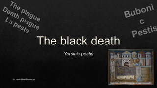
Considered as the Black Death for centuries, this infection still remains in our countries in small number and we do not think of Yersenia pestis as the main agent. Here is an view to this infection.
6. Dr. Leslie Millan Alvarez ppt 6
Y. pestis may be identified microscopically by examination of Gram, Wright, Giemsa, or
Wayson’s stained smears of peripheral blood, sputum, or lymph node specimen.Visualization
of bipolar-staining, ovoid, Gram-negative organisms with a “safety pin” appearance permits a
rapid presumptive diagnosis of plague.
https://www.cdc.gov/plague/healthcare/clinicians.html
Sailors were related with the disease. They were coming to towns and after that, the disease was found in those towns.
Religious thoughts: People at that time thought all their problems with the disease were part of God’s punishment for their sins.
Only looking at a sick person people could get sick and die.
Great Plague of London, epidemic of plague that ravaged London, England, from 1665 to 1666.
400 000 deaths
The genus Yersinia contains two other pathogenic species, Y. enterocolitica and Y. pseudotuberculosis. Molecular clock analysis suggests that Y. pestis emerged as a clone of Y. pseudotuberculosis about 20,000 years ag
Specimens of bubo aspirate, blood or sputum should be collected and appropriately plated onto blood and MacConkey agars and inoculated into nutrient broth for incubation at 37oC. Gram stain will reveal small Gram-negative bacilli. Characteristic colonies are small after 24 hours of growth and may require 48-72 hours to display lactose-negative colonies with irregular edges.
Enterotube:
nonmotility,
oxidase-negative,
catalase-positive,
lactose-negative,
indole-negative,
phenylalanine deaminase-negative,
rhamnose-negative,
urease-negative, and optimal growth rate at 28-300C.
Yersinia pestis that affects rodents (e.g., squirrels, prairie dogs, or mice), other mammals (e.g., rabbits or hares), and humans
Bubo Formation and Extracellular Growth
Y. pestis reaches a regional lymph node from the flea bite site by lymphatic flow. In experimental rat infection, extracellular bacteria appear in the marginal sinus of lymph nodes, and bacteria in macrophages and neutrophils arrive into germinal centers. Acute lymphadenitis develops with infiltration of the node by neutrophils. Additionally, the bubo contains thrombosed blood vessels, fibrin, hemorrhage, necrosis, and sheets of extracellular bacteria. Lymph node architecture is destroyed. Periglandular tissue also becomes involved with a sero-sanguinous exudate comprised of bacteria, blood cells, and fibrin.
Bacteria break out of the bubo into blood to seed other organs that contain macrophages, mainly other lymph nodes, spleen, and liver. There, the same progression follows as occurred in the bubo from intracellular multiplication to extracellular explosive growth with extensive inflammatory injury.