Call Girls Raipur Just Call 9630942363 Top Class Call Girl Service Available
Blood and Nerve supply to the Eye.pptx
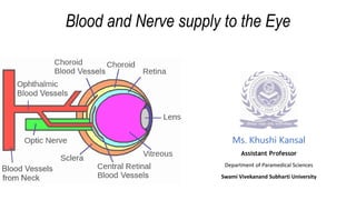
1. Blood and Nerve supply to the Eye
Ms. Khushi Kansal
Assistant Professor
Department of Paramedical Sciences
Swami Vivekanand Subharti University
2. • The optic nerve (CN II), which carries impulses from the retina to the
brain, as well as other cranial nerves, which transmit impulses to each
eye muscle, travel through the orbit.
• An ophthalmic artery and a central retinal artery (an artery that
branches off of the ophthalmic artery) provide blood to each eye.
• Ophthalmic veins (vortex veins) and a central retinal vein drain blood
from the eye. These blood vessels enter and leave through the back
of the eye.
4. • The eye is supplied by the ophthalmic artery,
which is the first branch of the internal carotid
artery.
• The ophthalmic artery has numerous branches
that supply the muscles that move the eye and
surround the eye, the eyelid and the eyeball
itself.
• The branches of the ophthalmic artery are
divided into the orbital (supply the orbit and
related structures) and the optical group (supply
the eye and its muscles).
7. Lacrimal Artery*
• This artery arises from the ophthalmic artery.
• It supply the lacrimal gland.
• The terminal branches of the artery pass through the lacrimal gland
and supply the eyelids and conjunctiva as the lateral palpebral
arteries, and pass medially to supply the upper and lower eyelids
respectively.
8. Supraorbital Artery*
• The supraorbital artery arises from the ophthalmic and supply the:
upper eyelid
frontal sinus
levator palpebrae superioris
part of the scalp
9. Posterior Ethmoidal Artery
• This artery supplies the posterior ethmoidal sinuses as well as
continuing to enter the skull and supply the meninges.
11. Medial Palpebral Artery*
• This artery has two branches i.e. the superior and inferior palpebral
arteries. They supply the upper and lower eyelids.
13. Long Posterior Ciliary Arteries
• These arteries branch from the ophthalmic artery.
• They run between the sclera and the choroid layers, and they run to
supply the ciliary muscle where they divide further and supply the:
choroid
ciliary body
iris
14. Short Posterior Ciliary Artery
• There are around 6-12 of these arteries for each eye.
• They pierce the back of the eye and run between the sclera (which
they supply) and choroid, and supply up to the ciliary processes.
• They also give off smaller branches that supply the optic disc. They
do this by forming an arterial ring known as the circle of Zinn-Haller.
15. Anterior Ciliary Arteries
• There are 7 of these arteries per eye, and they supply the sclera, and
rectus muscles.
• They branch from the ophthalmic artery. Medial, inferior and superior
rectus are supplied by two branches each, with the lateral rectus
receiving the remaining single branch.
16. Central Retinal Artery
• This artery runs underneath the optic nerve and lies within the dural
sheath of the nerve to reach the eyeball. It pierces the optic nerve
itself near the back of the eye, and sends numerous branches over
the internal aspect of the retina.
19. Sight
• The special sense of sight is transmitted by the optic nerve (CN II).
The pair of optic nerves are in fact a direct structure of the brain.
20. Lens Relaxation & Accommodation
• The short ciliary nerve innervate the ciliary muscle, which contract,
and release the tension on the zonular fibers, causing the lens to
become more convex (rounder) and hence focus on near objects.
When we need to focus on a distant object, the impulse to the ciliary
muscles is withdrawn, the suspensory ligaments become taut, and
the lens tenses and become broader.
21. Pupil Constriction/Miosis
• This occurs when the eye is exposed to light. This originates from the
Edinger Westphal nucleus which carries parasympathetic fibers that
run as the outer part of the oculomotor nerve, and eventually
synapse with the ciliary ganglion (which is a parasympathetic ganglion
that lies in the posterior orbit. This then gives off the short ciliary
nerves, which innervate the constrictor pupillae.
22. Pupil Dilation/Mydriasis
• This occurs when the eye is in the dark, when we are experiencing
sympathetic output i.e. adrenaline (fight, flight and fright response),
and in response to some drugs. Anatomically, the long ciliary nerves
(sympathetic nerves) mediate the reflex.
23. Movement
• The nerves of the orbit that enter through the superior orbital fissure
and supply the ocular muscles:
oOculomotor nerve (CN III), It innervates the inferior rectus, medial rectus and
inferior oblique.
oTrochlear Nerve (CN IV), innervates the superior oblique.
oAbducens Nerve (CN VI), innervates the lateral rectus muscle.
25. Sensation
• Sensation of the eye is possible via the Ophthalmic Nerve (V1)
division of the trigeminal nerve. It provides sensation to the:
eyeball
upper eyelid
ridge of the nose as far down as the nasal tip
• The ophthalmic nerve is one of the branches of the trigeminal nerve,
otherwise known as the fifth cranial nerve (CN V).