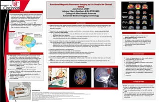
1. Functional Magnetic Resonance Imaging as it is Used in the ClinicalFunctional Magnetic Resonance Imaging as it is Used in the Clinical
SettingSetting
Julie Kohut, CNMTJulie Kohut, CNMT
Advisor: Barry Southers M.Ed RT(R)(MR)Advisor: Barry Southers M.Ed RT(R)(MR)
College of Allied Health SciencesCollege of Allied Health Sciences
Advanced Medical Imaging TechnologyAdvanced Medical Imaging Technology
BACKGROUND
INTRODUCTION
BENEFITS
FUTURE OF FMRI
Seiji Ogawa further developed MRI by discovering how to
use blood flow to map areas of the brain, providing more
information than regular MRI images and thus creating fMRI.
Oxygen-rich blood and oxygen-poor blood have a different
magnetic resonance. Since active areas of the brain use
more of the blood supply, fMRI concludes that activity is
occurring where there is more blood flow and oxygen. This is
referred to as BOLD signal.
BOLD stands for “blood oxygen level
dependent”.
The more oxygenated the blood, the more signal it
produces. Therefore, fMRI may sometimes also be referred
to as “BOLD imaging”.
Nicotine, cannabis, and acetazolamide are capable of
reducing the BOLD response.
Caffeine and theophylline have been shown to increase
BOLD response.
A functional MRI (fMRI) allows for the brain to be observed
in a whole new form. Brain mapping can help determine
how the brain is functioning in relation to certain stimuli
and actions. It can reveal which areas of the brain are
activated as it is exposed to visual stimuli, speech,
movement, and sensations.
MRI began in 1977 and fMRI developed in 1990.
It is different from a regular MRI in that it’s main purpose is
to demonstrate function and processes in thoughts rather
than only anatomy and pathology.
APPLICATIONS
Intensive studying of all different thought processes, emotions, and responses to stimuli are being researched to see
how we can further use fMRI. All sorts of stimuli ,from seeing a picture of someone’s face or tapping a finger, can be
detected in the brain.
Currently, it can be used to study how a brain could function or recover post stroke or cerebrovascular accident
Useful in evaluating brain tumors.
Surgeons can determine which areas are affected by the tumor and conclude which functions will be lost or
diminished after tumor resection.
FMRI to locate brain functions pre-surgery is much less invasive than other means such as subdural electrodes,
intracarotid amobarbital test, and intraoperative cortical stimulation.
Can be used for finding the location of a seizure and analyzing what areas of the brain are affected during that time.
Can be used along with EEG to generate even more detail.
“Spatial location of BOLD changes associated with myoclonic jerks of right foot helped to localize a focal cortical
dysplasia from left frontal lobe which was confirmed with intraoperative cortical mapping as seizure onset zone
and resected resulting in complete abolition of seizures” (Chaudhary et al., 2013, p.462).
Used to evaluate Alzheimer’s and depression.
Gender differences and homosexuality can be researched.
Even pleasure pathways and sexuality between couples has been seen on the brain.
Schizophrenia is being researched as well and could one day aid in treatment.
The brain’s response to prescribed drugs and illegal drugs can be seen.
Currently investigating fMRI’s use in lie detection. Different parts of the brain are activated when lying vs. telling the
truth.
FMRI was used in the Pepsi Challenge where it was revealed that more people’s brains prefer Pepsi however, the
branding makes them want to choose Coke. This is neuro marketing.
REFERENCES
Chaudhary, U. J., Duncan, J. S., & Lemieux, L. (2013). Mapping hemodynamic correlates of seizures using fMRI: A review. Human Brain Mapping, 34(2), 447-466. doi:10.1002/hbm.21448
Devlin, H. (2007). What is functional magnetic resonance imaging (fMRI)?
Feng, C., Narayana, S., Lancaster, J. L., Jerabek, P. A., Arnow, T. L., Zhu, F., Gao, J. (2004). CBF changes during brain activation: FMRI vs. PET. Neuroimage, 22(1), 443-446. doi:http://dx.doi.org/10.1016/j.neuroimage.2004.01.017
Fekete, T., Rubin, D., Carlson, J. M., & Mujica-Parodi, L. R. (2011). A stand-alone method for anatomical localization of NIRS measurements. Neuroimage, 56(4), 2080-2088. doi:http://dx.doi.org.proxy.libraries.uc.edu/10.1016/j.neuroimage.2011.03.068
Haller, S., & Bartsch, A. (2009). Pitfalls in fMRI.
Jezzard, P., & Buxton, R. B. (2006). The clinical potential of functional magnetic resonance imaging. Journal of Magnetic Resonance Imaging, 23(6), 787-793. doi:10.1002/jmri.20581
Kimberley, T., Khandekar, G., & Borich, M. (2007). fMRI reliability in subjects with stroke.
Lee, T. M. C., Liu, H., Tan, L., Chan, C. C. H., Mahankali, S., Feng, C., . . . Gao, J. (2002). Lie detection by functional magnetic resonance imaging. Human Brain Mapping, 15(3), 157-164. doi:10.1002/hbm.10020
Southers, G. B. (2013). MRI safety II & III. MRI Physics and Instrumentation 1. Cincinnati, OH.
Watson, S. (2013). How fMRI works. Retrieved November 13, 2013, from http://science.howstuffworks.com/fmri.htm
Wengenroth, M., Blatow, M., Guenther, J., Akbar, M., Tronnier, V. M., & Stippich, C. (2011). Diagnostic benefits of presurgical fMRI in patients with brain tumours in the primary sensorimotor cortex. doi:10.1007/s00330-011-2067-9
http://www.brandchannel.com/features_effect.asp?pf_id=201
http://www.newscientist.com/article/mg21028124.600-sex-on-the-brain-orgasms-unlock-altered-consciousness.html#.Uzn7xYVyXIU
www.Healthcare.siemens.com
http://psychcentral.com/fmri
No other imaging modality provides as much
spatial resolution and contrast as MRI.
No radiation is involved.
No fasting or prep and patient can be scanned as
many times as necessary with no negative results.
Only tool out there that can observe the brain as it
is functioning, thinking, and processing.
Non-invasive.
DISADVANTAGES
People with pacemakers and other unsafe objects in
their bodies may not have this test.
Limits who is safe to have scan.
Very sensitive to motion. Processing must be done
for epileptic patients.
One issue associated with fMRI is that the brain is
always active in some way, whether it is thinking or
responding to stimuli or sending signals to the body.
Because of this, it is often difficult to distinguish
between the baseline, or resting state, of the brain as
compared to when it is stimulated or appropriate for
the test.
Expensive.
FMRI will hopefully become a more common procedure.
While it is very expensive, the results are unlike any other
test and can offer great benefits.
There are endless possibilities for how fMRI could be
used.
Has the capability to provide such detailed information to
cure medical problems yet it can also play a role outside
the medical realm and influence law or marketing and
advertising.
.