From the workshop "High-Resolution Submillimeter Spectroscopy of the Interste...
ASM_Poster_2
1. References
Conclusion
Background Experimental Results
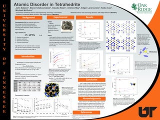
Atomic Disorder in Tetrahedrite
John Salasin1, Bryan Chakoumakos2, Claudia Rawn1, Andrew May3, Edgar Lara-Curzio3, Huibo Cao2,
Michael McGuire3
1 Department of Material Science and Engineering, University of Tennessee 3 Material Science and Technology Division; Oak Ridge National Laboratory
2 Quantum Condensed Matter Division, Oak Ridge National Laboratory
Introduction
Table 1: Atomic positions of Tetrahedrite
Atom site x y z Occ
Cu(2) 12e 0.2180 0 0 1.00
Cu(1) 12d 1/4 1/2 0 0.90
S(1) 24g 0.1144 0.1144 0.3635 0.99
S(2) 2a 0 0 0 0.89
Sb 8c 0.2683 0.2683 0.2683 1.00
Thermoelectrics (TE) are materials which turn
heat energy into electrical energy with
applications spanning multiple disciplines
including space exploration, Peltier cooling, and
engine efficiency.
Figure of Merit (zT):
zT = σS2T/κ (1-1)
T – Absolute temperature
S – Seebeck coefficient
σ – Electrical conductivity
k – Thermal conductivity
High efficiency TE are materials with a complex
unit cell and a balance between a phonon glass
(high k) and a electronic crystal (high σ).
Tetrahedrite is a natural copper sulfosalt with
the general formula:
Cu12-xMx(Sb,As)4S13
Where M denotes a Cu2+ site frequently replaced
in natural tetrahedrite with Zn, Fe, Hg, or Mn.
Structure:
It has a cubic structure with symmetry, a = 10.4 Å
(Figure 1), and only a handful of adjustable
parameters (Table 1)
Thermoelectric Properties:
Figure 2: a) Lattice electrical resistivity, b) Seebeck coefficient, c) lattice thermal conductivity for Cu12-xZnx(Sb,As)4S13 where (circles: x = 0;
squares: x = 0.5; triangles: x = 1.0; diamonds: x = 1.5).2
1: Snyder, G. J., & Toberer, E. S. (2008) Nature Materials, 7(2), 105–14.
2: Lu, X., Morelli, D. T., Xia, Y., Zhou, F., Ozolins, V., Chi, H., & Zhou, X. (2013) Advanced Energy Materials, 3,
342–348.
3:Lara-Curzio, E., May, A. F., Delaire, O., McGuire, M. A., Lu, X., Liu, C.-Y., … Morelli, D. T. (2014) Journal of
Applied Physics, 115(19).
Figure 1:Tetrahedrite crystal
structure with the formula
Cu12Sb4S13. Cu coordinations are
triangular (Cu(2),blue) and
tetrahedral (Cu(1),black). Sb has a
triangular pyramidal coordination
(brown shading)
Sample:
- Natural,
Tetrahderite, Sphalerite,
Quartz, Galena
-Casapalca District,
Huarochin Province,
Peru
Theory:
- Heat capacity shows an anomaly around 85K.3
- Calculated partial phonon density of states
shows harmonically unstable modes correlated
with the trigonally coordinate Cu sites.2
- Solving the Low-T structure will corroborate
both studies.
Low-T Single Crystal Diffraction:
X-ray, T = 28K-300K at ORNL
Neutron, T = 4.5K-450K at HFIR HB-3A: Four
Circle Diffractometer, ORNL
Refinements, Fullprof/Shelx
Figure 4: Experimental heat capacity data for natural
and synthetic tetrahedrite. (a) natural crystal; (b)
Cu12Sb4S13; (c) Cu12Sb4S13; (d) Cu11ZnSb4S13; (e)
Cu10Zn2Sb4S13.3
A) B)
C) D)
Figure 5: lattice parameter, a, as a function of
temperature. Red and Black scans are XRD both from
same parent single crystal and the blue is Neutrons.
Suggests multiple solid solution phases large enough
for an X-ray sample (80-100 µm). Neutron suggests an
averaging of the two phases with an anomalous
transition at 83K and continuation to 4K
Figure 6: Anisotropic thermal
ellipsoids clearly seen on the
Cu(2) site. All other atoms in the
UC appear to have more
isotropic representations. Very
Stark difference in Sb results
between neutron and X-ray.
A)28K X-ray; B)4K Neutron;
C)200k X-ray D) 200k Neutron.
Figure 7: Ueq as function of temperature. Showing the
increase in magnitude with Cu(2) in respect to other
atomic sites. Solid black lines are Neutron data, while
the red dashed lines are x-ray data. Cu(2) blue; Cu(1)
black; S(1) yellow; S(2) muddled yellow; Sb orange.
Low-T single crystal X-ray and Neutron diffraction
studies corroborate theoretical results predicting site
disorder at the Cu(2) trigonal planar sites and
provides further insight into the structural anomaly
around 85K. The static RMS displacement for the
Cu(2) site is 0.25Å. Further neutron studies as well as
continued sample characterization will be conducted
to understand the disorder in tetrahedrite structure
and how it correlates with the thermal conductivity.
Figure 9: Principle anisotropic direction of Cu(2) and
Cu(1) sites. The difference demonstrates the
anisotropic Cu(2) vs isotropic Cu(1) site. This confirms
the theoretical prediction of site disorder on the Cu(2)
site. Solid black line is neutrons and dashed red is X-
rays. Cu(2) blue; Cu(1) black. Static RMS displacement
for the Cu(2) sites is .25Å.
<ui>
<ui>
<uk>
<uk>
<uj> Figure 8: Ellipsoid
principle directions
Figure 10: Cu to S bond length comparison in Cu(1)
trigonal planar and Cu(1) tetrahedral coordination. X-
ray is represented by dash lines and neutrons by solid
lines. Cu(1) coordination is represented by black lines
and Cu(2) by blue lines. Cu to S(1) are red circle
markers and Cu to S(2) are green stars
markers.
U
N
I
V
E
R
S
I
T
Y
O
F
T
E
N
N
E
S
S
E
E
Aknowledgments
Many thanks to the ORAU HERE program and the
ESPN scholarship for supporting this research. This
research at ORNL's High Flux Isotope Reactor was
sponsored by the Scientific User Facilities Division,
Office of Basic Energy Sciences, U.S. Department of
Energy.
<ui>
Figure 3: Natural Crystal