
Recommended
More Related Content
What's hot
What's hot (20)
Similar to HEALTH ASSESSMENT AND DIAGNOSTIC TESTS OF EYE AND ENT DISORDERSDIAGNOSTIC TESTS FOR EYE DISORDERS.pdf
Similar to HEALTH ASSESSMENT AND DIAGNOSTIC TESTS OF EYE AND ENT DISORDERSDIAGNOSTIC TESTS FOR EYE DISORDERS.pdf (20)
More from JishaSrivastava
More from JishaSrivastava (10)
Recently uploaded
Recently uploaded (20)
HEALTH ASSESSMENT AND DIAGNOSTIC TESTS OF EYE AND ENT DISORDERSDIAGNOSTIC TESTS FOR EYE DISORDERS.pdf
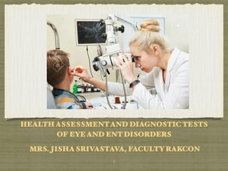
- 1. HEALTH ASSESSMENTAND DIAGNOSTIC TESTS OF EYE AND ENT DISORDERS MRS. JISHA SRIVASTAVA, FACULTY RAKCON  1
- 2. OBJECTIVES  2 At the end of the class students will be able to : 1.Describe the structures of the Eye and ENT. 2.Describe the functions of Eye and ENT. 3.Explain age affect on the Eye and ENT. . 4.Explain the techniques used in a physical examination of Eye and ENT. 5.List down the diagnostic tests for the disorders of the Eye and ENT. 6.Distinguish between normal and abnormal findings. 7.Explain the Nursing Interventions for diagnostic tests for the disorders of Eye and ENT.
- 3. ANATOMY AND PHYSIOLOGY OF EYE
- 4. ANATOMY AND PHYSIOLOGY OF EYE External structures The bony orbit (eye socket) The orbit is formed from portions of the frontal, lacrimal, ethmoid, maxillary, zygomaticus, sphenoid and palatine bones.  4
- 5. ANATOMY AND PHYSIOLOGY OF EYE External structures The eyeball is moved by six ocular muscles The four rectus muscles (the medial, lateral, superior, and inferior) move the eyes horizontally and vertically. The two oblique muscles (superior and inferior) rotate the eye in circular movements to allow vision at all angles. The upper and lower eyelids are folds of skin that close to protect the anterior eyeball. When the eyelids close, they distribute tear film  5
- 6. ANATOMY AND PHYSIOLOGY OF EYE External structures The elliptic space between the two open lids is the palpebral fissure . The corners of the fissure are called the canthi. The medial, or inner canthus is next to the nose; the lateral, or outer, canthus is the outside corner. Oil-secreting meibomian glands are embedded in both upper and lower lids.  6
- 7. ANATOMY AND PHYSIOLOGY OF EYE External structures The lacrimal gland, in the upper lid over the outer canthus, produces tears that reach the eyeball through secretory ducts. Tiny openings (puncti) in both the upper and lower lids at the inner canthus direct tears to the lacrimal sac. The nasolacrimal duct directs the flow of tears into the nose. The tear film is composed of lipids and dissolved salts, glucose, urea, protein, and lysozyme  7
- 8. Internal Structures The conjunctiva is a thin transparent layer of mucous membrane that lines the eyelids and covers the eyelid . The cornea is a transparent avascular structure with a brilliant, shiny surface. It is convex in shape, is about 0.5 mm thick, and acts as a powerful lens to bend and direct (refract) rays of light to the retina.  8 ANATOMY AND PHYSIOLOGY OF EYE
- 9. Internal Structures The cornea is composed of five layers. It derives oxygen from the atmosphere. A rich network of nerve fibers in the outer layer (epithelium) produce sensation of pain whenever the fibers are exposed or stimulated.  9 ANATOMY AND PHYSIOLOGY OF EYE
- 10. Internal Structures The sclera is the fibrous protective coating of the eye. It is white, dense, and continuous with the cornea. ln children, the sclera is thin and appears bluish because of the underlying pigmented structures. In old age, it may become yellowish from degeneration.  10 ANATOMY AND PHYSIOLOGY OF EYE
- 11. Internal Structures The uveal tract, the middle vascular layer of the eye furnishes the blood supply to the retina. The lens is a biconvex, avascular, colourless, and almost completely transparent structure, about 4 mm thick and 9 mm in diameter. The lens is surrounded by a transparent envelope (the capsule). The lens of the eye consists of about 65% water and 35% protein.  11 ANATOMY AND PHYSIOLOGY OF EYE
- 12. Internal Structures The vitreous body is a clear, avascular, jelly like structure, vitreous chamber. It helps maintain the shape and transparency of the eye.  12 ANATOMY AND PHYSIOLOGY OF EYE
- 13. Internal Structures Retina: The retina is a thin, semitransparent layer of nerve tissue that forms the innermost lining of the eye. It consists of 10 distinct layers of highly organized, delicate tissue. The retina contains all the sensory receptors for the transmission of light and is actually part of the brain. There are two types of retinal receptors: rods and cones. About 125 million rods are distributed in the periphery of the retina; they function best in dim light. Damage to these structures results in night blindness.  13 ANATOMY AND PHYSIOLOGY OF EYE
- 14. Internal Structures Optic Nerve and Neural Pathways The optic nerve is located at the posterior portion of the eye and transmits visual impulses from the retina to the brain. The head of the optic nerve (optic disc) can be Seen by ophthalmoscopic examination. The optic nerve* contains no sensory receptors (rods or cones) and represents a blind spot in the eye.  14 ANATOMY AND PHYSIOLOGY OF EYE
- 15. FUNCTION OF THE VISUAL SYSTEM Transmission of light Visual receptors of Retina: Cones and Rods Image processing  15
- 16. The major visual changes with ageing include decreases in (1) Visual acuity (2)Tolerance of glare (3) Ability to adapt to dark and lightPeripheral vision. (4)Each of these decreases is related to changes in the eye structure and each affects the quality and intensity of the light able to reach the retina. EFFECTS OF AGEING ON VISION  16
- 17. ANATOMY AND PHYSIOLOGY OF EAR
- 18. 2. AUDITORY SYSTEM 1. External Ear Auricle (Pinna): The auricle (pinna) is attached to the side of the head by skin at approximately a 20 to 30 degree angle. External Auditory Canal (Ear Canal): The ear canal extends from the concha of the pinna to the tympanic membrane. In adults, this slightly S shaped canal is approximately 2.5 cm (1 inch) in length .The sebaceous and ceruminous glands secrete a golden to black substance called cerumen (wax).  18
- 19. 2. AUDITORY SYSTEM 1. External Ear Tympanic Membrane: The tympanic membrane (eardrum) is an oval disk (approximately 1 cm in diameter); it covers the end of the auditory canal and separates the canal from the middle ear.  19
- 20. 2. AUDITORY SYSTEM 2. Middle Ear Ossicles:The outermost and largest ossicle is the malleus (hammer), which is firmly attached to the tympanic membrane. The innermost and smallest ossicle is the stapes (stirrup); its footplate occupies the oval window, in direct contact with the perilymph of the inner ear. The incus (anvil) lies between the other two and is shaped like a tooth with two roots.  20
- 21. 2. AUDITORY SYSTEM 2. Middle Ear Windows: The round window is an opening in the inner ear from which sound vibrations exit. The oval window is an opening in the inner ear into which sound vibrations enter. Eustachian Tube: The eustachian tube is a narrow channel approximately 35 mm (1% inches) long and only 1 mm wide at its narrowest end. This tube connects the middle ear to the nasopharynx.  21
- 22. 2. AUDITORY SYSTEM 2. Middle Ear Mastoid Bone: The mastoid section of the temporal bone includes the cone-shaped mastoid process; the mastoid antrum, a large cavity posteriorly continuous with the middle ear; and the mastoid air cells, which extend from the antrum and fill the temporal bone with air pockets. The mastoid bone is a bony protuberance behind the lower portion of the pinna.  22
- 23. 2. AUDITORY SYSTEM 3. Inner Ear (Labyrinth) The bony labyrinth is the rigid capsule (otic capsule) that surrounds and protects the delicate membranous labyrinth. The vestibule connects the cochlea (for hearing) to the three semicircular canals (for balance). The cochlea, which looks like a snail shell with 2% turns, is approximately 7 mm in diameter . The membranous labyrinth, lying within but not completely filling the bony labyrinth, is bathed in a fluid called perilymph, which communicates with the cerebrospinal fluid (CSF) via the cochlear duct. The membranous labyrinth consists of the utricle, the saccule, the semicircular canals, the cochleaar duct, and the organ of Corti (the end organ for division of the acoustic nerve to the brain).  23
- 24. EFFECTS OF AGEING ON HEARING Loss of auditory neurons in the organ of Corti and cochlear hair cell degeneration create an inability to hear high- frequency sounds. The hairs become coarser during the ageing process; thus retention of wax is more of a problem. Presbycusis, a gradual sensorineural loss caused by nerve degeneration in the inner ear or auditory nerve, even in people living in a quiet environment.  24
- 25. ANATOMY AND PHYSIOLOGY OF NOSE AND THROAT
- 26. External meatus. Triangular-shaped projection in the center of the face. External nostrils. Two chambers divided by the septum. Septum. Made up mainly of cartilage and bone and covered by mucous membranes. The cartilage also gives shape and support to the outer part of the nose. Nasal passages. Passages that are lined with mucous membranes and tiny hairs (cilia) that help to filter the air. NOSE  26
- 27. Ethmoid sinus. This sinus is located inside the face, around the area of the bridge of the nose. It is present at birth, and continues to grow. Maxillary sinus. This sinus is located inside the face, around the area of the cheeks. It is also present at birth, and continues to grow. Frontal sinus. This sinus is located inside the face, in the area of the forehead. It does not develop until around 7 years of age. Sphenoid sinus. This sinus is located deep in the face, behind the nose. It does not typically develop until the teen years. NOSE  27
- 28. THROAT The throat is a ring-like muscular tube. It is the passageway for air, food, and liquid. It also helps in forming speech. The throat is made up of: Voice box (larynx). The larynx is a cylindrical grouping of cartilage, muscles, and soft tissue that contains the vocal cords. The vocal cords are the upper opening into the windpipe (trachea), the passageway to the lungs. Epiglottis. A flap of soft tissue located just above the vocal cords. The epiglottis folds down over the vocal cords to prevent food and irritants from entering the lungs. Tonsils and adenoids. They are made up of lymph tissue and are located at the back and the sides of the mouth. They protect against infection. But they don't really have a function after childhood.  28
- 30. ASSESSMENT OF EYE •≈ Biographical and Demographic Data: • Age and gender. • The incidence of cataracts, dry eye, retinal detachment, glaucoma, esotropia (eyes turning inward), and exotropia (eyes turning outward) increases with age. • Hereditary color vision deficits are more common in men than in women.  30
- 31. ASSESSMENT OF EYE •≈ Current Health:There may be a fear of vision loss or uncorrectable visual manifestations  31
- 32. ASSESSMENT OF EYE: HISTORY •≈ Chief Complaint Change or loss of vision. Headache or eyestrain.  32
- 33. 1. ABDOMEN INSPECTION. Past Medical History : Diabetes mellitus, rheumatoid arthritis, and thyroid disorders, vaccinations, particularly for measles (rubella), hypertension, multiple sclerosis, and myasthenia gravis. If the client wears eyeglasses or contact lenses, ask when the last eye examination took place and when the prescription was last changed. History of head or eye trauma, must be assessed.  33 ASSESSMENT OF EYE
- 34. 1. ABDOMEN INSPECTION. Surgical History: Corrective vision surgery such as laser-assisted in-situ Keratomileusis (LASIK), radial keratotomy (RK), Cataract removal, glaucoma treatment, or eye muscle correction. Some eye surgeries, such as those for glaucoma, precipitate other eye issues (cataracts). History of brain or facial surgeries should also be assessed as there have potential to affect vision.  34 ASSESSMENT OF EYE
- 35. Allergies: Note any allergies to medications (eye drop) and other substances, such as inhalants (dust, chemicals or pollen) Medications: over-the counter (OTC), eye drops, as those with antihistamines and decongestants can dry the ocular surface. Record eye and systemic medications being used, and current and past ocular disorders.  35 ASSESSMENT OF EYE
- 36. 1. ABDOMEN INSPECTION. Dietary Habits: Herbal remedies and dietary supplements (vitamins). Some clients may consume large doses of vitamins, believing these substances will prevent the development of vision problems such as cataracts and macular degeneration. Studies have shown that diets rich in fruits, vegetables, and fish, or supplements of antioxidants C, E, and beta-carotene, have potential to reduce the incidence of visual problems like macular degeneration. harmful.  36 ASSESSMENT OF EYE
- 37. 1. ABDOMEN INSPECTION. Social History: Occupational hazards, leisure activities and hobbies, and health management behaviours. Assess the client's work and/or hobbies that may include exposure to harmful fumes, smoke or airborne particles. Assess participation in activities that increase risk for head trauma such as racquetball or baseball  37 ASSESSMENT OF EYE
- 38. 1. HEIGHT, WEIGHT, AND BODY MASS INDEX Family History: strabismus, glaucoma, myopia, hyperopia, glaucoma, cataracts, night blindness (retinitis pigmentosa), color blindness, diabetes mellitus, retinoblastoma, and macular degeneration also tend to appear in families  38 ASSESSMENT OF EYE
- 39. ASSESSMENT OF ORAL CAVITY Pain (Ophthalmalgia). Eyestrain, pulling, pressure, fullness, or generalized headache. The pain may be periocular, ocular, or retrobulbar (behind the globe). Deeper internal aching may indicate glaucoma, inflammation, muscle spasm, or infection.  39 CLINICAL MANIFESTATIONS:EYE
- 40. ASSESSMENT OF ORAL CAVITY Abnormal Vision. Refractive (focusing) error; lid ptosis (drooping eyelid); clouding lens, or aqueous or vitreous space and malfunction of the retina, optic nerve.  40 CLINICAL MANIFESTATIONS:EYE
- 41. ASSESSMENT OF ORAL CAVITY Abnormal Appearance. Lesions, edema, ptosis, and abnormal position. The most common abnormal appearance is a red eye . Abnormal Sensation. Reflex spasm of the ciliary muscle and iris sphincter that occurs with inflammation may produce brow ache and photophobia (sensitivity to light) or a constricted pupil (miosis). Itching is usually a sign of allergic reaction.  41 Clinical Manifestations:EYE CLINICAL MANIFESTATIONS:EYE
- 42. ASSESSMENT OF ORAL CAVITY Eye numbness: Diabetes nmellitus, peripheral neuropathy, Conjunctivitis, stroke EYE pain :Injury, infection, inflammation, glaucoma, foreign body, eye strain, sinusitis, corneal abrasions, hypertension, migraine, multiple sclerosis, tumor Flashes: Retinal detachment, blow to the head, ocular migraine  42 Clinical Manifestations:EYE CLINICAL MANIFESTATIONS:EYE
- 43. ASSESSMENT OF ORAL CAVITY Floaters :Red and white blood cells or gel-like clumps in the vitreous, retinal tear or detachment, normal part of aging. Foreign body: Object in eye, corneal scratches, abrasions or Foreign body, erosion, dry eye, conjunctivitis, contact lens issue, keratitis Itching :Blepharitis (eyelid infection), conjunctivitis, contact lens issues, ocular allergy  43 ASSESSMENT OF EYE CLINICAL MANIFESTATIONS:EYE
- 44. ASSESSMENT OF ORAL CAVITY Headache :Eyestrain, migraine headache, cluster headache, sinusitis, shingles, conjunctivitis, corneal abrasion or ulcer Light Sensitivity Tearing :Acute glaucoma, iritis, scleritis, corneal ulcer, thyroid disease, ectropion/entropion Tearing: Blocked tear duct, dry eye, infection of lacrimal sac, ectropion (everted lid) or entropion (inverted lid).  44 ASSESSMENT OF EYE CLINICAL MANIFESTATIONS:EYE
- 45. EXTERNAL EYE External Eye: eyebrows, eyelashes eyelids, the lacrimal apparatus, anterior portion of the eyeballs. Eye Position: symmetry and alignment. Eyebrows: eyebrows for symmetry, hair distribution, skin conditions, and movement. The eyebrows normally move up and down smoothly Eyelids and Eyelashes: placement and symmetry, lesions, I blink response. Blinking is an involuntary reflex that occurs bilaterally up to 20 times a minute. PHYSICAL EXAMINATION  45
- 46. EXTERNAL EYE Eyeballs and Lacrimal Apparatus : To palpate the eyeballs, instruct the client to close the eyes and look down. Place the tip of the index fingers on the upper eyelids, over the sclerae, and palpate gently. Normally, the eyeballs feel firm and symmetrical. Visualize the lacrimal apparatus by retracting the upper lid and having the client look down. The area should be free of swelling, edema, and excessive moisture, and there should be no regurgitation of fluid from the sac or puncta. Intraocular inflammation, iris adhesions, systemic or ocular medication side effects, or surgical alteration. PHYSICAL EXAMINATION  46
- 47. EXTERNAL EYE Conjunctivae and Sclerae: Inspect the conjunctivae and sclerae for color changes, texture, vascularity, lesions, thickness, secretions, and foreign bodies. The bulbar conjunctivae are colorless and transparent, allowing the sclerae to be seen. Small blood vessels may be visible. Healthy conjunctivae are pink to light red. PHYSICAL EXAMINATION  47
- 48. EXTERNAL EYE Cornea and Anterior Chamber: Inspect the cornea and anterior chamber from an oblique angle while shining a penlight on the corneal surface. The iris are easily visible. In older adults, a thin, greyish white ring around the edge of the cornea (arcus senilis) may be seen. The anterior chambers should appear clear and transparent with no cloudiness or shadows cast on the irides. The depth of the chamber between the cornea and iris is normally about 3 mm. PHYSICAL EXAMINATION  48
- 49. EXTERNAL EYE Iris and Pupil : Inspect the iris and the pupil with the same from the penlight. The iris should light up and have a consistent color. The light should also cause iris to constrict as the optic nerves are stimulated, using the pupil to become smaller. Neurologic disease, intraocular inflammation, iris adhesions, systemic or ocular medication side effects, or surgical alteration. PHYSICAL EXAMINATION  49
- 50. INTERNAL EYE  50 Direct Ophthalmoscopy In a darkened room, the instrument is held 1 to 2 inches away from the client's eye for examination Retinal veins radiate from the disc and are darker, and slightly thicker, than arteries. They should not be pulsating, although as a normal variation you may see an occasional spontaneous pulsation. The retinal background is pink in Caucasian people and heavily pigmented in people with a dark complexion. Choroidal vessels may appear as linear orange streaks. The presence of a cataract, or cloudy cornea, may impair examination. Abnormal findings include an altered arteriovenous ratio, narrowed arteries, widened veins, pinched off vessels, abnormal arterial light reflex, exudates, white patches, and focal hemorrhage.
- 51. INTERNAL EYE Indirect ophthalmoscopy The light source comes from a head-mounted light. The examiner holds a convex lens in front of the client's eye and, through a viewing device attached to the headband, sees an inverted reversed image. The indirect ophthalmoscope provides for binocular visual inspection with depth perception and permits a wider field of view compared with the direct method.  51
- 52. PHYSICAL EXAMINATION OF EYE INSPECTION Visual acuity 20/20. Eyebrows full, mobile. Eyelashes curve out and away from eyelids. Ptosis absent. Eyelids without Eyes moist. Palpebral conjunctivae pink, bulbar conjunctivae clear. Scleral color even, without redness. Corneal light reflection symmetrical. PERRLA,(pupils equal, round, reactive to light and accommodation) Cornea smooth; lens and anterior chamber clear, irides evenly coloured. EOMs (extra ocular movement)full, without nystagmus. Conjugate movement. No strabismus. Visual fields full to confrontation.  52
- 53. PHYSICAL EXAMINATION OF EYE PALPATION Eyeballs firm. Orbits without edema. No regurgitation from puncta. Tenderness absent over lacrimal apparatus. FUNDUSCOPIC EXAMINATION Red reflexes visualized. AV(Artery to vein) ratio approximately 2:3. Vessels without tortuosity, narrowing, pulsation. Disc margins clear, no cupping, cup-to-disc ratio 1:3. No evidence of retinal hemorrhage, patches, spots.  53
- 54. Corneal reflexes: Assesses function of fifth cranial nerve. Client looks straight ahead, bring sterile cotton wisp from behind and lightly touch cornea; may also use syringe to puff air gently across cornea. Blinking and tearing indicate intact (trigeminal) nerves. Corneal light reflex test (Hirschberg’s test): Determines eye alignment. Shine penlight 12-15 inches from bridge of nose with client staring straight ahead; observe light reflection from both corneas. Symmetrical reflection is normal Asymmetrical reflection can indicate strabismus, esotropia (deviates to nose), exotropia (deviates away from nose). hyperopia (vertical up), or hypotropia (vertical down). PHYSICAL EXAMINATION TEST OF THE EYE: REFLEXES AND MOTILITY  54
- 55. Ocular motility:Gathers information about extraocular muscles; orbit; oculomotor, trochlear, and abducens nerves; brain stem connections; and cerebral cortex. Client tracks target by both eyes as it is moved through the 6 cardinal directions of gaze . Eyes normally move parallel to each other in smooth unison. Involuntary , rapid oscillating movement of eyeball (nystagmus). Cover-uncover test: Client stares at fixed point 20 inches away; cover one eye with opaque card and observe uncovered eye for lateral or medial movement as it focuses on fixed point; remove cover and observe that eye for movement as it focuses on fixed point Repeat for other eye. No movement is a normal finding Test may need to be repeated several times to confirm abnormal findings PHYSICAL EXAMINATION TEST OF OF THE EYE:  55
- 56. ABNORMAL PHYSICAL EXAMINATION FINDING Eye position: Sunken or protruding eyes, such as protrusion of one eye or both eyes (exophthalmos). Lids: Sagging of upper lids that covers part of pupil (ptosis) Eyelids that turn inward (entropion) or outward (ectropion) Lid eversion and inversion. Blink: Rapid, infrequent, or asymmetrical blinking Asymmetrical blinking. Eyeball : Asymmetrical, hard, or soft lacrimal apparatus: Swelling, edema, excessive moisture, and regurgitation of fluid Conjunctiva: Paleness or a bright red color Cornea: Surface irregularity and cloudiness (opacity) Anterior chamber: Shallow or deep chambers (3 mm is normal) are abnormal Cloudy or nontransparent Iris: Bulging or uneven colouring Pupil: Light intolerance(photophobia). Light intolerance (photophobia), Irregular or unequal pupils that do not react to light  56
- 57. VISION TESTING Abnormal acuity implies uncorrected refractive error or pathology. Normal is 20/20. Myopia (nearsighted)is 20/30 or more. Farsighted is 20/15 or less, but may be greater than average acuity. 20/200 with corrected Visual acuity : Determines clarity of cornea, lens, and vitreous; determines function. Position client 20 ft in front of Snellen chart. Client sits with one eye covered. Ask client to read smallest line of print seen of visual pathway from retina to brain. Examples: 20/20 is normal; client reads at 20 ft what person with normal vision reads at 20 ft 20/60 means client reads at 20 ft what person with normal vision reads at 60 ft , 20/15 means client reads at 20 ft what person with normal vision reads at I5ft. Snellen chart generally placed at a distance of 20 feet, the distance at which rays of light from an object are practically parallel and little accommodation effort is needed. Sizes of symbols are identified according to the distances at which they are normally visible. For example, the largest symbols can be read 200 feet away by people with unimpaired vision.  57
- 58. VISION TESTING Visual fields To evaluate peripheral vision , use confrontational method or a computerised method. Gross visual field abnormalities can be detected. If found, refer for further exam. Visual field alteration may be caused by CNS disorders lesions, syphilis) or ocular disorders (glaucoma, retinal detachment). Client sits 2 ft away from you, looking into your eyes. Cover your right eye and client's left. To evaluate peripheral vision, you can use confrontational method (at right) or a computerized Hold a penlight equidistant between you and client, just out of view of peripheral visual field. Starting with superior field, bring object down until client states it is visible. Repeat at 45degree angles, through superior, temporal, inferior and nasal fields.  58
- 59. SELECTED EYE AND VISION DISORDERS INFLUENCED BY GENETIC FACTORS. Albinism Aniridia Color blindness Glaucoma Homocystinuria Isolated familial congenital cataracts Leber hereditary optic neuropathy Marfan syndrome Retinitis pigmentosa  59
- 62. BIOGRAPHICAL AND DEMOGRAPHIC DATA  62 Demographic data relevant to otologic assessment include the client's age. Hearing impairment may occur as a consequence of the ageing process
- 63. CURRENT HEALTH  63 Hearing problems can affect a client's ability to communicate as well as limit their social activities and job opportunities. A reduction in their physical, functional, and social activities, attributable to ear problems or hearing loss, can interfere with the client's independence, which can lead to isolation and depression. Hearing loss can result in feelings of frustration, embarrassment, and loneliness.
- 64. CHIEF COMPLAINT  64 When discussing ear complaints, ask about common clinical manifestations of the ear. The client may also complain of associated nausea or vomiting. Complete an analysis of clinical manifestations to determine the onset, duration, frequency, and precipitating and relieving factors. Explore the client's past health history to determine the chronicity of the problem and the probable cause.
- 65. CLINICAL MANIFESTATIONS Hearing Loss: Hearing loss may occur suddenly or gradually and can accompany the normal ageing process. The loss may be conductive, a result of damage to the middle ear or otosclerosis; sensory neural, a result of disease the inner car, nerve pathways or a result of loss of hair cells; or related to a CNS disorder. Vertigo. Vertigo is a sensation of motion while the person is not moving. A client may feel that either he or she or the room is moving. Dizziness is a sensation of unsteadiness and a feeling of movement within the head or lightheadedness.  65
- 66. CLINICAL MANIFESTATIONS Tinnitus. Tinnitus (ringing in the ears) may be reported as high- pitched or low-pitched, roaring, humming. This sensory neural form of auditory disorder occurs even in the absence of sound waves. Ear Drainage (Otorrhea). Ear drainage can be bloody (sanguineous), clear (serous), mixed (serosanguineous), or it may contain pus (purulent). Drainage may also be accompanied by odor and pain.  66
- 67. CLINICAL MANIFESTATIONS Infection. Ear infections in infants can impair their ability to hear spoken words and impede the development of the auditory nerve used for hearing and speech. Pain (Otalgia). Pain may be perceived as a feeling of fullness in the ear. It may be intensified by movement and relieved by holding the head still or by applying heat. Pain may occur in and around the ear and it may be intense. Fever, headache, nausea,  67
- 68. PAST MEDICAL HISTORY Infectious diseases with ear sequelae includes mumps, measles, and meningitis. Specifically inquire whether the Haemophilus influenza type b (Hib). In utero exposure to maternal influenza or rubella may result in congenital hearing loss in the child.  68
- 69. SURGICAL HISTORY History of t any surgical procedures that may have been performed. This may include a mastoidectomy, tympanoplasty, stapedectomy, or a labyrinthectomy, myringoplasty for repair of perforation of the eardrum.  69
- 70. PSYCHOSOCIAL HISTORY occupational hazards, environmental exposure, and leisure activities and hobbies. Leisure activities should include questions about swimming. Contaminated water can provoke an External car infection and, if the tympanic membrane is perforated, may lead to infection in the middle car.  70
- 71. ALLERGIES In addition to asking about allergies to medications and other substances, inquire about allergies resulting in nasal stuffiness and congestion. Close proximity of the eustachian tubes to the nasal mucosa may result in edema, which obstructs the flow of air between the middle ear and nose so that air pressure cannot be equalised.  71
- 72. MEDICATION HISTORY Aspirin is a common cause of tinnitus. Other drugs include aminoglycosides, analgesics, salicylates, quinine, chemotherapeutic agents, and antiprotozoal agents. Review the use of herbal remedies. Ginger (Zingiber Officinale) is known for its antinausea effect and can be used for the relief of motion sickness. Ginkgo biloba has been used for tinnitus and vertigo.  72
- 73. DIETARY HISTORY Adequacy of nutrient supply to local tissues should be considered. Inquire about dietary restrictions, use of food supplements (e.g.vitamins), ability to swallow and chew, and a history of any feeding problems. Ménière's disease may present with attacks of vertigo and tinnitus. Recent findings have indicated the use of a low-salt diet may be involved with the initial treatment of the disease.  73
- 74. FAMILY HISTORY Ask about a history of ear surgery or earring loss in the family. Determine the age at onset of hearing loss or changes in in hearing acuity  74
- 75. OTOTOXIC DRUGS AMINOGLYCOSIDE ANTIBIOTICS: Streptomycin, Neomycin ,Gentamicin, Tobramycin, Amikacin,Kanamycin ,Netilmicin OTHER ANTIBIOTICs: Vancomycin, Viomycin, Polymyxin B ,Polymyxin E, Erythromycin, Capreomycin , Chloramphenicol , Minocycline OTHER DRUGS: Chemotherapeutic agents (bleomycin, cisplatin, nitrogen, Salicylates )Quinine drugs Quinidine Chloroquine DIURETICS: Furosemide, Ethacrynic acid, Acetazolamide, Bumetanide, Mannitol CHEMICALS:Metals (lead, mercury, gold, arsenic), Alcohol,Aniline dyes,Caffeine, Carbon monoxide, Nicotine, Potassium bromate Povidine)  75
- 76. PHYSICAL EXAMINATION INSPECTION • Auricles symmetrical, superior portion level with outer canthus of eye. • Outer canals clear. • Pre auricular and post auricular areas without swelling, masses, or lesions. • Whisper heard at 3 feet. • Note any lumps, skin lesions, or cysts, and record approximate size and location. • Perform palpation and manipulation of the pinna to detect tenderness, nodules, or trophi (small, hard nodules in the helix that are deposits of uric acid crystals characteristic of gout).  76
- 77. PHYSICAL EXAMINATION PALPATION Tenderness over tragus and mastoid absent. No masses.  77
- 78. OTOSCOPIC EXAMINATION Soft cerumen present in canals. No discharge. TM intact, gray. Cone of light at 4:00 in right ear and at 7:00 in left ear. Landmarks visualized. No retraction or bulging. TM freely movable with pneumatic pressure.  78
- 79. THE EAR Extra cartilage tags/pre-auricular sinuses or pits. Signs of trauma to the pinna. Suspicious skin lesions on the pinna, including neoplasia. Skin conditions of the pinna and external canal. Infection/inflammation of the external ear canal, with discharge. Signs/scars of previous surgery.  79
- 80. EAR CANAL Direct Observation. Canal skin, and the presence of wax, foreign tissue, or discharge. The mobility of the eardrum can be evaluated using a pneumatic speculum, which attaches to the otoscope.  80
- 81. TESTS FOR AUDITORY ACUITY  81 The tuning fork: also provides a general estimate of hearing loss. A frequency of 512 Hz is recommended. The two major tuning fork tests date from the 19th century and are named after their originators: Weber and Rinne. Whisper Test: To exclude one ear from the testing, the examiner covers the untested ear with the palm of the hand. Then the examiner whispers softly from a distance of 1 or 2 feet from the unoccluded ear and out of the patient’s sight. The patient with normal acuity can correctly repeat what was whispered.
- 82. TESTS FOR AUDITORY ACUITY  82 Weber Test: The Weber test uses bone conduction to test lateralization of sound. A tuning fork (ideally, 512 Hz), set in motion by grasping it firmly by its stem and tapping it on the examiner’s knee or hand, is placed on the patient’s head or forehead. A person with normal hearing will hear the sound equally in both ears or describe the sound as centered in the middle of the head. In cases of conductive hearing loss, such as from otosclerosis or otitis media, the sound is heard better in the affected ear. In cases of sensorineural hearing loss, resulting from damage to the cochlear or vestibulocochlear nerve, the sound lateralizes to the better-hearing ear. The Weber test is useful for detecting unilateral hearing loss
- 83. TESTS FOR AUDITORY ACUITY  83 Rinne Test: In the Rinne test, the examiner shifts the stem of a vibrating tuning fork between two positions: 2 inchesfrom the opening of the ear canal (ie, for air conduction) and against the mastoid bone (ie, for bone conduction). As the position changes, the patient is asked to indicate which tone is louder or when the tone is no longer audible. Normally, sound heard by air conduction is audible longer than sound heard by bone conduction. The Rinne test is useful for distinguishing between conductive and sensorineural hearing losses. With a conductive hearing loss, bone-conducted sound is heard as long as or longer than air-conducted sound, whereas with a sensorineural hearing loss, air-conducted sound is audible longer than bone conducted sound. In a normal hearing ear, air-conducted sound is louder than bone-conducted sound.
- 84. TESTS FOR VESTIBULAR ACUITY ROMBERG TEST  84 A tandem Romberg test should also be performed. Instruct the client to walk forward and backward, heel to toe. A peripheral vestibular leision can cause marked swaying or falling.
- 85. TESTS FOR VESTIBULAR ACUITY ROMBERG TEST  85 A past pointing test can also indicate a labyrinthine disorder. While the client is seated, facing you with eyes open, hold out your index finger at the client's shoulder level. Instruct the client to touch your finger with the right index finger. Ask the client to lower the arm, close the eyes, and touch your finger again. A labyrinthine disorder can lead to past-pointing when the eyes are closed. Cerebral lesions are indicated when past- pointing occurs whether the eyes are open or closed.
- 86. TESTS FOR VESTIBULAR ACUITY ROMBERG TEST  86 Test for Nystagmus. Nystagmus is involuntary, rhythmic oscillation of the eyes associated with vestibular dysfunction. Nystagmus occurs normally when a client watches a rapidly moving object or looks beyond 30 degrees laterally (end-point nystagmus). To assess for gaze nystagmus, place your finger directly in front of the client at eye level. Ask the client to follow (track) the finger without moving the head.
- 87. SELECTED HEARING DISORDERS INFLUENCED BY GENETIC FACTORS • Autosomal dominant hearing loss • Autosomal recessive hearing loss (eg, connexin 26 gene) • Otosclerosis • Pendred syndrome • Usher syndrome • Waardenburg syndrome
- 89. A. LACTOSE INTOLERANCE BREATH History Smoking. Pets at home. Occupation. History of previous surgery. Previous trauma. General medical history. Seasonal or daily variation in symptoms.  89
- 90. EXTERNAL NOSE INSPECTION TEST Inspect the external surface of the nose from the front, side and behind the patient to identify any abnormalities. Inspect for skin lesions: Basal cell carcinoma: pearly lesions with telangiectasia and rolled edges. Squamous cell carcinoma: scaly lesions, sometimes with associated ulceration and hyperpigmentation. Keratoacanthoma: raised lesions with a core of scaly keratin.  90 • • Basal cell carcinoma 1 Squamous cell carcinoma 2 •
- 91. EXTERNAL NOSE INSPECTION Deviation in the nasal bones or cartilage suggestive of a fracture. This is best performed by standing behind the patient with their head tilted slightly backwards. Epistaxis is bleeding from the nose, caused by damage to the blood vessels of the nasal mucosa. Epistaxis can be caused by bleeding from anterior or posterior nasal structures.  91
- 92. BRIEF ASSESSMENT TEST 1. Sit facing the patient with your knees together and to one side of the patient’s legs. It is not pleasant for the patient to be straddled. 2. Ask the patient to look forward, keeping their head in a neutral position. 3. Carefully elevate the tip of the nose with your thumb, so that the nasal cavity becomes visible. Use a pen torch or otoscope as a light source to externally illuminate the cavity (elevating the tip of the nose will also assess for dislocated septal cartilage). 4. Inspect the nasal mucosa (including the septum) for any abnormalities. 5. Inspect and compare nasal cavity alignment (note any septal deviation). Further assessment Further assessment can be performed using an otoscope with a large speculum attached (inserting only the very tip into the nose) or using a nasal speculum (also known as Thudiculum’s speculum) which widens the nasal cavity to allow you to peer in using a light source.  92
- 93. NASAL AIRFLOW 1. Place your thumb over the nostril not being assessed to occlude airflow. 2. Ask the patient to breathe in through their nose and note the degree of airflow. 3. Repeat assessment on the other nostril, noting any difference in apparent airflow. Interpretation Reduced airflow through a particular nostril may indicate the presence of something blocking that air passage, such as a polyp, deviated nasal septum or foreign body  93
- 95. LIPS, ORAL MUCOSA AND GUMS  95 Evaluate the lips, noting color and moisture level. Scaliness or cracking may be indicative of pathology. Evaluate the oral mucosa for ulcers, color changes or nodules. Note the color of the normally pink gums, recognizing that in people of color, brown patches may be normal. Evaluate the dentition and note the presence of gum erythema or edema suggestive of gingivitis. Inspect the color and shape of the hard and soft palates. Normal mucosa is pink with a ridged hard palate. Torus palatinus may be present, and is a variation of normal.
- 96. TONGUE  96 Inspect for symmetry. The normal architecture of the tongue includes papillae that get bigger toward the rear of the tongue. Inspect the top, sides and undersurface of the tongue, noting any color variation, ulcerations or nodular lesions.
- 97. EXAMINATION OF THE NECK Inspection: Examination of the neck includes inspection for any scars, masses, glandular or nodal enlargement. Inspect the trachea, noting any deviation. Next inspect the thyroid gland as the patient swallows, noting any enlargement. Palpation: Evaluate by palpation the lymphatic chains as well as the presence of any masses in the neck. When evaluating lymph nodes for pathology, note their size, shape, consistency, mobility, and tenderness. Palpate the thyroid gland noting size, shape, consistency as well as presence of any nodules.  97
- 99. DIAGNOSTIC TESTS OF EYE  99
- 100. DIRECT OPHTHALMOSCOPY A direct ophthalmoscope is a hand-held instrument with various plus and minus lenses. The lenses can be rotated into place, enabling the examiner to bring the cornea, lens, and retina into focus sequentially. The examiner holds the ophthalmoscope in the right hand and uses the right eye to examine the patient’s right eye. The examiner switches to the left hand and left eye when examining the patient’s left eye. During this examination, the room should be darkened, and the patient’s eye should be on the same level as the examiner’s eye. The patient and the examiner should be comfortable, and both should breathe normally. The patient is given a target to gaze on and is encouraged to keep both eyes open and steady. When the fundus is examined, the vasculature comes into focus first. The veins are larger in diameter than the arteries. The retina of a young person often has a glistening effect, which is sometimes referred to as a cellophane reflex.  100
- 101. INDIRECT OPHTHALMOSCOPY It produces a bright and intense light. The light source is affixed with a pair of binocular lenses, which are mounted on the examiner’s head. The ophthalmoscope is used with a hand-held, 20-diopter lens. This instrument enables the examiner to see larger areas of the retina, although in an unmagnified state.  101
- 102. SLIT-LAMP EXAMINATION The slit lamp is a binocular microscope mounted on a table. This instrument enables the user to examine the eye with magnification of 10 to 40 times the real image. Cataracts may be evaluated by changing the angle of the light.  102
- 103. COLOUR VISION TESTING Color vision deficits can be inherited. For example, red/green color deficiencies are inherited in an X- linked manner, affecting approximately 8% of men and 0.4% of women. Acquired color vision losses may be caused by medications (eg, digitalis toxicity) or pathology such as cataracts. Changes in the appreciation of the gradations of the color red can indicate macular or optic nerve disease.. The most common color vision test is performed using Ishihara polychromatic plates. Patients with diminished color vision may be unable to identify the hidden shapes. Patients with central vision conditions (eg, macular degeneration) have more difficulty identifying colors than those with peripheral vision conditions (eg, glaucoma) because central vision identifies color.  103
- 104. AMSLER GRID The Amsler grid is a test often used for patients with macular problems, such as macular degeneration. It consists of a geometric grid of identical squares with a central fixation point. The grid should be viewed by the patient wearing normal reading glasses. Each eye is tested separately. The patient is instructed to stare at the central fixation spot on the grid and report any distortion in the squares of the grid itself. For patients with macular problems, some of the squares may look faded, or the lines may be wavy. Patients with age-related macular degeneration are commonly given these Amsler grids to take home.  104
- 105. D. COMPUTED TOMOGRAPHY Tonometry measures IOP by determining the amount of force necessary to indent or flatten (applanate) a small anterior area of the globe of the eye. The three most common types of tonometers are indentation, applanation, and noncontact. The procedure is noninvasive and is usually painless. A topical anesthetic eye drop is instilled in the lower conjunctival sac, and the tonometer is then used to measure the IOP.  105
- 106. GONIOSCOPY Gonioscopy visualizes the angle of the anterior chamber to identify abnormalities in appearance and measurements. The gonioscope uses a refracting lens that can be a direct or indirect lens.  106
- 107. PERIMETRY TESTING Perimetry testing evaluates the field of vision. A visual field is the area or extent of physical space visible to an eye in a given position. Its average extent is 65 degrees upward, 75 degrees downward, 60 degrees inward, and 95 degrees outward when the eye is in the primary gaze (ie, looking directly forward). It is most helpful in detecting central scotomas (ie, blind areas in the visual field) in macular degeneration and the peripheral field defects in glaucoma and retinitis pigmentosa. The two methods of perimetric testing are manual and automated perimetry.  107
- 108. FUNDUS PHOTOGRAPHY Special retinal cameras are used to document fine details of the fundus for study and future comparison. Photographs are compared over time to identify subtle changes in disc shape and color  108
- 109. MRI MRI allows multidimensional views to be obtained without repositioning client. MRI is used to image the areas of demyelination, and vascular lesions.  109
- 110. ULTRASONOGRAPHY High-frequency sound waves transmitted through a probe placed directly on the eyeball. A-scan measures axial length (distance from cornea to retina) to determine the refractive power of an intraocular lens in cataract surgery. B-scan US is used to evaluate lesions and their growth over time, or the presence o foreign body  110
- 111. EXOPHTHALMOMETERY It is designed to measure forward protrusion of the eye. An exophthalmometer is an instrument used for measuring the degree of forward displacement of the eye in exophthalmos.  111
- 112. X-ray study, tomography, and computed tomography (CT) are useful in evaluation of orbital and intracranial conditions, as well as detection of foreign bodies. OPHTHALMIC RADIOGRAPHY  112
- 113. Is a test that exerts pressure on the sclera with a spring plunger while retinal vessels emerging from the disc are observed. This instrument gives an approximate measuring the relative pressures in the central retinal arteries and indirectly assesses carotid arterial Flow. OPHTHALMODYNAMOMETRY  113
- 114. ELECTRORETINOGRAPHY (ERG) ERG measures the change in electrical potential of the eye caused by a diffuse flash of light through incorporated onto a contact lens that is placed directly on the eye. To assess the status of the retina in eye diseases in human patients and in laboratory animals used as models of retinal disease.  114
- 115. VISUAL EVOKED RESPONSE (VER) VER is similar to ERG in that it also measures the electrical potential resulting from a visual stimulus. Visual pathway from the retina to the cortex can be evaluated through placement of electrodes on the scalp  115
- 116. DIAGNOSTIC TESTS FOR EAR DISORDERS  116
- 117. AUDIOMETRY Audiometric testing is of two kinds: pure- tone audiometry, in which the sound stimulus consists of a pure or musical tone (the louder the tone before the patient perceives it, the greater the hearing loss) speech audiometry, in which the spoken word is used to determine the ability to hear and discriminate sounds and words. When evaluating hearing, three characteristics are important:frequency, pitch, and intensity. The normal human ear perceives sounds ranging in frequency from 20 to 20,000 Hz.  117 LOSS IN DECIBELS INTERPRETATION 0–15 Normal hearing >15–25 Slight hearing loss >25–40 Mild hearing loss >40–55 Moderate hearing loss >55–70 Moderate to severe hearing loss >70–90 Severe hearing loss >90 Profound hearing loss
- 118. TYMPANOGRAM A tympanogram, or impedance audiometry, measures middle ear muscle reflex to sound stimulation an compliance of the tympanic membrane by changing the air pressure in a sealed ear canal. Compliance is impaired with middle ear disease.  118
- 119. PURE TONE AUDIOMETRY During this test, an electrical machine produces sounds at different volumes and pitches in ear, usually while wearing some type of earphones. This test is commonly used for children, and the child is simply asked to respond in some way when the tone is heard in the earphone.  119
- 120. EVOKED POTENTIAL (EP) EP testing uses two sets of electrodes to assess hearing or sight, especially in infants and children.  120
- 121. AUDITORY BRAIN STEM RESPONSE The auditory brain stem response is a detectable electrical potential from cranial nerve VIII and the ascending auditory pathways of the brain stem in response to sound stimulation. Electrodes are placed on the patient’s forehead. Acoustic stimuli, usually in the form of clicks, are made in the ear. The resulting electrophysiologicmeasurements can determine at which decibel level a patient hears and whether there are any impairments along the nervepathways (eg, tumor on cranial nerve VIII).  121
- 122. ELECTRONYSTAGMOGRAPHY Electronystagmography is the measurement and graphic recording of the changes in electrical potentials created by eye movements during spontaneous, positional, or calorically evoked nystagmus. It is also used to assess the oculomotor and vestibular systems and their corresponding interaction. It helps in diagnosing conditions such as Ménière’s disease and tumors of the internal auditory canal or posterior fossa. Any vestibular suppressants, such as sedatives, tranquilizers, antihistamines, and alcohol are withheld for 24 hours before testing. Prior to the test the procedure is explained to the patient.  122
- 123. Platform posturography is used to investigate postural control capabilities. The integration of visual, vestibular, and proprioceptive cues (ie, sensory integration) with motor response output and coordination of the lower limbs is tested. The patient stands on a platform, surrounded by a screen, and different conditions such as a moving platform with a moving screen or a stationary platform with a moving screen are presented. The responses from the patient on six different conditions are measured and indicate which of the anatomic systems may be impaired. Preparation for the testing is the same as for electronystagmography. PLATFORM POSTUROGRAPHY  123
- 124. Sinusoidal harmonic acceleration, or a rotary chair, is used to assess the vestibulo-ocular system by analyzing compensatory eye movements in response to the clockwise and counterclockwise rotation of the chair. SINUSOIDAL HARMONIC ACCELERATION  124
- 125. MIDDLE EAR ENDOSCOPY To evaluate suspected perilymphatic fistula and new- onset conductive hearing loss, the anatomy of the round window before transtympanic treatment of Ménière’s disease, and the tympanic cavity before ear surgery to treat chronic middle ear and mastoid infections. The tympanic membrane is anesthetized topically for about10 minutes. Then, the external auditory canal is irrigated with sterile normal saline solution. With the aid of a microscope, a tympanotomy is created with a laser beam or a myringotomy knife, so that the endoscope can be inserted into the middle ear cavity. Video and photo documentation can be accomplished through the scope.  125
- 126. DIAGNOSTIC TESTS OF NOSE AND THROAT  126
- 127. A narrow tube with a lighted magnifying lens or tiny camera (nasal endoscope) enables to perform a detailed examination inside nose and sinuses. NASAL ENDOSCOPY  127
- 128. This imaging method uses X-rays and CT scan technology to give a clear look at sinus cavities SINUS COMPUTER TOMOGRAPHY (CT OR CAT) SCANS  128
- 129. Images obtained with computerized tomography (CT) can help to pinpoint the size and location of polyps in deeper areas of sinuses and evaluate the extent of swelling and irritation (inflammation). These studies may also help to rule out other possible blockages in nasal cavity, such as structural abnormalities or another type of cancerous or noncancerous growth. 3. IMAGING STUDIES.  129
- 130. ALLERGY TESTS With a skin prick test, tiny drops of allergy-causing agents (allergens) are pricked into the skin of your forearm or upper back. Your doctor or nurse then observes your skin for signs of allergic reactions.If a skin test can't be performed, your doctor may order a blood test that screens for specific antibodies to various allergens.  130
- 131. TEST FOR CYSTIC FIBROSIS A child diagnosed with nasal polyps, may be suggested testing for cystic fibrosis, an inherited condition affecting the glands that produce mucus, tears, sweat, saliva and digestive juices. The standard diagnostic test for cystic fibrosis is a noninvasive sweat test, which determines whether the child's perspiration is saltier than most people's sweat is.  131
- 132. Low levels of vitamin D, which are associated with nasal polyps. BLOOD TEST  132
- 133. It is used to diagnose certain infections of the respiratory system. The flu COVID-19 Respiratory syncytial virus (RSV) Whooping cough Meningitis MRSA (methicillin- resistant Staphylococcus aureus) NASAL SWAB  133
- 134. A laryngoscope is a thin, flexible lighted scope that can be inserted into the throat to look at the larynx (voice box). Laryngoscopy is used check for masses, vocal cord irregularities, pooling of secretions and to see if the vocal cords are working normally.  134 FLEXIBLE LARYNGOSCOPY
- 135. It is a swallowing test that uses X-Rays to monitor the swallowing process as you eat and drink food that contains barium. A series of X-Rays then show the progress of the substance through the throat, allowing the doctor to identify any problems that may affect swallowing. VIDEO FLUOROSCOPIC SWALLOW STUDY (VFSS)  135
- 136. If a suspicious mass or unusual- looking bit of tissue in the throat, a small sample – a throat biopsy – and send it to the lab for analysis. C. ENDOSCOPIC RETROGRADE  136 THROAT BIOPSIES
- 137. This involves a series of X-rays taken while you swallow a chalky substance called barium. The barium coats the inside of your throat so any swallowing changes can be seen on the X-rays.  137 BARIUM SWALLOW OR ESOPHAGOGRAM
- 138. This test measures electrical activity in the muscles of the throat to help identify nerve problems. A thin needle is put into some of the neck muscles while electrodes send signals from the muscles to a computer. C. ENDOSCOPIC RETROGRADE  138 LARYNGEAL ELECTROMYOGRAPHY (EMG)
- 139. C. ENDOSCOPIC RETROGRADE  139 STROBOSCOPY OR VIDEOSTROBOSCOPY This test uses a strobe light and a video camera to see how the vocal cords are vibrating during speech.
- 140. Understanding the complexity of ocular structures and the physiology of vision is essential to providing comprehensive nursing care for clients with Ocular disorders. Hearing and balance are vital to a person's safety and independence. Understanding the physiology of hear ing and balance is essential to providing comprehensive nursing care for clients with ear disorders.Ophthalmic and otolaryngology registered nurses perform the roles of educator, technician, counselor, and coordinator in the diagnostic setting. SUMMARY AND CONCLUSION  140
- 141. RESEARCH IOSR Journal of Dental and Medical Sciences (IOSR-JDMS) e-ISSN: 2279-0853, p-ISSN: 2279-0861.Volume 11, Issue 4 (Nov.- Dec. 2013), PP 87-92 www.iosrjournals.org www.iosrjournals.org 87 | Page Ophthalmic Manifestations in ENT Diseases & Surgical Procedures Vikas Sikarwar1 , R.S Bisht1 , Dharmendra Kumar1 , R.K Shukla2 1Department of E.N.T and Head & Neck Surgery; Veer Chandra Singh Garhwali Govt. Medical Science and Research Institute, Srinagar, UK, India. 2Department of E.N.T and Head &Neck Surgery NSCB Medical College, Jabalpur , MP , India. Abstract Objective: A prospective study was conducted in the department of ENT to see the incidence, Prevalence, Symptoms, signs and etiopathogenesis in relation to ophthalmic manifestations in ENT diseases and various ENT surgical procedures (iatrogenic). Material and methods: 35 cases were selected from IN and OUT patients of the department between July 1st 2008 to June 30th of 2009. 26 cases of orbital complications were due to various ENT diseases and 9 cases were of ENT surgical procedures with orbital complications. The standard otolaryngological and ophthalmological examination was carried out and thereafter proper hematological, radiological and histopathological examinations were done. After reaching the diagnosis, proper management was carried out. Result :35 cases of orbital complications were reported in ENT Department. Out of which, 29 cases were due to various ENT diseases and 6 cases were iatrogenic, caused by various ENT surgical procedures. Commonest ENT disease responsible for orbital complication is sinonasal tumours. Carcinoma maxilla was the most common tumour responsible for the orbital complications. Sinonasal tumours involving eye were found in wide age group range of 11 to 75 years, with higher incidence in males. Incidence of sinusitis complicating orbit is decreasing. Variety of ENT surgical procedures can involve orbital . Total maxillectomy has higher incidence of orbital complications. Conclusion: A variety of ENT diseases & surgical procedures can present with orbital complications due to anatomical association of orbit with the surrounding head and neck structures. Orbital involvement must be ruled out whenever an ENT patient presents with orbital complaints .Rapid diagnosis and treatment is necessary for preserving vision and life in these patients. Teamwork between ophthalmologist and the otolaryngologist is required for the appropriate management of such lesions.
- 142. BIBLIOGRAPHY Brunner & suddarth’s, Medical-Surgical nursing,(2015) 10th edition, Page1743-47 Joyce.M.Black, Medical-Surgical nursing,(2020) 6th edition, Page 1687-1708 Williams.S. Linda,Understanding Medical Surgical Nursing, 3rd edition(2007), page 678-690 Taylor Carol, Fundamentals of nursing(2010), 5th edition Chintamani, Lewis Medical-surgical nursing,( 2011), 2nd edition,CBS Publications Priscilla Lemonne, Medical Surgical Nursing, 2nd edition https://accesspharmacy.mhmedical.com/data/interactiveguide/physexam/ heent/neckexamination.html https://pubmed.ncbi.nlm.nih.gov/21250067/ https://www.ncbi.nlm.nih.gov/pmc/articles/PMC3858710/