
Recommended
More Related Content
What's hot
What's hot (20)
Similar to Ectopic pregnancy pannel discussion
Similar to Ectopic pregnancy pannel discussion (20)
Recently uploaded
Recently uploaded (20)
Ectopic pregnancy pannel discussion
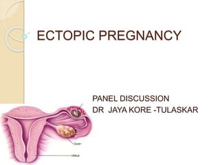
- 1. ECTOPIC PREGNANCY PANEL DISCUSSION DR JAYA KORE -TULASKAR
- 3. Moderator
- 4. Under the guidance of
- 5. Context Pregnancy in fallopian tube is a black cat in a dark night. It makes its presence felt in subtle ways and leap at you or it may slip unobserved. Although its difficult to distinguish from cats of other colours in darkness. Illumination clearly identifies it. Mc Fadyen ,1981.
- 6. DEFINITION ‘Any pregnancy where fertilized ovum gets implanted and develops In a site other than Normal uterine cavity’ Also called eccysis
- 7. Incidence The rate of ectopic pregnancy is about 1 and 2% that of live births in developed countries. 4% among those using assisted reproductive technology The risk of death among those in the developed world is between 0.1 and 0.3 percent while in the developing world it is 1-3 %., 10 times higher than those reported in developed countries The prevalence of ectopic pregnancy first trimester bleeding, pain, or both ranges from six to 16 percent in world.
- 8. An extrauterine gestation is 50 times more likely to result in a maternal death than a first-trimester abortion and 10 times more likely than delivery in the third trimester(FIGO 2011) The pregnancy rate after an ectopic pregnancy may be decreased by 40– 70%(FIGO 2011)
- 9. HISTORY History Ectopic pregnancies were initially described in the 10th century (Albucasis in 963 A.D.) For a long time were universally fatal events for the mother Initial treatments (in the old days) were desperate primitive attempts designed to destroy the growing pregnancy
- 10. Starvation Bleeding Administration of strychnine Administration of electricity into the growing gestational sac
- 11. Lawson Tait, THE FATHER OF GYNECOLOGIC SURGERY, reported the first successful operation for ectopic pregnancy in 1883. His main difficulty lay in establishing the diagnosis.
- 12. HOW DO YOU CLASSIFY ECTOPIC PREGNANCY ?
- 14. SITES OF ECTOPIC PREGNANCY
- 15. WHAT ARE RISK FACTOR ?
- 16. PATHOPHYSIOLOGY FACTORS Anatomic obstruction to the passage of the zygote Abnormal conceptus Abnormalities in the mechanisms responsible for tubal motility Transperitoneal migration of the zygote.
- 17. 1.ANATOMIC OBSTRUCTION TO THE PASSAGE OF THE ZYGOTE a.Major causes Pelvic inflammatory diseases Most important cause Chlamydial infection leads to EP Pelvic TB is another cause Post abortal & puerperal sepsis
- 18. o b.Congenital factors Tubal tortuosity , accessory ostia , diverticula & partial stenosis In utero exposure to diethyl stilboesterol ‘DES daughters’ c. Salpingitis isthimica nodosa of the tube {SIN} Tubal epithelium invades myosalpinx, forming a diverticulum
- 19. d.SURGICAL PROCEDURES ◦ Tubectomy,tubal recanalisation,tuboplasty ◦ ventrosuspension ◦ Laproscopic cauterization ◦ 1/3 rd pregnancies after tubal sterilisation turns to be ectopic (FIGO 2011)
- 20. II .AN ABNORMAL CONCEPTUS - Rapid development of trophoblast leads to premature implantation in the tube.. III .TUBAL MOTILITY - influenced by the hormonal milieu.- high estrogen levels - in cases of hyperstimulation with human menopausal gonadotropins interfere with tubal transport.) - In contrast, subnormal estrogen levels.
- 21. IV.Transperitoneal migration of the zygote. SART data from 2011 show that ectopic pregnancy occurs in 1.8% of recipients of embryo transfer during in vitro fertilization and up to 4.3% in patients undergoing zygote intrafallopian transfer (ZIFT).Indeed, the first pregnancy reported in humans with this technique was an ectopic pregnancy.
- 22. V.OTHERS; CONTRACEPTIVE METHODS IUCD prevents intrauterine pregnancy more effectively than tubal pregnancy Progesterone containing IUCD and progesterone only pills-delay tubal peristalsis and motility PREVIOUS ECTOPIC - chance of second ectopic – 12% AGE - Elderly age-more at risk
- 23. ASSISTED REPRODUCTIVE TECHNOLOGIES- IVF - IVF involves multiple egg transferred with fluid medium. - leads to flushing of one egg into tubular lumen - can also lead to implantation in uterus along with tubal implantation-heterotopic pregnancy INDUCTION OF OVULATION - by gonadotrophins - multiple pregnancy and ectopic pregnancy SMOKING
- 24. The trophoblast develops in the fertilized ovum and invades deeply into the tubal wall. Following implantation, the trophoblast produces hCG which maintains the corpus luteum. Produces oestrogen and progesterone. Which change the secretory endometrium into decidua. The uterus enlarges up to 8 weeks and becomes soft
- 25. The tubal pregnancy does not usually proceed beyond 8-10weeks due to: > lack of decidual reaction in the tube > the thin wall of the tube > the inadequacy of tubal lumen > bleeding in the site of implantation as trophoblast invades.
- 26. Changes in uterus enlarged – myohyperplasia & hypertrophy endometrium shows typical histological pattern – arias stella phenomenon –Hyperplasia of glands with loss of polarity,cytoplasmic vacuolisation,hyperchromatic nucleus. absence of chorionic villi in the endometrial curettage arias stella reaction along with absence of chorionic villi ectopic pregnancy
- 27. WHAT IS FATE OF TUBAL ECTOPIC PREGNANCY?
- 28. Separation of the gestational sac from the tubal wall leads to • Its degeneration, and fall of hCG level, • Regression of the corpus luteum and • Subsequent drop in the oestrogen and progesterone level. This leads to separation of the uterine decidua with uterine bleeding. Fate of tubal pregnancy Tubal mole Tubal abortion Tubal rupture
- 29. Fate of tubal pregnancy 1- Tubal mole: The gestational sac is surrounded by a blood clot and retained in the tube. may remain for long period in the tube- chronic ectopic pregnancy may be gradually absorbed- involution May be expelled out through the ostia-tubal
- 30. 2-Tubal abortion: Common in ampullary pregnancy Complete expulsion blood collected in pouch of douglas- pelvic hematocele Incomplete expulsion diffuse intraperitoneal haemorrhage
- 31. 3-Tubal rupture: More common in isthmic and interstitial implantation Isthmic rupture---6-8 weeks Ampullary rupture---8-12 weeks Interstitial rupture---4 months Rupture may occur in the anti- mesenteric border of the tube→ intraperitoneal haemorrhage. If rupture occurs in the mesenteric border of the tube, broad ligament haematoma →intraligamentous pregnancy
- 32. CASE…. A 22-year-old G2P1L1 was admitted with mild vaginal bleeding after 7 weeks of amenorrhoea. She had had a positive home pregnancy test. Ultrasound scan showed an empty uterus, with an adnexal mass around 2 cm. quantitative β-hCG was 2000 iu/ml On examination p-84/m and bp-120/70 mm Hg
- 33. CASE…. An 33-year old woman G4P3L3, was brought In emergency collapsed with lower abdominal pain. On admission she was in shock with blood pr. Of 80/60, a pulse of 120 bpm and tender rigid abdomen. Vaginal exam. Revealed a slight red loss, bulky uterus and marked cervical excitation with a tender mass in the right fornix.
- 34. CASE 2 A 22-year-old woman G2P1L1, was admitted with vaginal bleeding after 8 weeks of amenorrhoea. She had had a positive home pregnancy test, and described passing some tissue per vaginum. Ultrasound scan showed an empty uterus, although urinary B-hCG was still positive. A presumptive diagnosis of incomplete abortion was made, and evacuation of the uterus carries out uneventfully. She was discharged the following day. Was readmitted that night with lower abdominal pain;
- 35. WHAT ARE SIGNS AND SYMPTOMS OF TUBAL ECTOPIC PREGNANCY? WHAT ARE SIGNS AND SYMPTOMS OF TUBAL ECTOPIC PREGNANCY?
- 36. Presentation Early symptoms are either absent or subtle. CLINICAL TRIAD OF 3A’s 1.Ammenorrhoea 2.Abdominal pain 3.Abnormal uterine bleeding
- 37. Symptoms 1.Pain and discomfort Mainly due to intraperitoneal bleeding In the Lower back , abdomen, or pelvis. Acute agonizing/colicky Usually unilateral Shoulder pain – accumulation of blood in subdiaphramatic regions → stimulate phrenic nerve→shoulder tip pain Pain while urinating and passing bowels
- 38. 2.Bleeding Vaginal bleeding usually mild. Withdrawal bleeding due to decreased progesterone from corpus luteum in the failing ectopic pregnancy Internal bleeding (haemaoperitoneum) is due to hemorrhage from the affected tube. Dizziness, headache, weakness, fainting all may happen due to bleeding 3.Amenorrhea Not always present 4.Retention of urine 5.Fever,vomiting,fainting attacks
- 39. Irregular bleeding in a sexually active women should always suggestive of ectopic, until proved otherwise
- 40. Signs General examination: Weakness, pallor, hypotension,thready pulse with tachycardia, tachypnea,cold extremities-features of shock Signs of early pregnancy (breast tenderness, nausea and vomiting, change of apettite …) Abdominal examination: Lower abdominal tenderness and rigidity especially on one side may be present. No mass felt Shifting dullness Distended bowels Muscle guarding-usually absent
- 41. Vaginal examination: 1.RUPTURED Vaginal spotting with blanched white mucosa Bluish vagina and bluish soft cervix. Uterus is slightly enlarged and soft. Extreme tenderness on fornix palpation or on movement of cervix No mass usually felt Uterus floats as in water 2.UNRUPTURED Ill-defined mass with arterial pulsations
- 42. In a woman of child bearing age with pelvi- abdominal pain and/ or vaginal bleeding …… ALWAYS….think ECTOPIC PREGNANCY
- 43. WHAT IS EPAC ?
- 44. Early Pregnancy Assessment Clinic {EPAC}: . “ONE-STOP CLINIC” for women who have complications of pregnancy before 20 weeks’ gestation First established at North York General Hospital in August 2005 To offer women with earlypregnancy complications prompt diagnosis, options for
- 45. The clinic is run by a -team of dedicated gynaecologists and -experienced obstetrical nurses, with on-site ultrasound (both transabdominal and transvaginal) services performed by the gynaecologists, -easy access to laboratory services, -readily available operating services and blood bank In UK almost all hospitals have EPAC
- 46. DIAGNOSIS D/t widespread introduction of diagnostic tests and an increased awareness of the serious nature of this disease. This has resulted in early diagnosis and effective treatment. Now the rate of tubal rupture is as low as 20%. AIM has changed from " saving the mother's life " to recently " saving the woman's fertility "
- 47. METHODS OF EARLY DIAGNOSIS Immunoassay utilising monoclonal antibodies to beta HCG Ultrasound scanning – Abdominal & Vaginal including Colour Doppler Laparoscopy Serum progesterone estimation not helpful A combination of these methods may have to be employed.
- 48. Diagnosis of ruptured ectopic o Patient may be in shock with pallor , tachycardia , hypotension & cold clammy extrimities o Abdominal examination - all signs of intra abdominal haemorrhage o cullens sign may be present o Abdomen – distended with tenderness , guarding , rigidity& shifting dullness o Vaginal examination – normal or bulky uterus with tenderness on moving the cervix
- 49. Culdocentesis Determines if there is blood in the space behind the uterus, The results of a culdocentesis can be classified A negative culdocentesis - by the presence of clear fluid. A positive - free flow of nonclotting blood- intraperitoneal haemorrhage is diagnosed. But if not, ectopic pregnancy cannot be excluded. Nondiagnostic -No fluid Hematocrit of the aspirate is helpful. -Hematocrits of more than 15% -a/w ectopic -Lower hematocrits frequently indicate the presence of cystic fluid
- 50. Diagnosis of unruptured ectopic pregnancy test is +ve 1. - Pregnancy test. a) Urinary B-hCG… sensitive, detects 25-50 ml I.U/ml.. Positive before missing the next period b) Serum B-hCG…… Mainly used for quantitative rather than qualitative purposes TVS β hCG Curettage laproscopy
- 51. 1.TVS Intrauterine gestational sac with a yolksac and double decidual sign---INTRAUTERINE PREGNANCY Psuedosac---ECTOPIC PREGNANCY Transvaginal. A wk earlier than abdo… empty bladder Diagnosis made by 1. An empty uterus 2. An empty uterus with adnexal mass 3. Bagel sign 4. Presence of a gestational sac in adnexa with fetal heart
- 52. 2.Serum β-hCG If the test is negative (generally less than 5 IU/L), normal and abnormal pregnancy including ectopic are excluded. Test positive with 1500IU/L WITH 1. and an intrauterine gestational sac seen— intrauterine pregnancy 2. w/o any intrauterine sac---ectopic pregnancy If β-hCG < 1500IU/L, second assay after 48hrs 1. If doubling after 48hrs---intrauterine pregnancy 2. No doubling---failing/ectopic pregnancy
- 53. 3.Curettage Curettage of the uterus Flotation test---floating of chorionic villi in water Confirmed by microscopic examination of presence of villi CHORIONIC VILLI ABSENT IN ECTOPIC PREGNANCY 4-Laparoscopy Allows you to see the fallopian tubes and other organs Gold standard
- 54. An 33-year old woman G4P3L3, was brought In emergency collapsed with lower abdominal pain. On admission she was in shock with blood pr. Of 80/60, a pulse of 120 bpm and tender rigid abdomen. Vaginal exam. Revealed a slight red loss, bulky uterus and marked cervical excitation with a tender mass in the right fornix. WHAT WILL BE YOUR MODEL ACTION ?
- 55. • Patient usually in shock-resusciation done • Immediate arrangements of laparotomy with necessary arrangements like blood • If tubal rupture-immediate salpingectomy • If rupture at isthmial region –segmental resection of ruptured site • Cornual rupture—hysterectomy
- 57. INDICATIONS 1. Clinically stable asymptomatic women 2. Initial ß hCG < 1000IU/L and subsequent falling levels 3. Gestational sac size <4cm 4. No fetal heartbeat on TVS 5. No evidence of rupture/bleeding • Proper monitering of ß hCG twice weekly
- 58. INDICATIONS ◦ Asymptomatic women no evidence of rupture or hemodynamic instability ◦ less than 100 ml fluid in the pouch of Douglas ◦ hCG less than 1000 iu/l at initial presentation ◦ Adnexal mass less than 3cm ◦ they should objective evidence of resolution, such as declining bhCG levels. ◦ They must be fully compliant - willing to accept the potential risks of tubal rupture.
- 59. ◦ Initial follow up twice weekly with serial hCGmeasurements weekly by transvaginal examinations ◦ By the first week drop in HCG level Adnexal mass size Otherwise reassess the options (Medical/Surgical) ◦ If the fallof HCG & reduction in size of adnexalmass satisfatory weekly hCG andtransvaginal ultrasound examinations Till the HCG falls less than <20 IU MONITORING
- 60. Selection criteria ◦ Minimal symptoms &Thepatient must be hemodynamicallystable ◦ no signs or symptoms of active bleeding orhaemoperitoneum. ◦ Absence of foetal heart beat ◦ NormalFBC,U&E(urea&electrolytes),LFT(liver functiontests) Exclusion criteria ◦ Anyhepatic dysfunction, thrombocytopenia (platelet count<100,000),blood dyscrasia(WCC <2000cells cm3). ◦ Difficulty or unwillingness of patient for prolonged follow-up (average follow-up 35days). ◦ Ectopic mass >3.5cm ◦ Thepresence of cardiac activity in an ectopic pregnancy CRITERIA for MEDICAL MANAGEMENT
- 61. METHOTREXATE THERAPY WHAT IS METHOTREXATE -Methotrexate is a folic acid antagonist that interferes with the synthesis of DNA. -The use of drug therapy for ectopic pregnancy was first reported in 1982, in a patient with an interstitial pregnancy who refused surgery WHEN TO GIVE Hemodynamically stable NO cardiac activity in an ectopic pregnancy Desire future fertility.
- 62. Methotrexate – destroys actively growing tissues such as the placental tissues , (in non ruptured ectopic) Side effects include abdominal pain for 3 – 7 days in 50% of cases and mild symptoms of nausea, mouth dryness and soreness and diarrhoea, ◦ Methotrexate-Intramuscular(buttock or lateral thigh) ◦ Dose calculated from body surface area ◦ Usual dose ranges between 75-95 mg ◦ HCG checked on day 4 & day 7 If fall is less than 15 % consider second dose of methotrexate Anti-D should also be given if required METHOTREXATE
- 64. ADVICES Patient should be given information on(preferably written) ◦ Need for further treatment ◦ Adverse effects Women should be able to return easily for assessment at any time during follow-up Advice ◦ avoid sexual intercourse during treatment ◦ to maintain fluid intake ◦ use reliable contraception for three months after methotrexate has been given, barrier or hormonal) ◦ Avoid exposure to sunlight. “- Avoid alcohol and vitamin preparations containing folic acid until the hormone level is back to zero. - Avoid aspirin or drugs such as Ibuprofen for one week after treatment.
- 65. ◦ 90% successful treatment with single dose regime. ◦ Recurrent ectopic pregnancy rate 10 – 20%. ◦ Tubal patency approximately 80%. ◦ 14 % of medical management second dose of methotrexate ◦ 75% would experience abdominal pain- separation pain. This usually occurs between day 3-7 ◦ 10% would finally require surgical management OUTCOME
- 66. ADVICE 1 Avoid sexual intercourse during treatment 2 Maintain ample fluid intake 3 To use contraception for three months after methotrexate has been given, because of a possible teratogenic risk.
- 67. Day 1 is considered the day of administration of methotrexate. Follow-up hCG levels should be obtained on days 4 and 7, with an expected 15% drop between the two latter values. Thereafter, hCG values should be followed weekly until negative. Treatment failure - -less than 15% drop in hCG values between days 4 and 7 -worsening abdominal pain concerning for rupture -increasing or plateauing hCG values after the first week of therapy *Large uncontrolled studies have reported that about 14% of women will require more than one dose of methotrexate and less than 10% of women treated with this regimen will require surgical intervention(RCOG ,2014)
- 68. Contraindications TO MEDICAL THERAPY A ruptured ectopic Ectopic mass greater than 3.5 cm Fetal cardiac activity High level hCG value (10,000 IU) Breastfeeding Immunodeficiency Elevated creatinine or liver function tests Alcoholism Active pulmonary or gastrointestinal disease.
- 69. SIDE EFFECTS OF MEDICAL THERAPY Nausea and vomiting, abdominal pain, diarrhea, stomatitis, dizziness, and rarely neutropenia or reversible alopecia.
- 70. 1.Conservative surgery Indicated when woman not completed her family 5%cases—persistant ectopic noted hCG monitoring and single dose methotrexate continued after surgery Includes--1.linear salpingostomy 2.segmental resection 3.milking of the tube 2.Radical surgery—salpingectomy Indications- When the tube is not salvageable Recurrent ectopic Childbearing completed Previous sterilisation
- 71. Surgical Management Conservative, Open vs laparoscopic….. Linear salpingotomy vs milking of the tube Radical, laparoscopic vs open ……. salpingectomy
- 73. Salpingectomy versus Salpingo-oophorectomy In 1955, Jeffcoate suggested that in conjunction with a salpingectomy an oophorectomy on the ipsilateral side be done as well. The theory behind this is that all ovulations would be into the good tube; this discounts the importance of transmigration.
- 74. MANAGEMENT OF RUPTURED ECTOPIC CALL FOR HELP ABC of resuscitation ◦ give facial oxygen ◦ Site two IV lines , commence IV fluids (crystalloid) ◦ Send blood for FBC, Clotting screen and cross-match at least 4 units of blood. insert indwelling catheter arrange theatre for laparotomy whilst awaiting transfer to theatre continue fluid resuscitation and ensure intensive monitoring of haemodynamic state do not wait for BP and pulse to normalise prior to transfer-resuscitation and surgery need to go hand in hand. Pfannensteil incision, salpingectomy and wash out of abdomen assess bloods /consider CVP record operative findings including the state of the remaining tube/pelvis Anti – D immunoglobulin (250 IU)to be given to Rhesus negative women
- 75. Laparascopy OR laparatomy?? Laparoscopy has become the recommended approach in most cases. Laparotomy is usually reserved for patients: who are hemodynamically unstable patients with cornual ectopic pregnancies. for surgeons inexperienced in laparoscopy and in patients where laparoscopic approach is difficult
- 76. Laparoscopy • Less intraoperative blood loss • Shorter operation time • Shorter hospital stay • Lower analgesic requirement • Future intrauterine pregnancy rate same • Lower repeat ectopic pregnancy rate Laparotomy • Future intrauterine pregnancy rate same • Preferable in the haemodynamically unstable patient
- 77. Salpingectomy OR Salpingotomy ?? Salpingectomy Salpingectomy (tubal removal) is the principle treatment especially where there is tubal rupture Salpingotomy Conservative surgical management may be employed when the ectopic has not ruptured and where the tube appears normal Total salpingectomy is the procedure of choice: In a patient who has completed childbearing and no longer desires fertility in a patient with a history of an ectopic pregnancy in the same tube. in a patient with severely damaged tubes,
- 78. Salpingectomy Salpingotomy • There may be a higher subsequent intrauterine pregnancy rate associatedwith salpingotomy but the magnitude of this benefit may be small • Trend towards higher subsequent ectopic pregnancy • small risk of tubal bleeding in the immediate postoperative period • potential need for further treatment for persistent trophoblast
- 79. 1. Heterotopic pregnancy ◦ ectopic pregnancy coexist with intra uterine pregnancy ◦ incidence has ↑sed due to ART ◦ Surgical management with continuation of intrauterine pregnancy
- 80. 2.Interstitial pregnancy /Cornual angular preganancy ◦ implantation – interstitial part of tube ◦ pregnancy advance to a later date – myometrium ◦ abdominal pain & collapse – rupture of uterine wall ◦ In some cases, the pregnancy is expelled into the uterus and rupture does not occur. ◦ TREATMENT-immediate laprotomy with salpingectomy wedge resection of cornua reconstruction of uterine wall if severe uterinewall damage- hysterectomy
- 81. Pregnancy in a rudimentary horn Pregnancy occurs in the blind rudimentary horn of a bicornuate uterus. As such a horn is capable of some hypertrophy and distension, rupture usually does not occur before 16-20 weeks.
- 82. 3. Intraligamentous pregnancy ◦ Rare ◦ due to penetration of tubal wall by the trophoblast & its advancement b/w the two layers of broad ligament ◦ 2º to tubal pregnancy ◦ clinical findings are similar to abdominal pregnancy
- 83. Cervical pregnancy Implantation in the substance of the cervix below the level of uterine vessels. May cause severe vaginal bleeding. Can be diagnosed by trans vaginal ultrasound
- 84. Ovarian pregnancy Spiegelberg criteria for diagnosis of ovarian pregnancy: * The gestational sac is located in the region of the ovary, * the ectopic pregnancy is attached to the uterus by the ovarian ligament, * ovarian tissue in the wall of the gestational sac is proved histologically, * the tube on the involved side is intact. www.freelivedoctor.com
- 85. Abdominal (peritoneal) pregnancy Types: Primary: implantation occurs in the peritoneal cavity from the start. Secondary: usually after tubal rupture or abbortion. Intraligamentous pregnancy: is a type of abdominal but extraperitoneal pregnancy. It develops between the anterior and posterior leaves of the broad ligament after rupture of tubal pregnancy in the mesosalpingeal border or lateral rupture of intramural (in the myometrium) pregnancy.
- 86. Abdominal (peritoneal) pregnancySpecial investigations: Plain X-ray: shows abnormal lie. In lateral view, the foetus overshadows the maternal spines . Ultrasound: shows no uterine wall around the foetus Magnetic resonance imaging (MRI): has a particular importance in preoperative detection of placental anatomic relationships. www.freelivedoctor.com
- 87. PREGNANCY OF UNKNOWN LOCATION When serum hCG levels are below the discriminatory zone(intra- or extrauterine) visible on transvaginal ultrasound scan, the pregnancy can be described as being of unknown location.(RCOG 2010) Using an initial upper level of serum hCG of 1000– 1500 iu/l to diagnose pregnancy of unknown location, women with minimal or no symptoms at risk of ectopic pregnancy should be managed expectantly with 48–72 hours of follow-up and should be considered for active intervention if symptoms of ectopic pregnancy occur, serum hCG levels rise above the discriminatory level (1000 iu/l) or levels start to plateau
- 88. 45–70% of pregnancies of unknownlocation resolve spontaneously with expectant management Ectopic pregnancy was subsequentlydiagnosed in 14– 28% of cases of pregnancy of unknownlocation Interventionhas been shown to be required in 23–29% of cases.
- 89. Persistent trophoblast - detected by the failure of serum hCG levels to fall as expected after initial treatment. - occurring after salpingotomy rather than following salpingectomy. Although, even in the presence of persistent trophoblast, Acc to DATA persistent trophoblast has been seen in 8.1–8.3% after laparoscopic salpingotomy and 3.9–4.1% after open salpingotomy. .
- 90. Factors that have been suggested as increasing the risk of developing persistent trophoblast - higher preoperative serum hCG levels (>3000 iu/l), - a rapid preoperative rise in serum Hcg - the presence of active tubal bleeding. Following the elimination of all trophoblastic tissue, serum hCG levels will fall a predictable clearance curve, but the proportion of women treated for persistent trophoblast will in part depend upon the frequency of postoperative measurement and the cut off used for its definition
- 91. persistent trophoblast was initiated if the serum hCG was greater than 10% of the preoperative level ten days after surgery. Another study has suggested initiating treatment if hCG levels are above 65% of their initial level at 48 hours after surgery Methotrexate at a dose of 50 mg/m2 - used as a single dose instead of a repeat surgical procedure. The use of prophylactic methotrexate at the time of laparoscopic salpingotomy has also been reported and in one randomised trial
- 92. TAKE HOME MESSAGE Incidence of ectopic pregnancy is rising while maternal mortality from it is falling. ALWAYS suspect ectopic pregnancy in a woman of a child-bearing age c/o pain and/or p.v. bleeding Early diagnosis of ectopic is the key to less invasive treatment. Nowadays ,trend –towards conservative treatment. Careful monitoring and proper counselling of patients is mandatory. Ruptured ectopic should be unusual with complaint patients and appropriate medical care.
Editor's Notes
- without sacrificing the mother's life.
- -(hoping that the fetus would starve before the mother) -(intentional exsanguination of the mother in the hope that the fetus would die and the mother could be spared) -(to preferentially destroy the fetus)
- Often the intrauterine pregnancy is discovered later than the ectopic, mainly because of the painful emergency nature of ectopic pregnancies.
- Scarring of the endosalpinx could lead to diverticuli formation, in which the zygote could be trapped, or to simple obstruction of the tubal passage. . Elias and co-workers26 found that the incidence of chromosomal abnormalities in ectopic pregnancies is no different from that in intrauterine pregnancies. The bias with this type of study, however, is that a significant proportion of ectopic pregnancies cannot be adequately karyotyped because of the nonviability of their cells in culture. Similarly, Fedele and colleagues,27 in a case-control study, reported the risk of ectopic pregnancy (after adjustment for maternal age and parity) to be fourfold greater in women with a history of recurrent spontaneous abortion. The suspicion that some cases of ectopic pregnancy may be due to endocrine abnormalities stems from clinical observations that have suggested an association in patients using a progesterone-only pill, an IUD,13, 15, 16 or human menopausal gonadotropins for ovulation induction. An alternative explanation is that an increased number of eggs are released (superovulation) , resulting in an increased risk of ectopic implantation Abnormal progesterone levels in the luteal phase of the cycle could theoretically lead to impaired motility levels subsequent to vigorous exercise and dietary fads have been hypothesized to contribute to increased ectopic rates in today's more health-conscious society
- Synthetic nonsteroidal Estrogen drug has many bad medical effects and female babies of women who used it were at risk of developmental abnormalities of the genital system Their tubes are more likely to be abnormal and predispose to ectopic pregnancy, these females were known as DES daughters
- -partial stenosis of the tube -kinking at the isthmic portion of tube -fistulous opening in the medial end of tube
- SOCIETY FOR ASISTED REPRODUCTIVE TECHNOLOGY,SART
- it can be concluded that an IUD affords some protection against ectopic gestation for 2 years of use, after which the risk approaches that for women who are not currently using contraception. When accidental pregnancy occurs in a woman using an IUD, there is an increased likelihood that the pregnancy will be an ectopic one.
- Smoking — Cigarette smoking in the periconceptional period increases the risk of ectopic pregnancy in a dose-dependent manner, thus it can be either a low or moderate risk factor depending on the patient's habits [10,23]. This may be the result of impaired immunity in smokers, thus predisposing them to pelvic inflammatory disease, or to impairment in tubal motility.
- Separation of the gestational sac is followed by its expulsion into the peritoneal cavity through the tubal ostium with variable amount of haemorhage
- a ruptured ampullary ectopic was found at laparotomy
- Retention of urine d/t pelvic hematocel pushing bladder anteriorly
- Speculum or bimanual examination should not be performed unless facilities for resuscitation are available, as this may induce rupture of the tube
- With Advance in diagnosis and improvement in patient awareness ectopic pregnancy is more and more being diagnosed in its early stages. So, to reduce the incidence of maternal mortality and serious morbidity .
- In recent years, inspite of an increase in the incidence of ectopic pregnancy there has been a fall in the case fatality rate. s
- A needle is inserted into the space at the top of the vagina, behind the uterus and in front of the rectum to aspirate fluid -When bloody fluid is obtained -Other causes of positive culdocentesis included ruptured ovarian cysts, retrograde menstruation, endometriosis, torsion of the fallopian tube, and bleeding of unknown etiology.
- RUPTURED ECTOPIC
- AMERICAN SOCIETY FOR REPRODUCTIVE MEDICINE
- Adverse Effects Associated with Methotrexate Treatment it should not be given to women with blood dyscrasias or active gastrointestinal and respiratory disease it should not be used in women with liver or kidney disease. Methotrexate morbidity usually is dose and treatment duration dependent. Because methotrexate affects rapidly dividing tissues, gastrointestinal side effects, such as nausea, vomiting, and stomatitis, are the most common. Therefore, women treated with methotrexate should be advised not to use alcohol and nonsteroidal anti-inflammatory drugs (NSAIDs). Elevation of liver enzymes usually is seen only with multidose regimens and resolves after discontinuing methotrexate use or increasing the rescue dose of folinic acid. Alopecia is a rare side effect with the doses used to treat ectopic pregnancy. It is not unusual for women treated with methotrexate to experience abdominal pain 2–3 days after administration, presumably from the cytotoxic effect of the drug on the trophoblast tissue, causing tubal abortion. Because methotrexate affects all rapidly dividing tissues within the body, including bone marrow, the gastrointestinal mucosa, and the respiratory epithelium, . Methotrexate is directly toxic to the hepatocytes and is cleared from the body by renal excretion
- procedure involves an antimesenteric incision over the ectopic pregnancy, excising the products of conception and closing the tube in either one or two layers with fine suture material after hemostasis is achieved. The sutures used should be interrupted.
- In the management of suspected ectopic pregnancy there is a serum hCG level at which it is assumed that all viable intrauterine pregnancies will be visualised by transvaginal ultrasound. This is referred to as the discriminatory zone.