1. Malarial Parasite - Part 1 - M P, Plasmodium, Life Cycle And Diagnosis
Sample
1. Malarial parasite ( MP ) may be diagnosed from a blood smear of a patient with fever.
1. Best time to make smear is during shivering.
2. Make thick and thin blood smears.
2. Serum needed for a Serological method and for PCR.
Indication
The diagnosis of the malarial parasite.
Parasitology
1. This word malaria is the Italian word means mala, aria means bad air.
2. The plasmodium grows in the swamp bred mosquito.
3. Malaria is caused by protozoan parasites called Plasmodia and belonging to Plasmodiidae
family.
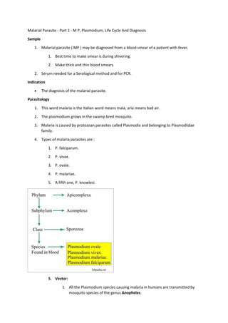
4. Types of malaria parasites are :
1. P. falciparum.
2. P. vivax.
3. P. ovale.
4. P. malariae.
5. A fifth one, P. knowlesi.
5. Vector:
1. All the Plasmodium species causing malaria in humans are transmitted by
mosquito species of the genus Anopheles.
2. 6. Spread:
1. Sporozoites from the saliva of a biting female mosquito are transmitted to either the
blood or the lymphatic system of the recipient.
2. Malaria spreads through:
1. Mosquito bite.
2. Transfusion malaria from the contaminated transfused blood.
3. By sharing contaminated needle and syringes mostly in the drug abusers.
4. Congenital malaria which is rare.
Plasmodium different shapes are discussed before starting the actual life cycle.
1.
1. Ring form is early trophozoites.
1. This is a ring-like malarial parasite following the invasion of RBCs.
2. Giemsa stain shows it as blue stain cytoplasmic circle connected to
red chromatin dot.
3. The space inside the ring is known as vacuole.
2. Trophozoites: The shape varies according to the type of malarial parasite.
1. There are a cytoplasmic circle and the chromatin dot.
2. More space is taken by the developing trophozoites.
3. Pigments are brown in color.
3. 3. Immature schizonts: There is active chromatin replication.
1. Visible cytoplasmic material surrounds the growing chromatin.
2. Pigments are often brown.
3. It occupies more space in the RBC.
4. Mature Schizonts: These are characterized by the presence of merozoites.
5. Microgametocytes: In Plasmodium falciparum is crescent-shaped, in others
is typical round shape.
1. There is large diffuse chromatin mass which stains pink to purple.
2. Chromatin mass is surrounded by colorless to pale halo.
3. Pigments are usually visible.
6. Macrogametes: These are round to oval in shape with exception of
P.falciparum which is the crescent shape.
1. Chromatin is surrounded partially or completely by the cytoplasm.
4. 2. Pigments are also present.
Lifecycle:
1. Asexual cycle:
2. The majority of sporozoites migrate to the liver and invade hepatocytes.
1. Initially, elongated sporozoite has transformed into a rounded form.
2. This rounded form then matures within the hepatocyte to a schizont containing
many merozoites.
3. This cycle takes 5 to 16 days.
4. Merozoites leave the liver and enter the blood, this is an asexual cycle.
3. The sexual cycle:
4. It starts when the mosquito sucks the blood from the patients.
1. Micro and macrogametocytes in the stomach of mosquito combine and form zygote.
2. This forms oocyst and sporozoites.
3. Sporozoites are injected into humans containing merozoites.
RBCs cycle (Erythrocytic cycle) Asexual cycle:
1. Now Merozoites enter the RBCs and start the Asexual cycle.
1. produce more merozoites. This cycle repeats 1 to 3 days.
2. This multiplication can result in thousands of parasite-infected cells in the host
bloodstream.
3. The patient may develop sign and symptoms of illness and complications of malaria.
4. The complication of malaria parasite if not treated then it may last for months.
5. Some of the merozoites transform into a sexual form called as male and female
gametocytes.
6. These gametocytes circulate in the blood are taken up by the biting mosquito.
5. The life cycle in the mosquito (Sexual cycle):
1. When the mosquito bites the infected humans, then suck the blood and these gametocytes
go into along the blood.
1. In mosquito RBCs, burst and gametocytes are released These will get into more
mature form Gametes.
2. Male and female gametes fuse to form diploid zygotes.
3. Zygotes develop into actively moving ookinetes that burrow into the mosquito
midgut wall and form oocysts.
4. Growth and division of each oocyst produce thousands of active haploid forms
called sporozoites.
6. 5. After 8-15 days, the oocyst bursts, releasing sporozoites into the body cavity of the
mosquito, from which they travel to and invade the mosquito salivary glands.
6. Mosquito is ready to infect the humans.
7. Clinical presentation
1. The Typical patient is initially asymptomatic following the mosquito bite.
2. Once the erythrocytic phase starts there is a large number of RBCs rupture, leading to
release of merozoites and toxic material.
1. This is the time when patients get the first attack of malaria as:
1. First chills for 10 to 15 minutes and then fever 2 to 6 hours or more.
2. When fever settles to normal then patient get profuse sweating.
3. This cycle may vary with the type of malaria.
3. The patient may have a headache, lethargy, anorexia, nausea, and vomiting.
4. There may be diarrhea.
5. There is anemia.
6. There may be involvement of the central nervous system.
7. Kidney involvement may lead to nephrotic syndrome.
Complications of Malaria
1. The patient may have :
8. 2. Cerebral malaria.
3. Anemia.
4. Organ failure.
5. Some patient may have respiratory difficulty.
6. Rarely there may be low blood sugar level.
Diagnosis
1. History of the patient in suspected areas.
2. Blood smear:
1. Make a blood smear when the patient has a fever. Thin and Thick smears are made.
2. Thick smear is more helpful to find M.Parasites.
1. Thin smear is good to identify the type of malarial parasite.
3. Collect blood 6 to 8 hourly till 48 hours to declare negative for malaria.
4. Giemsa stain is the best choice.
3. Serologic methods are based on immunochromatic techniques. Tests most often use a
dipstick or cassette format and provide results in 2-15 minutes.
4. Polymerase chain reaction (PCR): Parasite nucleic acids are detected using the
PCR technique. This is more sensitive than smear microscopy. This is of limited value for the
diagnosis of acutely ill patients because of the time needed for this procedure.
Mosquito control
1. Try to eliminate the breeding places:
1. Fill the vacant land and pump out the water.
2. Remove the junk and water retaining debris.
2. Destroy the larvae:
1. Clean the drains.
2. Try to remove algae from the ponds.
3. Add larva-eating fish to the ponds.
3. Use of the insecticide:
1. The best example is DDT.
4. Use of mosquito repellent:
1. Pyrethroid repellent.
2. N, N- diethyl meta tolbutamide.
5. Use of mosquito nets.
9. 6. Use of clothes to prevent the mosquito bites.
7. Train people for the malaria prevalence.
8. Train the people for the detection of malaria, treatment, and follow-up.
Prevention
1. No vaccine is available.
2. Give prophylactic anti-malarial drugs.
3. Use mosquito net.
4. Use mosquito repellant.
Treatment
1. Antimalarial drugs used are quinidine, chloroquin, primaquin, pyrimethamine, sulfadoxine,
mefloquin, tetracyclines and proguanil.
2. Chloroquine is the drug of choice and best for P. falciparum.
1. This is effective for the erythrocytic stage and not for the liver stage.
2. Must use primaquine to eradicate P. ovale and P. vivax.
3. there are chloroquine resistant cases of P. falciparum.
3. Amodiaquin, piperaquin and pyronaridine are close to chloroquin.
1. Amodiaquine is less toxic, cheap and in some areas effective against chloroquine-
resistant P. falciparum.
4. Mefloquine is effective against choloquin resistant P. falciparum.
5. Quinine and quinidine are still the first line of therapy against P.falciparum.
6. Primaquine is a synthetic drug and is the drug of choice for the eradication of liver stage
from P. vivax and P. ovale.
7. Antibiotics and Inhibitors of folate synthesis are slow acting antimalarial drugs.
8. Halofantrine and Lumefantrine are related to quinine and effective against the erythrocytic
stage.
9. Malria drug resistant strains are emerging.
Characteristic features of various forms of the Malarial parasite
Malaria type Stages Infected RBC
size
Trophozoite Scuffer
stippling
GAMETOCYTES
P. vivax All enlarged small and may become
amoeboid
present rounded
P. Ovale All enlarged small, with large chromatin present rounded
10. P. malariae All normal small with dense cytoplasm,
rarely band form
absent rounded
P. falciparum ring
form
normal smallest, sometimes
multiple, often double
chromatins
absent banana shaped
Typical features of Malarial parasite
P. falciparum P. vivax P. ovale
Incubation period 8 to 11 days 7 to 10 days 7 to 10 days
Fever pattern Continuous, quotidian or
remittent
Irregular or quotidian Irregular or qu
Prodromal symptoms Influenza-like Influenza-like Influenza-like
The periodicity of symptoms 36 to 48 hours 44 to 48 hours 48 to 50 hours
The severity of the initial attack Severe 16 to 36 hours moderate to severe, 10
hours
mild for 10 ho
Duration without treatment 2 to 3 weeks 3 to 8 + weeks 2 to 3 weeks
CNS involvement very severe ++++ ± ±
Anemia very severe ++++ ++ +
Kidney involvement + ± -
Red blood cell normal size larger than the normal,
Schuffner's dots are seen
larger than the
schuffner's do
seen
Early trophozoite about 1/5 diameter of RBC,
chromatin is a small dot
about 1/3 diameter of RBC
with heavy chromatin dots
Like vivax and
Trophozoite mature usually not seen in peripheral
blood
as a large mass of
chromatin, fine brown
pigments
11. Schizont 8 to 24 or more merozoites 12 to 24 merozoites 4 to 16 but mo
merozoites
Macrogametocyte size like microgametocyte.
chromatin more compact
Cytoplasm stains dark blue,
chromatin more compact
Microgametocyte Crescent-shaped, chromatin is
diffuse, pink, cytoplasm pale blue
Round or oval, almost fill the
RBC, chromatin is diffuse in
large masses, pink, no
vacuoles
Like vivax but
Stages development in the
mosquito
10 to 12 days at 27 °C 10 days at 25 °C to 30 °C 14 days at 27