
Recommended
More Related Content
Similar to Anemia.pptx
Similar to Anemia.pptx (20)
Recently uploaded
Recently uploaded (20)
Anemia.pptx
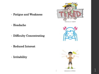
- 1. • Fatigue and Weakness • Headache • Difficulty Concentrating • Reduced Interest • Irritability 1
- 2. 2
- 3. • According to the World Health Organization (WHO), an estimated 1.62 billion people worldwide had anemia in which represents about 25% of the global population. • According to the WHO, approximately 47% of preschool children in developing countries were estimated to be anemic. 3
- 4. • According to the “National Nutrition Survey” 2018, The prevalence of anemia among children aged 6 to 59 months in Pakistan is 62.3%. Among women of reproductive age (15-49 years), the prevalence of anemia is 52.1%. Among pregnant women, it is 62.8%. 4
- 5. Anemia 5
- 6. Learning Objectives To define “Anemia”. To evaluate patients presenting with anemia. To manage patients diagnosed with anemia. 6
- 8. Anemia Anemia is a common complaint in primary care. Anemia is defined as low hemoglobin concentration or a low hematocrit/RBC count. According to the World Health Organization, anemia is diagnosed with: oa hemoglobin concentration <13 g/dL in men oA hemoglobin concentration <12 g/dL in women 8
- 10. 10
- 12. Classification of Anemia Anemia is classified as macrocytic, microcytic or normocytic anemia based on the MCV. oMicrocytic anemia: when the MCV is less than 80. oMacrocytic anemia: when the MCV exceeds 100. oNormocytic anemia: when MCV is between 80 and 100. 12
- 13. Types of Anemia Microcytic Anemia Normocytic Anemia Macrocytic Anemia Iron deficiency anemia Thalassemia & other hemoglobinopathies Sideroblastic anemia Lead poisoning Copper deficiency Porphyria Anemia of inflammation (anemia of chronic disease) Acute bleeding Early iron deficiency Anemia of chronic disease/inflammation Bone marrow suppression (cancer, aplastic anemia, infection) Chronic renal insufficiency Hypothyroidism Hypopituitarism Excess alcohol Vitamin B12 deficiency Folate deficiency Alcohol abuse Liver disease Hypothyroidism Medications that interfere with nuclear maturation (hydroxyurea, methotrexate, some chemotherapy agents) Conditions associated with abnormal RBC maturation such as myelodysplastic syndrome and acute leukemia 13
- 16. 16
- 17. 17 SxS of Anemia CBC MCV < 80 Iron Studies Ferritin TIBC %Sat Serum Iron Iron Deficiency Anemia ACD Thalassemia Sidroblastic Anemia
- 18. 18 SxS of Anemia CBC MCV > 100 Peripheral Smear Vit B12 Deficiency Folate Deficiency Liver Disease Medication Alcohol Megaloblastic Anemia Non-Megaloblastic Anemia > 5 lobes PMNs
- 20. 20
- 21. Red Blood Cells Indices Red blood cell (RBC) count: the number of RBCs contained in a specified volume of whole blood. Mean corpuscular volume (MCV): the average volume or size of the RBCs. Mean corpuscular hemoglobin (MCH): the average hemoglobin content in a RBC. A low MCH indicates decreased hemoglobin content per cell. Mean corpuscular hemoglobin concentration (MCHC): the average hemoglobin concentration per RBC. 21
- 22. Red cell distribution width (RDW): measure of the variation in RBC size. (always high in Nutritional Cause of Anemia) 22
- 23. 23 Retic Count ; Differentiates between Production and destruction anemia. Retic Count > 2 % = Destructive Anemia Retic Count < 2 % = Productive Anemia
- 24. History Check the duration of anemia Check for general symptoms of anemia if present such as fatigue, dizziness, pica, craving for ice, or dyspnea Check for constitutional symptoms (loss of appetite, weight loss, fever, and/or night sweats), which might indicate an infection or a malignancy. Check for other associated symptoms such as abdominal discomfort, hematochezia, and bright red rectal bleeding. Check for medical conditions that might be associated with anemia such as chronic kidney disease, rheumatoid arthritis, peptic ulcer etc. Check medications intake: Macrocytosis can be caused by Valproic acid, Trimethoprim/ sulfamethoxazole, Biguanides. Check family history of anemia, which can indicate hemoglobinopathies. Check nutrition, eating habits, alcohol consumption. Check exposure to toxins (occupational or environmental exposure to toxins such as Lead) 24
- 26. Physical Examination Check vital signs namely heart rate, respiratory rate, temperature, and blood pressure (look for postural hypotension). Check for the presence of PALLOR (nail bed, palm crease, conjunctiva) , jaundice. Conduct a comprehensive physical examination to detect signs of ORGAN INVOLVEMENT (such as splenomegaly) and assess the severity of the condition. 26
- 28. Microcytic Anemia- Causes MAIN MICROCYTIC ANEMIA Iron deficiency anemia Thalassemia & other hemoglobinopathies Sideroblastic anemia Anemia of inflammation (anemia of chronic disease) OTHERS Lead poisoning Copper deficiency Porphyria 28
- 30. Approach to Microcytosis History Tests o CBC o MCV+RETIC COUNT IRON STUDIES o FERRITIN o TIBC o Transferrin o % Saturation 30
- 32. Definition Iron deficiency anemia is due to diminished red blood cell production as a result of low iron stores in the body. It is the most common nutritional disorder worldwide. It accounts for approximately one-half of anemia cases. 32
- 33. Iron Journey Before discussing causes, let’s review iron journey 33
- 34. Copyrights apply
- 35. 35
- 36. 36
- 37. 37
- 38. SUMMARY OF RBC INDICES in iron deficiency anemia 38
- 39. 39
- 40. Causes of Iron deficiency anemia 40
- 41. Causes 1. Inadequate iron intake (Nutrition Problem) 2. Decreased iron absorption like in celiac disease, gastrectomy, prolong usage of PPIs 1. Increased iron demand 2. Increased iron loss: can be benign (menstrual blood loss) or malignant (occult gastrointestinal malignancy), or Peptic Ulcer 41
- 42. 42
- 43. 43
- 44. 44
- 45. 45
- 46. 46
- 47. 47
- 48. Symptoms Many patients have no symptoms (asymptomatic) (Already discussed) Symptoms include: oWeakness oHeadache oDecreased exercise tolerance oFatigue, Vertigo oIrritability oDepression oPica and pagophagia (ice craving) 48
- 49. Physical Examination Pallor Dry or rough skin Blue/Yellow sclerae Atrophic glossitis with loss of tongue papillae, which may be accompanied by tongue pain or dry mouth Cheilosis (also called angular cheilitis) Koilonychia (spoon nails) Esophageal web, which may be accompanied by dysphagia (Plummer-Vinson or Patterson-Kelly syndrome) Alopecia (rare) in especially severe cases Chlorosis (pale, faintly green complexion; extremely rare) Dermatitis herpetiformis in celiac disease Bloody stool or melena 49
- 50. Treatment Iron Supplements Dose is 120 mg of elemental iron for 3 months, continue for additional 3 months to replenish iron stores + Stool Softeners NOTE With oral iron : Reticulocytosis should be seen within 7 days correction of anemia in 6 weeks. In 1 month, increase of 1g/dl of Hb is the expected response Vitamin C may enhance absorption of oral iron if administered 30 minutes prior to iron. 50
- 51. Copyrights apply
- 52. IV iron Treatment IV iron is appropriate for patients who are unable to tolerate gastrointestinal side effects of oral iron. Examples include older individuals, pregnant women (who already have gastrointestinal symptoms related to the pregnancy), and individuals with existing gastrointestinal disorders that may exacerbate oral iron side effects. IV iron may be needed for those with severe/ongoing blood loss (eg, telangiectasias, varices). Gastric surgery (bypass, resection) that reduces gastric acid may severely impair intestinal absorption of oral iron. Malabsorption syndromes (celiac disease, Whipple's disease, bacterial overgrowth) may limit absorption of oral iron. In the second trimester of pregnancy, if the Hb is less than 10.5 g, or at any time in the third trimester, at which oral iron is unlikely to rapidly supply adequate iron to the developing fetus. 52
- 53. Copyrights apply
- 54. 54
- 56. 56
- 57. 57
- 58. diagnosis 58
- 59. 59
- 60. 60
- 61. treatment 61
- 62. 62
- 64. 64
- 65. 65
- 66. causes 66
- 67. 67
- 68. 68
- 70. 70
- 71. Thalassemia 71
- 72. Definition of Thalassemia A group of inherited autosomal recessive hematologic disorders that cause hemolytic anemia due to decreased or absent synthesis of a globin chain. oAlpha thalassemia is caused by reduced or absent synthesis of alpha globin chains, resulting in excess beta globin chains. oBeta thalassemia is caused by reduced or absent synthesis of beta globin chains, resulting in excess beta chains. Imbalances of globin chains cause hemolysis and impair erythropoiesis. 72
- 73. 73
- 74. Prototypical Forms of Alpha Thalassemia 74
- 75. Prototypical Forms of Beta Thalassemia 75
- 76. 76
- 77. Thalassemia Trait Thalassemia Trait is asymptomatic. Majority of cases of Thalassemia Trait are discovered incidentally in the presence of microcytic anemia. Microcytic anemia can be caused by iron deficiency, thalassemia, lead poisoning, sideroblastic anemia, or anemia of chronic disease. RBC count may be increased despite the presence of anemia 77
- 78. diagnosis 78
- 79. Diagnosis of Thalassemia The hemoglobin Electrophoresis is the diagnostic test. Beta thalassemia o reduced or absent HbA o elevated levels of HbA2 o increased HbF. Alpha Thalassemia: Hemoglobin electrophoresis is usually normal in adults. Molecular Diagnosis is required and not Hemoglobin Electrophoresis In the newborn period, if the electrophoresis shows Hb 79
- 80. 80
- 81. Beta Thalassemia Major Persons with beta thalassemia major are diagnosed during infancy. Pallor, irritability, growth retardation, abdominal swelling, and jaundice appear during the second six months of life. Persons with a microcytic anemia but milder symptoms that start later in life have beta thalassemia intermedia. 81
- 82. Thalassemia Trait Most persons with thalassemia trait are found incidentally when their CBC shows a mild microcytic anemia. The MCV is usually less than 75 with thalassemia. The RDW may assist in differentiating iron deficiency and sideroblastic anemia from thalassemia. The RDW will be elevated in more than 90% of persons with iron deficiency, but in only 50% of persons with thalassemia. Supplemental tests include serum ferritin, the peripheral smear, hemoglobin electrophoresis, serum lead level, and rarely bone marrow aspirate. 82
- 83. treatment 83
- 84. 84
- 85. Complications Complications are related to: ooverstimulation of the bone marrow oineffective erythropoiesis oIron overload from regular blood transfusions. Iron is deposited in visceral organs (mainly the heart, liver, and endocrine glands). Most patient deaths are caused by cardiac complications. 85
- 86. Important Persons with anemia from thalassemia trait should not take iron supplements unless they have coexistent iron deficiency. Persons with beta thalassemia major require periodic lifelong blood transfusions to maintain hemoglobin levels higher than 9.5 g per dL and sustain normal growth. Persons with beta thalassemia major require chelation therapy for iron overload. Folic acid deficiency has been reported in thalassemia major and intermedia as a result of increased erythropoiesis. Therefore, daily oral supplementation with 1 mg of folic acid is recommended for persons with evidence of folate deficiency. 86
- 88. Normocytic Anemia- Causes Acute bleeding Early iron deficiency Anemia of chronic disease/inflammation Bone marrow suppression (cancer, aplastic anemia, infection) Chronic renal insufficiency Hypothyroidism Hypopituitarism Excess alcohol 88
- 89. Approach to Normocytic Anemia History o Symptoms of acute blood loss o Symptoms of chronic diseases o Medications intake Tests o BUN, Creatinine o TSH, Free T4 o Peripheral blood smear o SPEP/UPEP to rule out multiple myeloma o PTH 89
- 91. Macrocytic Anemia- Causes Vitamin B12 deficiency Folate deficiency Alcohol abuse Liver disease Hypothyroidism Medications: anticonvulsants (valproate, phenytoin), antimicrobials (valacyclovir, trimethoprim sulfa), antiretrovirals (zidovudine, stavudine), chemotherapeutic agents (hydroxyurea, methotrexate) Myelodysplastic syndrome and acute leukemia 91
- 92. Approach to Macrocytosis History oCheck diet and nutrition oCheck for thyroid disease oEvaluate for alcohol intake oCheck medications intake oCheck for chronic diseases namely liver diseases Tests oVitamin B12 level oOther tests as indicated: TSH, Free T4, Liver function tests oNo need to do folate level. If deficiency is suspected, empiric treatment with folic acid is indicated. oPeripheral smear : if it shows signs of myelodysplastic disease, a bone marrow biopsy is indicated. 92
- 94. Clinical Manifestations of Vitamin B12 Cutaneous: Hyperpigmentation, Jaundice, Vitiligo Gastrointestinal: Glossitis Hematologic: Anemia (macrocytic, megaloblastic), Leukopenia, Pancytopenia, Thrombocytopenia, Thrombocytosis Neuropsychiatric: Areflexia, Cognitive impairment (including dementia-like symptoms and acute psychosis), Gait abnormalities, Irritability, Loss of proprioception and vibratory sense, Olfactory impairment, Peripheral neuropathy. 94
- 95. History Paresthesia Memory loss Dementia Ataxia History of malabsorption or gastrectomy 95
- 96. Physical exam Glossitis Cheleitis Decreased proprioception Decreased vibratory sense 96
- 97. Screening It is not recommended to screen persons at average risk of vitamin B12 deficiency. It is recommended to screen persons with risk factors. 97
- 98. Risk Factors Decreased ileal absorption: such as in Crohn disease, Ileal resection,Tapeworm infection Decreased intrinsic factor: Atrophic gastritis, Pernicious anemia, Postgastrectomy syndrome Genetic: Transcobalamin II deficiency Inadequate intake: Alcohol abuse, Patients older than 75 years, Vegans or strict vegetarians (including exclusively breastfed infants of vegetarian/vegan mothers) Prolonged medication use: Histamine H2 blocker use for more than 12 months; Metformin use for more than four months; Proton pump inhibitor use for more than 12 months. 98
- 99. Diagnosis Diagnostic testing is recommended in persons with suspected clinical manifestations. Testing includes CBC and serum vitamin B12 level. oA level of 150-399 pg per mL (111-294 pmol per L) is considered as low-normal. oA level < 150 pg per mL (<111 pmol per L) is diagnostic for vitamin B12 deficiency. 99
- 100. Artificial Elevation of Vitamin B12 Serum vitamin B12 levels may be artificially elevated in patients with oAlcoholism oliver disease ocancer This is due to decreased hepatic clearance of transport proteins and resultant higher circulating levels of vitamin B12. 100
- 101. Additional Tests Methylmalonic acid level: in case of a normal or low-normal serum vitamin B12 level and macrocytosis in CBC. Anti-intrinsic factor antibodies: to test for pernicious anemia in patients with vitamin B12 deficiency and without evidence of dietary or malabsorptive causes by history and physical exam. In case, these antibodies are negative, serum gastrin level can be requested to check further for presence of pernicious anemia. 101
- 102. Treatment Intramuscular injections of cyanocobalamin or oral vitamin B12 therapy. Injectable therapy leads to more rapid improvement and should be considered in patients with severe deficiency or severe neurologic symptoms. Injections three times per week for two weeks in patients without neurologic deficits. If neurologic deficits are present, injections should be given every other day for up to three weeks or until no further improvement is noted. 102
- 103. Prevention Patients who have had bariatric surgery should receive 1 mg of oral vitamin B12 per day indefinitely. Patients older than 50 years may not be able to adequately absorb dietary vitamin B12 and should consume food fortified with vitamin B12. Vegans and strict vegetarians should be counseled to consume fortified cereals or supplements to prevent deficiency. 103
- 104. After initiation of treatment Homocysteine or methylmalonic acid level, or reticulocyte count: improve in 1 week Neurologic symptoms improve in 6 weeks to three months Anemia, leukopenia, mean corpuscular volume, or thrombocytopenia improve in 8 weeks. 104
- 105. References 105