1. ISOLATED RUPTURE OF THE BRACHIALIS
MUSCLE
DR.HAFIZ-UR-REHMAN, M.S.(ORTHOPAEDICS SURGERY)
A/P.GHULAM MUSTAFA, Professor.M.A.QURAISHI,
Department of Orthopaedics Surgery Unit-I Dow University of Health Sciences/ Civil
Hospital Baba -e-Urdu Road KARACCHI-PAKISTAN.
The brachialis muscle is rarely seen with isolated tear that has not been well
documented. A most common traumatic muscular injury in the upper arm is rupture of the
biceps brachii; 2,4, We report the case of a young patient with h/o stab wound injury right
upper arm who had an isolated tear of the brachialis muscle that was treated
conservatively, only skin closure was done with silk. After 4 months had a return to full
function with full range of movements.
Case Report
A twenty four years old right-hand-dominant young machinist presented with
history of trauma as alleged assault with knife stab injury, bleeding wound anterio medial
aspect of left upper arm three inches superior to elbow joint, margins of the wound were
sharp cutting 2.5cm x 2 mm deep to muscles. In wound management, after thorough
irrigation only skin closure was done with silk. Sutures removed on eleventh day.
On presentation patient c/o pain, tiredness on working and numbness of the right
upper extremity.
On examination the patient reported no erythema or tenderness and muscles power
of grade 4. Active passive movements were pain free with full range of motion. The skin
scar was slightly puckered, adherent to underling soft tissue. There was firm, mobile in
transverse plane mass measuring 0.5X 3 cm on the aneriomedial side of the distal aspect
of the arm. Neurovascular examination revealed normal. No sensory deficits were noted;
motor examination showed strength of 4 of 5 throughout, excess for flexion and
supination of the elbow, which were rated 4. The remainder of the physical examination
revealed normal findings,
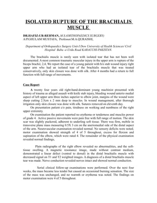
Plain radiographs of the right elbow revealed no abnormalities, and the soft-
tissue swelling. A magnetic resonance image, made without contrast medium,
demonstrated a linear defect (ventral to dorsal) in the distal brachialis muscle with
decreased signal on T1 and T2 weighted images. A diagnosis of a distal brachialis muscle
tear was made. Nerve conduction revealed nerves intact and showed normal conduction.
Serial clinical follow-up examinations were performed. Over the next four
weeks, the mass became less tender but caused an occasional burning sensation. The size
of the mass was unchanged, and no warmth or erythema was noted. The findings on
motor examination were 4 of 5 throughout.
2. Six weeks after the injury, magnetic resonance imaging revealed increased
signal within, and thickening of, the distal brachialis muscle. The plane of cleavage and
the retracted muscle fibers were consistent with a partial rupture of the brachialis muscle.
Eight months after the initial presentation, the mass was smaller and non-tender
and the findings on physical examination were otherwise normal.
Discussion
Isolated rupture of the brachialis muscle appears to be a rare injury that has not
been well documented. The current case (resident of Sher Shah, Karachi) involved a
partial distal brachialis tear that responded to nonoperative treatment. Muscle injuries are
common and can usually be diagnosed on the basis of the medical history and the
physical examination. On examination, localized tenderness and pain with muscle
activation are usually present. Our patient had a muscle tear just proximal to the
musculotendinous junction that presented as a recommended only when a diagnosis
cannot be made on the basis of the history and the physical examination3. Magnetic
resonance imaging can demonstrate both acute and chronic muscle tears. T1-weighted
images may show disruption of the normal architecture of the muscle belly or the
tendinous junction. These areas of abnormal signal can have a varied appearance ranging
from linear to more mass-like4.
Referenc
1-De Smet AA, Fisher DR. Magnetic resonance imaging of muscle tears, Skeletal Radiol.
1990;19;283-6.
2-Le Huec JC, Zipoll B, Chauveaux D, Le Robeller A. Distal rupture of the tendon of
biceps brachii, Evaluation by MRI and the results of repair, J Bone Joint Surg Br 1996;
78; 767-70.
3-Noonan TJ, Garrett, Muscle strain injury; Diagnosis and Treatment. J Am Acad Orthop Surg, Keene JS,
Magnetic resonance imaging of muscle tears, Skeletal Radiol. 1990; 19; 283-6.
4-Seller JG 3rd, Parker LM, Chamberland PD. The distal biceps tendon.two potential
mechanisms involved in its rupture; arterial supply and mechanical impingement. J
Shoul. 1999; 7; 262
AUTHOR:- Dr.Hafiz-ur-Rehman M.S.(Orthopaedics Surgery)
Cell:0301-2575144 Phone Residence +92-021-9216055
E.mail: hafeezorthochk@hotmail.com / ortho1chk@yahoo.com
Mail address:- Room no.14 Second Floor Taj Medical Complex
M.A.Jinnah Road Karachi, Pakistan.