
Recommended
More Related Content
Similar to SKIN DISEASES LECTURE.pptx
Similar to SKIN DISEASES LECTURE.pptx (20)
Recently uploaded
Recently uploaded (20)
SKIN DISEASES LECTURE.pptx
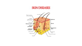
- 1. SKINDISEASES
- 2. DESCRIBING SKIN LESIONS PRIMARY MORPHOLOGY >FLAT VS RAISED >SIZE >CONSTITUENCY SECONDARY MORPHOLOGY >COLOR >SHAPE >TEXTURE >LOCATION
- 4. RAISED LIQUID FILLED PUS FILLED SOLID CONTITUENCY Pustule: a circumscribed, elevated lesion filled with purulent fluid, less than 1 cm in size (e.g. erythema toxicum neonatorum, acne). Vesicle: a circumscribed, elevated, fluid- filled lesion up to 1 cm in size NODULE - A palpable, solid lesion that is greater than 10 mm* in diameter. Nodules are usually found in the dermal or subcutaneous tissue, and the lesion may be above, level with, or below the skin surface. Example: Dermatofibroma. TUMOR - A solid, firm lesion that is typically greater than 20 mm in diameter.
- 5. SKIN BLEEDING(non-blanchable) Purpura is small, flat spots on your skin. They look red or purple on lighter skin tones but appear brown or black on darker skin tones. Purpura is commonly referred to as a blood spot under your skin. Purpura usually consists of smaller dots that cluster in a specific area but may appear as one larger patch.May be palpable. 3-10mm Ecchymosis is usually caused by an injury, such as a bump, blow, or fall. This impact may cause a blood vessel to burst open leaking blood under the skin, creating a bruise. While bruises are very common and affect almost everyone, women tend get them more easily than others do.More than 10mm Petechiae are pinpoint, round spots that appear on the skin as a result of bleeding. The bleeding causes the petechiae to appear red, brown or purple. Petechiae commonly appear in clusters and may look like a rash. Usually flat to the touch, petechiae don't lose color when you press on them.They are Non-Palpable
- 6. Telangiectasias are small, widened blood vessels on the skin. They are usually harmless, but may be associated with several diseases Urticaria – also known as hives, weals, welts or nettle rash – is a raised, itchy rash that appears on the skin. It may appear on one part of the body or be spread across large areas. The rash is usually very itchy and ranges in size from a few millimetres to the size of a hand. Skin ulcers are open sores caused by poor blood circulation. If you have poor blood circulation, then minor wounds (that otherwise would heal quickly) may not heal properly leaving that injury to develop into a painful skin ulcer. These sores and ulcers often become infected if not properly treated.
- 7. “SCRATCH MARKS”EROSION OF THE SKIN THAT IS LINEAR IN SHAPE EXCORIATION Scales are a visible peeling or flaking of outer skin layers(EPIDERMAL THICKENING. These layers are called the stratum corneum. This view shows the red, scaly patches called plaques that are characteristic of atopic dermatitis. An actinic keratosis is a thick, scaly, or crusty skin patch that's typically less than 2 centimeters (cm), or about the size of a pencil eraser. It appears on parts of the body that receive a lot of sun exposure (the hands, arms, face, scalp, and neck).
- 8. SECONDARY MORPHOLOOGY SHAPE ROUND LINE COIN-SHAPED Annular skin lesions are figurate lesions characterized by a ring-like morphology. Although plaques represent the most common presentation of annular lesions, lesions may also be macular, nodular, or composed of grouped papules, vesicles, or pustules. Linear lesions act as diagnostic clues in many disorders. They also help in elucidating the pathogenesis as they give a clue to the pathway of spread of the disease. Koebner phenomenon indicates the presence of active disease and helps to decide the line of management.(eg:CONTACT DERMATITIS) Nummular eczema is a skin condition that causes circular, raised spots on your skin. Nummular comes from a Latin word for “coin,” and the patches are coin- shaped. The lesions are often itchy, sometimes ooze clear fluid and may become crusty on top. The condition is chronic.
- 9. OTHER SHAPES TARGET WEB-LIKE SNAKE LIKE A target lesion is a round skin lesion with three concentric colour zones: =A darker centre with a blister or crust =A ring around this that is paler pink and raised due to oedema (fluid swelling) =A bright red outermost ring. Target lesions typically occur in erythema multiforme. They can arise on any body site, including face, upper chest, back, arms, legs, hands, feet and mucous membranes (such as the lips). A target lesion is also called a bulls-eye lesion or a cockade (a rosette pattern of concentric rings). RETICULAR “reticulate” is used for clinical description of skin lesions that are configured in a net- like pattern. “LAZY NETWORK”also known as Livedo reticularis Serpiginous “Squiggly”branching lesion.(Parasitic Infection) Cutaneous larva migrans (CLM) is a serpiginous eruption that can occur anywhere on exposed body parts but is usually confined to the skin of the feet. It is most often caused by dog and cat hookworms, which are types of nematodes (roundworms).
- 10. HERPETIFORM IS CLUSTER OF PAPULES OR VISICLES Dermatitis herpetiformis (DH) is a chronic, intensely itchy, blistering skin manifestation of gluten- sensitive enteropathy, commonly known as celiac disease. DH is a rash that affects about 10 percent of people with celiac disease. Zosteriform vesicular lesions of the skin could be due to either herpes zoster (caused by varicella zoster virus) or zosteriform herpes simplex (caused by herpes simplex virus).
- 11. SECONDARY MORPHOLOOGY TEXTURE Hyperkeratosis is the skin's response to rubbing or irritation. A corn or callus on your hands or feet. A bump or patch of thickened skin is known as a hyperkeratotic lesion. TINEA PEDIS Verrucous lesions are defined as “pertaining to or marked by wart like growth pattern.” SEBORRHEIC KERATOSIS =WARTS Lichenification is a secondary skin lesion that is characterized by hyperpigmentation, thickening of the skin and exaggerated skin lines. These skin lesions usually appear from constant scratching or rubbing in areas, such as the elbows.
- 12. SECONDARY MORPHOLOOGY TEXTURE Xanthomas are lesions on the skin containing cholesterol and fats. They are often associated with inherited disorders of lipid metabolism (inherited problems with the way that fats are broken down and used). Xanthomas are raised, waxy-appearing, frequently yellowish-colored skin lesions. Induration refers to the thickening and hardening of soft tissues of the body, specifically the skin, and is the result of an inflammatory process caused by various triggering factors. Indurated areas commonly appear on the hands and face but can also be found on the chest, back, abdomen, breasts, or buttocks. UMBILICTED Molluscum contagiosum virus causes characteristic skin lesions consisting of single or, more often, multiple, rounded, dome-shaped, pink, waxy papules that are 2-5 mm (rarely up to 1.5 cm in the case of a giant molluscum) in diameter. The papules, or bumps, are umbilicated and contain a caseous plug.
Editor's Notes
- due to thrombocytopenia