
Recommended
More Related Content
What's hot
What's hot (20)
Similar to CT guided CTO PCI
Similar to CT guided CTO PCI (20)
More from Euro CTO Club
More from Euro CTO Club (20)
Recently uploaded
Recently uploaded (20)
CT guided CTO PCI
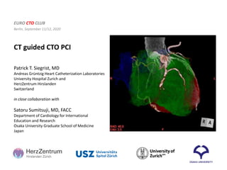
- 1. CT guided CTO PCI EURO CTO CLUB Berlin, September 11/12, 2020 Patrick T. Siegrist, MD Andreas Grüntzig Heart Catheterization Laboratories University Hospital Zurich and HerzZentrum Hirslanden Switzerland in close collaboration with Satoru Sumitsuji, MD, FACC Department of Cardiology for International Education and Research Osaka University Graduate School of Medicine Japan
- 2. Concept of Imaging Guided PCI Complex/CTO PCI Planning → CT Procedure → IVUS NOT CALIBRATED
- 3. Commonly used imaging methods Unfamiliar image for interventional cardiologists. Operator dependent presets. Curved MPR
- 4. The Present Commonly Used Imaging Methods Volume Rendering Time-consuming computation required. Limited to a superficial observational view.
- 5. A Recommended MSCT Coronary Angiography Imaging Method for Interventional Cardiologists “Sliding Slab MIP” method
- 6. Sliding Slab MIP method
- 7. Slab MIP image Maximum Intensity Projection (MIP) is a volume rendering technique which is used to visualize high-intensity structures within a volumetric data. At each pixel, the highest data value, which is encountered along a corresponding viewing ray is depicted.
- 8. MIP; Maximum Intensity Projection Slab Thickness Maximum intensity projection (MIP) is a method for 3D data that projects in the visualization plane the voxels with maximum intensity that fall in the way of parallel rays traced from the viewpoint to the plane of projection.
- 9. Move the slab back and forth. Rotate the Heart. Change the slab thickness. Only “3 Actions” !!
- 10. Move the slab back and forth Find out the intended coronary tree.
- 11. Move the slab back and forth. Rotate the Heart. Change the slab thickness. Only “3 Actions” !!
- 12. Rotating the Heart Visualizes any intended projection of the coronary tree.
- 13. Slab MIP image Visualizes any intended projection of the coronary arteries
- 14. Move the slab back and forth. Rotate the Heart. Change the slab thickness. Only “3 Actions” !!
- 15. Changing the Slab Thickness Excludes the extra enhanced structures of less interest around the vasculature.
- 16. Slab MIP and CAG image
- 18. Slab MIP Images of Normal Coronary Arteries RCA (LAO) (CRA)
- 19. RAO 50 CAU 10 RAO 51.2 CAU 11.8 RAO 40 A B C Siegrist PT, Sumitsuji S. Cardiovascular Medicine 2014;17(12):347-56 Determine optimal projection
- 20. Case 1: RCA CTO
- 21. Case 1: antegrade approach
- 22. RCA CTO
- 23. RCA CTO
- 24. RCA CTO
- 25. RCA CTO
- 26. RCA CTO
- 27. RCA CTO
- 28. RCA CTO
- 29. Case 2 How to Predict Subintimal Tracking
- 30. Case 2: How to Predict Subintimal Tracking
- 31. Case 2: How to Predict Subintimal Tracking 3.0mm Sigmoid shape can be a sign of subintimal tracking. Compare the width of the “S” to the vessel size. 3.0mm 3.0mm
- 32. RAO CRA LAO CRA Case 3: Calcium as Landmark
- 33. Case 3: Calcium as Landmark
- 34. Landmark calcification Case 3: Calcium as Landmark
- 35. Landmark calcification Case 3: Calcium as Landmark
- 36. Case 3: Calcium as Landmark
- 37. Case 3: Calcium as Landmark
- 38. Case 3: Calcium as Landmark
- 39. Plaque characterization on CT – Cross Sectional View (Color mapping) -1000-0 1-50 51-250 251-350 700-2000 Thin slice, cross-sectional view CT value (HU)
- 40. Assessment of Calcified Plaques by MSCT bean stone rock C-type ring multi full moon Predictive of PCI procedural difficulty (provided by Yamasaki K MD; Osaka University)
- 41. Classification of calcium Center/Full Moon Difficult Partial Depends on size Outside Depends on thickness PCI
- 42. Classification of calcium Partial Depends on size Outside Depends on thickness Center/Full Moon Difficult PCI
- 43. Case 4: antegrade success with CT support NOT CALIBRATED
- 44. Case 4: antegrade success with CT support NOT CALIBRATED
- 45. NOT CALIBRATED NOT CALIBRATED NOT CALIBRATED NOT CALIBRATED NOT CALIBRATED NOT CALIBRATED NOT CALIBRATED NOT CALIBRATED NOT CALIBRATED NOT CALIBRATED NOT CALIBRATED NOT CALIBRATED NOT CALIBRATED Case 4: antegrade success with CT support
- 46. NOT CALIBRATED NOT CALIBRATED NOT CALIBRATED NOT CALIBRATED Case 4: antegrade success with CT support
- 47. The RCA was occluded from the mid segment (J-CTO score: 3) Unique collateral channel: originating from sinus node artery, running along the left atrium, descending to the AV-groove and then connecting to the distal RCA. Case 5: channel visualization
- 48. Case 5: Unique collateral channel
- 49. Case 5: Unique collateral channel Unique collateral channel: originating from sinus node artery, running along the left atrium, descending to the AV-groove and then connecting to the distal RCA.
- 50. Case 5: Antegrade approach An antegrade approach using wires with increasing stiffness failed. Rotational fluoroscopy showed an unnatural course of the wire. The course of the RCA remained unclear
- 51. Case 5: Retrograde approach – channel negotiation After a Finecross microcatheter was not able to follow the tortuous channel it was exchanged to the new Caravel. Finally a SUOH guidewire followed by the Caravel microcatheter could cross the channel and reach the distal CTO entry point at the RCA bifurcation.
- 52. Case 5: Final angiogram Final angiogram displayed successful revascularization of the right coronary artery and no significant vascular complications. Procedure time: 3 hrs 50 min, Contrast: 260 ml Fluoro time: 102 min, Air kerma: 4164.57 mGy, DAP: 32.9 mGy.m2
- 53. Novel Angio-CT (XACT) Toshiba INFX-8000C/AquilionONE Vision Edition Treat and verify in the same room – on a single system
- 54. CT Fusion Baseline CT scan (i.c. contrast) Wire position CT scan Non-rigid image registration Fusion image radiopaque wire tip guiding catheter LAD LAD
- 55. Color Coded Cross Sectional Images -3000 - 0 1 - 50 51 - 250 > 251 Color code CT value (HU) Low density (e.g. fatty tissue) Low-intermediate desity (e.g.fatty-fibrous) Intermediate density (e.g. fibrous) High density (e.g. contrast, calc, metal) LAD CT gray scale Cross sectional view (gray scale) Cross sectional view (color coded) Fusion image cross section superimposed wire
- 56. Case example 1: Subintima (RCA CTO) Initial Angiogram Antegrade Wiring The wire could be advanced into the CTO, however the course of the RCA was unclear. The RCA was occluded from the mid segment (J-CTO score: 3). Ipsilateral collateral channel
- 57. Case example 1: Subintima (RCA CTO) CT Fusion ① ① ② ② ③ ③ ④ ④ ⑤ ⑤ ⑥ ⑥ The CT revealed, that the wire runs in the center of the vessel up to position ①. After that the wire migrates off the center (position ②) and circles the vessel border clockwise (position ③ー⑥); i.e. a sign of subintimal tracking. Cross Section
- 58. Conclusions CT is an ideal tool for preparation of CTO PCI because it… • … shows the entire course of occluded vessel • … provides information on plaque characteristics • … helps to find landmarks • … helps to predict subintimal tracking • … helps to find the best view/projection
- 59. Thank you Patrick T. Siegrist, MD Attending Physician / Interventional Cardiologist Andreas Grüntzig Catheterization Laboratories University Hospital Zurich and HerzZenztrum Hirslanden Zurich Email: patrick.siegrist@usz.ch