1. VISION THERAPY: The patient began vision therapy per request of his parents. Our
initial prognosis was very guarded. The parents’ goals were to improve fixation and
tracking of faces and objects. Our goals were to improve our patient’s visual attention and
to increase his ability to fixate and make pursuit and saccadic eye movements.
Activities With Positive Response:
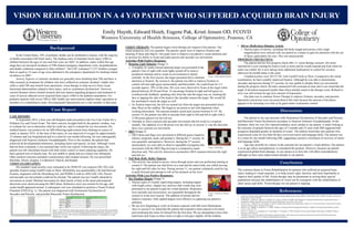
• Rotating Light Stimulus (Image 1)
o A brightly lit, multi-colored spinning target was presented in the
patient’s line of sight with room lights turned off to remove
peripheral stimulus and to create an environment to attend
centrally. At the first session, the target presented led to minimal
attention or fixation. By session 6, the patient was able to improve fixation in
central, left and right gazes monocularly and binocularly. He could fixate for 2-3
seconds approx. 50% of the time. His eyes were observed to fixate at the target when
placed between 30.50 and 45cm. To encourage fixation in right and left gazes we
would provide feedback, speaking only from the side the target was on, stroking the
face or tapping the side of the head or the shoulder nearest the target. We would lift
his arm/hand to touch the target as well.
o As fixation improved, the left eye turned out when the target was presented closer
than 30cm on the midline. We began to use prism to aid with alignment when
working binocularly. The amount needed varied each session between 8-14∆. By
session 10, the patient was able to saccade from right to left and left to right with a
12 BI prism placed over his left eye.
o Attempts to illicit a pursuit and saccadic movements did not result in a response
initially. The addition of a 6 BI prism over the left eye in session 11 was the first time
there was a pursuit eye movement.
• OKN (Image 2)
o OKN drum and flags were presented in different gazes (superior,
inferior, temporal, nasal, and primary). During the 1st session, he
was generally unresponsive. However, during the 3rd session,
monocularly, we were able to observe repeatable nystagmus-like
movement with the OKN flag moving in a temporal to nasal
direction only. This activity showed an asymmetric OKN response between the right
and left eye.
• Full Body Rolls, Reflex Patterns
o This activity was aimed to access vision through motor and was performed starting at
session 9. Our patient was laid down on a mat and his entire body was rolled moving
his right and left sides for him. During session 11, our patient voluntarily used his leg
to push forward and attempt to roll on his stomach on his own!
Activities With Less Positive Responses:
• Toy Fixation Targets (Image 3)
o Various types of visually captivating targets, including targets
with bright colors, clapper toys and toys that would sing were
presented to our patient to gain his visual attention. Responses
were sporadic and inconsistent, not repeatable throughout the
session or at the next session. The addition of prisms did not
improve response. Only lighted targets were effective in capturing our patient’s
attention.
o We are now beginning to work on fixation response with full room illumination.
Also in session 8 we noted that the patient did respond to items placed in his hand,
and would grip the items for himself for the first time. We are attempting to have him
hold items and fixate on these items in right or left gaze slightly off the midline.
CASE HISTORY:
In September 2014, a four year old Hispanic male presented to the Eye Care Center for a
Pediatric Eye and Vision Exam. The chief concern, brought forth by the patient’s mother, was
to evaluate her son to determine what he could see, and to evaluate his eye movements. His
medical history was positive for an ABI following asphyxiation from choking on a piece of
candy in January 2014. At the time of the injury, he was deprived of oxygen for approximately
15 minutes and was hospitalized and in a coma for one month following. He was subsequently
diagnosed with Hypoxic Ischemic Encephalopathy. Prior to the incident, the patient had
achieved all developmental milestones, including motor and speech, on time. Although vision
had not been evaluated, it was assumed that vision was typical. Following the injury, the
patient was left wheelchair bound with little motor control or motor planning capability. He
had a general loss of muscle tone. He was unable to speak and eye contact was infrequent.
Other medical concerns included a tracheostomy and residual seizures. He was prescribed
Baclofan, Diocto, Keppra, Levalbuterol, Pepcid, and Reglan.
VISION EXAMINATION:
His unaided visual acuities were estimated to be 20/80 OU (no response OD, OS) with
sporadic fixation using Cardiff Cards at 50cm. (Reliability was questionable.) He had bifoveal
fixation, alignment with the Hirschberg test, and PERRLA with no APD (OD, OS). Pursuit
and saccadic eye movements could not be elicited. The patient was not visually attracted to
movement or sound. Minimal movement for short bursts of time in the nasal and temporal
directions were observed using the OKN drum. Refractive error was normal for his age, and
ocular health appeared normal. A subsequent visit was scheduled to perform a Visual Evoked
Potential (VEP) (Fig. 1). The patient was diagnosed with Oculomotor Dysfunction of
Saccades and Pursuits, and possible Binocular Vision Dysfunction.
Emily Huynh, Edward Hsieh, Eugene Pak, Kristi Jensen OD, FCOVD
Western University of Health Sciences, College of Optometry, Pomona, CA
Background
VISION REHABILITION IN A 4 Y/O PATIENT WHO SUFFERED ACQUIRED BRAIN INJURY
References
Conclusion
The common theme in Vision Rehabilitation for patients who suffered an acquired brain
injury leading to visual sequelae, is to help restore sight, function, and most importantly to
improve their quality of life. Vision therapy may be paramount in serving these special
populations because the rehabilitation of vision can be synergistic with the rehabilitation of
other senses and skills. Vision therapy for our patient is ongoing.
1. Centers for Disease Control and Prevention (2002). Nonfatal choking-related episodes among children--United States, 2001. MMWR: Morbidity and Mortality
Weekly Report, 51(42), 945-948.
2. Ciuffreda, K. J., Rutner, D., Kapoor, N., Suchoff, I. B., Craig, S., & Han, M. (2008). Vision therapy for oculomotor dysfunctions in acquired brain injury: a
retrospective analysis. Optometry-Journal of the American Optometric Association, 79(1), 18-22.
3. Faul, M., Xu, L., Wald, M. M., & Coronado, V. (2010). Traumatic Brain Injury in the United States. Atlanta, GA: Centers for Disease Control and Prevention,
National Center for Injury Prevention and Control.
4. Johnson, A. R., DeMatt, E., & Salorio, C. F. (2009). Predictors of outcome following acquired brain injury in children. Developmental disabilities research
reviews, 15(2), 124-132.
5. Kriel, R. L., Krach, L. E., Luxenberg, M. G., Jones-Saete, C., & Sanchez, J. (1994). Outcome of severe anoxic/ischemic brain injury in children. Pediatric
neurology, 10(3), 207-212.
6. Tinsworth, D. K., & US Consumer Product Safety Commission (2001, April). Special study: Injuries and deaths associated with children's playground
equipment. Washington, DC: US Consumer Product Safety Commission.
Case Report Discussion
The patient in our case presents with Oculomotor Dysfunction of Saccades and Pursuits,
and Binocular Vision Dysfunction secondary to Hypoxic Ischemic Encephalopathy. In the
literature, there are very few reported pediatric cases similar to our patient. A study looking at
the outcome of patients between the ages of 2 months and 14 years with ABI suggested that
prognosis depended greatly on duration of coma5. The authors noted that only patients who
experienced coma for less than 60 days recovered motor and language skills. Our patient was
in a coma for one month following ABI, suggesting a positive correlation of recovery of motor
and language skills.
Age may possibly be a factor in the outcome for our patient’s visual abilities. Our patient
is at an age where neuroplasticity is considered the greatest. However, because our patient
experienced global brain damage, we are unsure as to how this will affect neuroplasticity
although we have seen improvement already in our patient.
In the United States, 50% of pediatric deaths can be attributed to trauma, with the majority
of deaths associated with brain injury. The leading cause of traumatic brain injury (TBI) in
children between the ages of zero and four years are falls6. In addition, males within this age
range have an increased incidence of TBI-related emergency department visits, hospitalizations,
and deaths combined compared to older children3. The CDC estimated 17,537 children younger
than or equal to 14 years of age were admitted to the emergency department for choking-related
incidences in 20011.
Anoxic, hypoxic or ischemic incidents are generally more disabling than TBI and there is
little research on treatment for children who have suffered an ischemic incident4. Adults who
suffer a mild TBI may benefit from optometric vision therapy to help recover from visual
functional abnormalities related to their injury, such as oculomotor dysfunction2. However,
current literature shows limited research and case reports regarding prognosis and treatment in
cases of anoxia-induced severe acquired brain injury (ABI) in the pediatric population. For
pediatric patients with severe TBI or ABI, health care interventions implore many specialists as
members of a rehabilitative team. Developmental Optometrists are a vital member of that team.
Image 1. Rotating light stimulus.
• Mirror (Reflection) Stimulus Activity
o Various types of mirrors, including full-body length and mirrors with a high
magnification were utilized with our patient as a means to gain his attention with the use
of faces, particularly his own. This was unsuccessful.
PROGRESS CHECK(3/2015):
The patient had his first progress check after 11 vision therapy sessions. His mom
reported he is now turning his head to look at items and he would respond and look when his
name was called. He is now taking some additional medications to control his seizures,
otherwise his health status is the same.
Unaided acuities were 20/127 OU with Cardiff Cards at 50cm. Compared to the initial
examination, he had a notably improved fixation. Although he was able to demonstrate
saccades and pursuits during VT sessions, he was unable to display these eye movements
during the progress check. He had a measured 8 left exotropia at near and it was noted that the
angle of deviation measured smaller than when initially noted in the therapy room. Refractive
error was still normal for age (low amount of hyperopia).
Our plan was to continue Vision Therapy to improve fixation, saccades, and pursuits.
Spectacles and prisms were not prescribed at this time because the amount of deviation
appears to be lessening over time as he gains better oculomotor control.
Figure 1. VEP OD & OS. Monocular Snellen acuity potential was 20/264. Binocular Snellen
acuity potential was 20/128. The VEP findings demonstrated a diminished response to visual
stimulus OU, however, the results also illustrated that the primary visual pathway is intact
and that there is sight.
Image 2. OKN flags.
Image 3. Various toy fixation
targets.