SlideShare a Scribd company logo
1 of 54
4- Crown I Root Fractures:
Diagnosis:
Clinical a mobile coronal segment
attached to the gingival with or without pulp
exposure. Radiographic a radiolucent
oblique line that comprises the crown & root in
a vertical direction.
Treatment objectives
1- To maintain pulp vitality.
2- To restore normal esthetics & function.
3-To induce apical closure of incomplete roots of
permanent teeth.
Treatment:
1- Primary teeth:
If the tooth cannot be or should not be restored,
Extraction is the treatment of choice unless
removal of the apical fragment would result in
damage to the successor tooth bud.
II- Permanent teeth:
•Emergency treatment: stabilize (reattach) the
coronal fragment.
•Definite treatment: removal of the coronal
fragment followed by a supra-gingival
restoration.
a) If the fracture extends deeper sub-
gingivally, gingivectomy, osteotomy or
surgical or orthodontic extrusion is done
to prepare for restoration.
b) If the pulp is exposed, alternatives
treatment include; direct pulp capping,
pulpotomy or root canal treatment.
Prognosis:
Fractures extending deeply below the
gingival margin may not be restorable.
Closed apex
Open apex
RCT Apexification
Class IV complicated fracture
5-Root Fractures:
Diagnosis:
Clinical : mobile coronal fragment
attached to the gingival that may be
displaced.
Radiographic : 1 or more, radiolucent lines
that separate the tooth fragment in
horizontal fractures. Multiple radiographic
exposures may be required for diagnosis
of other types of fractures.
Treatment objectives:
1. Reposition as soon as possible
2. Stabilize the coronal fragment in its
anatomically correct position to optimize
healing of the periodontal ligament &
neurovascular supply while maintaining
esthetic & functional integrity.
Treatment:
I- Primary teeth:
1. Extraction of the coronal fragment
without insisting on removing apical
fragment.
2. Wait & See is considered as alternative
treatment.
II- Permanent teeth:
A) Apical root fractures
 No treatment is required, just follow-up
by x-rays up to 6 months.
 Instruct the patient not to overload the
tooth
•1 month later, union may occur by a
calcific or fibrous tissue.
•If the fracture line increases in width,
this indicates failure of union RCT
followed by surgical removal of the
apical fragment.
B) Middle-third root fractures:
•Splinting is the treatment because the root part
attached to the crown isn't enough to stabilize the
tooth.
*Types of splints:
1- Composite splint: it is used when the adjacent
teeth are structurally healthy & well-aligned.
2- Light stainless steel wire splint.
3- Arch bar splint.
4- acrylic resin splint.
Teeth with root fractures should be splinted for 4-6
weeks.
* Ideal requirements of a splint:
1- Quick & easy construction.
2- Stability throughout the healing period.
3- Should allow physiologic response of the
teeth.
4- Should allow easy access for Endodontic
treatment.
5- Should be passive & atraumatic.
6- Should be esthetically pleasant.
7- Shouldn't enhance plaque accumulation.
C) Cervical-third root fractures:
•If the fracture occurs at the bone level, the
coronal fragment is removed, RCT is
performed.
•If the fracture occurs 1-2 mm infrabony,
localized osteoplasty may be performed to
enable treatment followed by RCT.
•If the fracture is too far infrabony, then
extraction is the treatment of choice.
•Requisites of successful treatment of root
fractures:
1. The fragments must be in close contact.
2. The fragments must be immobilized.
3. Absence of infection.
*Patterns of healing of root fractures:
1. Healing by calcified tissue.
2. Healing by connective tissue.
3. Healing by bone & connective tissue.
4. Healing by granulation tissue.
Prognosis:
The best prognosis occurs in
1- Young age.
2- Immature roots.
3- Positive pulp response at the time of
injury.
4- Approximating the dislocation within I
mm.
Generally, apical fractures have better
prognosis than cervical ones.
6. Concussion
Diagnosis:
Clinical : The tooth is tender to pressure &
percussion due to inflammation of the PDL
without mobility or sulcular bleeding.
Radiographic
There may or may not be thickening of the
periodontal ligament space.
Treatment objectives:
To optimize healing of the PDL & maintain
pulp vitality.
Treatment:
I- Primary teeth:
Unless associated infection exists, no pulpal
therapy is indicated.
II- Permanent teeth:
Although there is, a minimal risk of pulp
necrosis teeth with closed apices may undergo
pulpal necrosis due to associated injury to blood
vessels at the apex, & therefore must be
carefully followed-up.
Soft diet & analgesics .
Prognosis:
The younger the patient the better the
chances for tooth remaining.
Concussion may result in:
1- Asymptomatic pulp necrosis.
2- Discoloration of teeth.
3- Internal or external resorption.
4- No change & complete recovery.
7. Subluxation:
Diagnosis:
C. P.
Mobile tooth without displacement which may or
may not have sulcular bleeding.
Radiographic
No change.
Treatment objectives:
To enhance healing of the PDL & neurovascular
supply.
Treatment:
I- Primary teeth:
Followed-up .
II- Permanent teeth:
Tooth stabilization & relieve any occlusal
interference.
For comfort, a flexible splint can be used for less
than 2 w.
Prognosis:
Primary tooth usually returns to normal within 2
weeks.
Mature permanent teeth may undergo pulpal
necrosis, so must be carefully followed-up.
8. Lateral Luxation
Diagnosis
C.P.
-The tooth is displaced laterally with the crown
usually in a palatal or lingual direction & may
be locked firmly into this new position.
- The tooth is usually not mobile nor tender to
touch.
Radiographic: Increase in the periodontal
ligament space & displacement of the apex.
Treatment
I- Primary teeth
Allow passive repositioning or actively reposition
& splint for 2 weeks to allow for healing.
If injury is severe or the tooth is nearing
exfoliation Extraction.
II- Permanent teeth
•Reposition and stabilization as soon as possible
in its anatomically correct position to optimize
healing of the PDL & neurovascular supply,
while maintaining esthetic & functional integrity.
•Repositioning of the tooth is done with
digital pressure & little force.
•The tooth may extruded to free apical
lock in the cortical bone.
Splinting an additional 2-4 weeks may
be needed with breakdown of marginal
bone.
Prognosis:
Active repositioning increased risk of
pulp necrosis compared to teeth that are
left for passive repositioning.
In mature permanent teeth with closed
apices, pulp necrosis & obliteration are
common healing complications while
progressive root resorption is less likely to
occur.
9. Intrusion:
C.P.
 The tooth appears to be shortened or missed in
severe cases (more than 6 mm intrusion).
 The tooth apex is usually displaced labially
towards or through the labial cortical plate of
bone in primary teeth or driven into the alveolar
process in permanent teeth
 The tooth isn't mobile or tender to touch.
Radiographic
•Tooth appears to be displaced apically & the
PDL isn't continuous.
•The relationship of the intruded primary tooth
and the successor's tooth follicle should be
determined;
•If the apex is displaced labially, the apical tip can
be seen radiographically with the tooth appearing
shorter than its contralateral.
•If the apex is displaced palatally towards the
permanent tooth germ, the apical tip won't be
seen radiographically & the tooth appears
elongated.
Extra-oral lateral radiograph may be used to
detect displacement of the apex towards or
through the labial plate of bone.
•An intruded young permanent tooth may
mimic an erupting tooth.
Treatment:
I- Primary teeth:
Spontaneous re-eruption (within 3-4W.) unless it
endangering the developing tooth germ, otherwise
Extraction is indicated.
II- Permanent teeth:
 Passive repositioning.
 Active repositioning (Traction) or surgical
repositioning if intrusion is greater than 6mm.
 Tooth stabilization to allow PDL healing & maintain
esthetic & function
Intrusion greater than 6 mm
indicates surgical
repositioning
•In mature teeth, reposition the tooth via
surgical or orthodontic extrusion & initiate
endodontic treatment within 3 weeks of the
traumatic incidence.
•In teeth with immature root formation, the
objective is to allow spontaneous eruption.
Prognosis:
1- Primary teeth:
90% re-erupt spontaneously (either partially or
completely) in 2-6 months. Even in cases of complete
intrusion & displacement of primary teeth through the
labial plate, re-eruption may occurs for more than 36
months.
Ankylosis may occur if the periodontal ligament was
severely damaged, thereby delaying or altering the
eruption of the permanent successor.
Complications following intrusion of primary teeth:
1-Dilacerations:
It occurs in permanent successors of intruded primary
teeth where the calcified portion is twisted providing a
new direction of growth. It may lead to a sharp angle
between the crown and the root.
2-Gemination:
Partial duplication of the affected teeth in that part of the tooth
formed after injury.
3.Turner's hypoplasia:
Localized small pigmented hypoplastic areas.
4. Appearance of additional cusps or denticles.
II- Permanent teeth:
• Mature teeth with closed apices have a risk for
pulp necrosis, pulp canal obliteration, and
progressive root resorption.
• Immature teeth that are allowed to reposition
spontaneously show the lowest risk for
complications.
• An intrusion of 7 mm or more have a negative
influence on healing.
Primary dentition
0pen apex
Closed apex /
less than 5
mm intrusion
Allow 6
months for
spontaneous
re-eruption &
advise the
parents about
the potential
the damage to
the tooth
Extract the tooth if
root tip is displaced
into the permanent
tooth bud
Permanent dentition
Closed
apex/greater than
6 mm intrusion
INTRUSION INJURIES
Consider
slight surgical
Luxation &
allow for
spontaneous
re-eruption or
orthodontic
repositioning.
-surgical
repositioning
&physiological
splinting for 1-2
weeks of injury
-Ca(OH)2
pulpectomy
within tow weeks
of the injury.
-surgical or
orthodontic
repositioning.
-Ca(OH)2
pulpectomy
within two
weeks of the
injury.
Follow up is recommended to monitor signs
of pathology, via radiographs.
-Follow up for 4 weeks.
-Advise the parents about
possible.
10. Extrusion:
(partial Avulsion)
Diagnosis:
C.P.
The tooth appears elongated & is mobile.
Radiographic findings:
An increase in the width of the periodontal ligament space
apically.
Treatment:
I- Primary teeth:
Reposition and splinting the tooth for 1-2 weeks & allow for
healing, except when there are indications for extraction
(e.g.: sever injury or exfoliation).
II- Permanent teeth:
•Reposition the tooth as soon as possible & then
stabilize the tooth in its anatomically correct position
to optimize healing of the periodontal ligament &
neurovascular supply while maintaining esthetic &
functional integrity.
•Repositioning may be done with slow & steady
apical pressure to gradually displace the coagulum
formed between the apex & the floor of the socket
•Splint the tooth for up to 3 weeks.
Prognosis:
•There is a lack of clinical evidence
evaluating repositioning of extruded
primary teeth ( NO DATA).
•In mature permanent teeth with closed
apices, there is a considerable risk for
pulp necrosis & pulp canal obliteration,
therefore these teeth must be followed-up
carefully.
AVULSION
11. Avulsion:
Diagnosis:
Clinical & radiographic features:
Reveal that the tooth is not present in its socket or
that the tooth already has been replanted.
Treatment Objectives:
I- Primary teeth:
To prevent further injury to the developing successor,
Avulsed primary teeth should not be replanted.
II- Permanent teeth:
Replant & stabilization the tooth as soon as
possible in its anatomically correct position to
optimize healing of the periodontal ligament
and neurovascular supply while maintaining
esthetic and functional integrity, except when
replantation is contraindicated.
Contraindications of tooth replantation:
1-The child's stage of dental development (the risk of
ankyloses where considerable alveolar growth has to take
place)
2-Compromising medical condition; such as:
Immunocompromise, severe congenital cardiac anomalies,
severe uncontrolled seizure disorder, severe mental disability
& severe uncontrolled diabetes.
3-Compromised integrity of the avulsed tooth or the
supporting structures.
Treatment:
The treatment strategies are directed towards avoiding the
inflammation that may occur as a result from the tooth's
attachment damage and/or pulpal infection.
Technique of replantation:
1- Patient preparation:
Inform the parents -and if possible, the patient-
about: - The need for splinting, for 1-2 weeks.
•The need for RCT, 1-2 weeks after replantation.
•The uncertain prognosis of tooth replantation.
the cementum.
2- Tooth preparation:
•The tooth is held from the crown.
•The dentist should avoid using any brush or sharp
instrument on the root surface as it might remove
remnants of the periodontal fibers and exposed the
cementum.
3- Socket preparation:
•The socket and the surrounding tissues should
be carefully inspected and cleaned with sterile
saline.
•Use a soft tissue curette to remove any
coagulated tissue from the socket, then
carefully irrigate the socket.
4- Tooth replantation:
The tooth is placed into the socket carefully using
gentle pressure.
5- Checking occlusion:
The replanted tooth should be free of contact
with the opposing teeth to avoid overloading the
tooth, which may interfere with proper
reattachment.
6- Splinting:
Flexible splinting for 1-2 weeks is indicated.
Prognosis:
depends on:
A) The stage of root development:
The chance for revascularization is better in case
of open apices than in case of closed apices.
B) The extra-oral dry time:
The tooth has the best prognosis if replanted
immediately. If the tooth cannot be replanted
within 5 minutes, it should be stored in a medium
that will help to maintain vitality of the periodontal
ligament fibers.
Transportation media for avulsed teeth:
1- Viaspan.
2-Hank's balanced salt solution-HBSS-
(tissue culture medium).
3- Cold milk.
4- Saliva (buccal vestibule or floor of the
mouth).
5- Physiologic saline or water.
Limited tooth storage in a cell-compatible medium
prior to replantation has produced similar healing
results as compared with immediately replanted
teeth.
The risk of ankylosis increases with an extra-oral dry
time of 15 minutes. An extra-oral dry time of 60
minutes is considered as the crtical point where
survival of the periodontal cells is unlikely to occur..
In permanent anterior teeth, there is a considerable
risk for pulp necrosis, root resorption, ankylosis, and
subsequent infra-occlusion during adolescent
growth.
First aid for avulsed teeth
1-Keep the patient calm
2-Find the tooth & pick
it up by the crown
3-Clean the tooth 4-Place the tooth in a
suitable storage medium
Seek emergency dental treatment immediately
Immediate Delayed
Reactions of tooth to trauma
1. Pulpal Hyperemia
2. Internal Hemorrhage
3. Pulp Calcification
4. Internal Resorption
5. External Resorption
6. Pulp Necrosis
7. Ankylosis
1. Infraction
2. Uncomplicated crown
fracture
3. Complicated crown fracture
4. Crown /root fracture
5. Root fracture
6. Concussion
7. Subluxation
8. Lateral luxation
9. Extrusion
10.Intrusion
11.Avulsion
Instructions to the patient on Telephone

More Related Content

Similar to 2. TRAUMATIC_INJURIES.pptx

Preventive and interceptive orthodontics
Preventive and interceptive orthodonticsPreventive and interceptive orthodontics
Preventive and interceptive orthodontics
Ayesha Abbas
 
Extractionsinorthodontics ug-130701154008-phpapp02 (1)
Extractionsinorthodontics ug-130701154008-phpapp02 (1)Extractionsinorthodontics ug-130701154008-phpapp02 (1)
Extractionsinorthodontics ug-130701154008-phpapp02 (1)
Moosa Ahmed
 
Immediate denture
Immediate dentureImmediate denture
Immediate denture
dukeheart
 

Similar to 2. TRAUMATIC_INJURIES.pptx (20)

Traumatic injuries
Traumatic injuriesTraumatic injuries
Traumatic injuries
 
Management of crossbite in mixed dentition
Management of crossbite in mixed dentitionManagement of crossbite in mixed dentition
Management of crossbite in mixed dentition
 
Preventive and interceptive orthodontics
Preventive and interceptive orthodonticsPreventive and interceptive orthodontics
Preventive and interceptive orthodontics
 
Extractionsinorthodontics ug-130701154008-phpapp02 (1)
Extractionsinorthodontics ug-130701154008-phpapp02 (1)Extractionsinorthodontics ug-130701154008-phpapp02 (1)
Extractionsinorthodontics ug-130701154008-phpapp02 (1)
 
Pulp therapy
Pulp therapyPulp therapy
Pulp therapy
 
Single denture construction technique
Single denture construction technique Single denture construction technique
Single denture construction technique
 
Management of traumatic dental injury of primary teeth
Management of traumatic dental  injury of primary teethManagement of traumatic dental  injury of primary teeth
Management of traumatic dental injury of primary teeth
 
avulsion
avulsionavulsion
avulsion
 
Class i orthodontics Dentistry
Class  i orthodontics DentistryClass  i orthodontics Dentistry
Class i orthodontics Dentistry
 
Adult Orthodontics.pptx
Adult Orthodontics.pptxAdult Orthodontics.pptx
Adult Orthodontics.pptx
 
Paediatric Endodontics.pdf
Paediatric Endodontics.pdfPaediatric Endodontics.pdf
Paediatric Endodontics.pdf
 
Root Canal Treatment in India
Root Canal Treatment in IndiaRoot Canal Treatment in India
Root Canal Treatment in India
 
Single complete denture
Single complete dentureSingle complete denture
Single complete denture
 
Pedodontics ii lecture 05
Pedodontics ii lecture 05Pedodontics ii lecture 05
Pedodontics ii lecture 05
 
Ped ii 05
Ped ii 05Ped ii 05
Ped ii 05
 
Management of displaced tooth
Management of displaced toothManagement of displaced tooth
Management of displaced tooth
 
MANAGEMENT OF AVULSED TEETH-converted.pptx
MANAGEMENT OF AVULSED TEETH-converted.pptxMANAGEMENT OF AVULSED TEETH-converted.pptx
MANAGEMENT OF AVULSED TEETH-converted.pptx
 
Occlusion of single denture (Management of Maxillary and Mandibular Single C...
Occlusion of single denture  (Management of Maxillary and Mandibular Single C...Occlusion of single denture  (Management of Maxillary and Mandibular Single C...
Occlusion of single denture (Management of Maxillary and Mandibular Single C...
 
Immediate denture
Immediate dentureImmediate denture
Immediate denture
 
Ped ii 06
Ped ii 06Ped ii 06
Ped ii 06
 

Recently uploaded

Gabriel_Carter_EXPOLRATIONpp.pptx........
Gabriel_Carter_EXPOLRATIONpp.pptx........Gabriel_Carter_EXPOLRATIONpp.pptx........
Gabriel_Carter_EXPOLRATIONpp.pptx........
deejay178
 
Call Girls Bidadi Just Call 👗 7737669865 👗 Top Class Call Girl Service Bangalore
Call Girls Bidadi Just Call 👗 7737669865 👗 Top Class Call Girl Service BangaloreCall Girls Bidadi Just Call 👗 7737669865 👗 Top Class Call Girl Service Bangalore
Call Girls Bidadi Just Call 👗 7737669865 👗 Top Class Call Girl Service Bangalore
amitlee9823
 
0425-GDSC-TMU.pdf0425-GDSC-TMU.pdf0425-GDSC-TMU.pdf0425-GDSC-TMU.pdf
0425-GDSC-TMU.pdf0425-GDSC-TMU.pdf0425-GDSC-TMU.pdf0425-GDSC-TMU.pdf0425-GDSC-TMU.pdf0425-GDSC-TMU.pdf0425-GDSC-TMU.pdf0425-GDSC-TMU.pdf
0425-GDSC-TMU.pdf0425-GDSC-TMU.pdf0425-GDSC-TMU.pdf0425-GDSC-TMU.pdf
ssuserded2d4
 
Virgin Call Girls Delhi Service-oriented sexy call girls ☞ 9899900591 ☜ Rita ...
Virgin Call Girls Delhi Service-oriented sexy call girls ☞ 9899900591 ☜ Rita ...Virgin Call Girls Delhi Service-oriented sexy call girls ☞ 9899900591 ☜ Rita ...
Virgin Call Girls Delhi Service-oriented sexy call girls ☞ 9899900591 ☜ Rita ...
poojakaurpk09
 
Call Girls Btm Layout Just Call 👗 7737669865 👗 Top Class Call Girl Service Ba...
Call Girls Btm Layout Just Call 👗 7737669865 👗 Top Class Call Girl Service Ba...Call Girls Btm Layout Just Call 👗 7737669865 👗 Top Class Call Girl Service Ba...
Call Girls Btm Layout Just Call 👗 7737669865 👗 Top Class Call Girl Service Ba...
amitlee9823
 
Dombivli Call Girls, 9892124323, Kharghar Call Girls, chembur Call Girls, Vas...
Dombivli Call Girls, 9892124323, Kharghar Call Girls, chembur Call Girls, Vas...Dombivli Call Girls, 9892124323, Kharghar Call Girls, chembur Call Girls, Vas...
Dombivli Call Girls, 9892124323, Kharghar Call Girls, chembur Call Girls, Vas...
Pooja Nehwal
 
Call Girls Hoodi Just Call 👗 7737669865 👗 Top Class Call Girl Service Bangalore
Call Girls Hoodi Just Call 👗 7737669865 👗 Top Class Call Girl Service BangaloreCall Girls Hoodi Just Call 👗 7737669865 👗 Top Class Call Girl Service Bangalore
Call Girls Hoodi Just Call 👗 7737669865 👗 Top Class Call Girl Service Bangalore
amitlee9823
 
Call Girls In Kengeri Satellite Town ☎ 7737669865 🥵 Book Your One night Stand
Call Girls In Kengeri Satellite Town ☎ 7737669865 🥵 Book Your One night StandCall Girls In Kengeri Satellite Town ☎ 7737669865 🥵 Book Your One night Stand
Call Girls In Kengeri Satellite Town ☎ 7737669865 🥵 Book Your One night Stand
amitlee9823
 
Call Girls Brigade Road Just Call 👗 7737669865 👗 Top Class Call Girl Service ...
Call Girls Brigade Road Just Call 👗 7737669865 👗 Top Class Call Girl Service ...Call Girls Brigade Road Just Call 👗 7737669865 👗 Top Class Call Girl Service ...
Call Girls Brigade Road Just Call 👗 7737669865 👗 Top Class Call Girl Service ...
amitlee9823
 
reStartEvents 5:9 DC metro & Beyond V-Career Fair Employer Directory.pdf
reStartEvents 5:9 DC metro & Beyond V-Career Fair Employer Directory.pdfreStartEvents 5:9 DC metro & Beyond V-Career Fair Employer Directory.pdf
reStartEvents 5:9 DC metro & Beyond V-Career Fair Employer Directory.pdf
Ken Fuller
 

Recently uploaded (20)

Gabriel_Carter_EXPOLRATIONpp.pptx........
Gabriel_Carter_EXPOLRATIONpp.pptx........Gabriel_Carter_EXPOLRATIONpp.pptx........
Gabriel_Carter_EXPOLRATIONpp.pptx........
 
Call Girls Bidadi Just Call 👗 7737669865 👗 Top Class Call Girl Service Bangalore
Call Girls Bidadi Just Call 👗 7737669865 👗 Top Class Call Girl Service BangaloreCall Girls Bidadi Just Call 👗 7737669865 👗 Top Class Call Girl Service Bangalore
Call Girls Bidadi Just Call 👗 7737669865 👗 Top Class Call Girl Service Bangalore
 
Hyderabad 💫✅💃 24×7 BEST GENUINE PERSON LOW PRICE CALL GIRL SERVICE FULL SATIS...
Hyderabad 💫✅💃 24×7 BEST GENUINE PERSON LOW PRICE CALL GIRL SERVICE FULL SATIS...Hyderabad 💫✅💃 24×7 BEST GENUINE PERSON LOW PRICE CALL GIRL SERVICE FULL SATIS...
Hyderabad 💫✅💃 24×7 BEST GENUINE PERSON LOW PRICE CALL GIRL SERVICE FULL SATIS...
 
Dubai Call Girls Kiki O525547819 Call Girls Dubai Koko
Dubai Call Girls Kiki O525547819 Call Girls Dubai KokoDubai Call Girls Kiki O525547819 Call Girls Dubai Koko
Dubai Call Girls Kiki O525547819 Call Girls Dubai Koko
 
Dubai Call Girls Demons O525547819 Call Girls IN DUbai Natural Big Boody
Dubai Call Girls Demons O525547819 Call Girls IN DUbai Natural Big BoodyDubai Call Girls Demons O525547819 Call Girls IN DUbai Natural Big Boody
Dubai Call Girls Demons O525547819 Call Girls IN DUbai Natural Big Boody
 
0425-GDSC-TMU.pdf0425-GDSC-TMU.pdf0425-GDSC-TMU.pdf0425-GDSC-TMU.pdf
0425-GDSC-TMU.pdf0425-GDSC-TMU.pdf0425-GDSC-TMU.pdf0425-GDSC-TMU.pdf0425-GDSC-TMU.pdf0425-GDSC-TMU.pdf0425-GDSC-TMU.pdf0425-GDSC-TMU.pdf
0425-GDSC-TMU.pdf0425-GDSC-TMU.pdf0425-GDSC-TMU.pdf0425-GDSC-TMU.pdf
 
Solution Manual for First Course in Abstract Algebra A, 8th Edition by John B...
Solution Manual for First Course in Abstract Algebra A, 8th Edition by John B...Solution Manual for First Course in Abstract Algebra A, 8th Edition by John B...
Solution Manual for First Course in Abstract Algebra A, 8th Edition by John B...
 
Virgin Call Girls Delhi Service-oriented sexy call girls ☞ 9899900591 ☜ Rita ...
Virgin Call Girls Delhi Service-oriented sexy call girls ☞ 9899900591 ☜ Rita ...Virgin Call Girls Delhi Service-oriented sexy call girls ☞ 9899900591 ☜ Rita ...
Virgin Call Girls Delhi Service-oriented sexy call girls ☞ 9899900591 ☜ Rita ...
 
Résumé (2 pager - 12 ft standard syntax)
Résumé (2 pager -  12 ft standard syntax)Résumé (2 pager -  12 ft standard syntax)
Résumé (2 pager - 12 ft standard syntax)
 
Call Girls Btm Layout Just Call 👗 7737669865 👗 Top Class Call Girl Service Ba...
Call Girls Btm Layout Just Call 👗 7737669865 👗 Top Class Call Girl Service Ba...Call Girls Btm Layout Just Call 👗 7737669865 👗 Top Class Call Girl Service Ba...
Call Girls Btm Layout Just Call 👗 7737669865 👗 Top Class Call Girl Service Ba...
 
Dombivli Call Girls, 9892124323, Kharghar Call Girls, chembur Call Girls, Vas...
Dombivli Call Girls, 9892124323, Kharghar Call Girls, chembur Call Girls, Vas...Dombivli Call Girls, 9892124323, Kharghar Call Girls, chembur Call Girls, Vas...
Dombivli Call Girls, 9892124323, Kharghar Call Girls, chembur Call Girls, Vas...
 
Joshua Minker Brand Exploration Sports Broadcaster .pptx
Joshua Minker Brand Exploration Sports Broadcaster .pptxJoshua Minker Brand Exploration Sports Broadcaster .pptx
Joshua Minker Brand Exploration Sports Broadcaster .pptx
 
Call Girls Hoodi Just Call 👗 7737669865 👗 Top Class Call Girl Service Bangalore
Call Girls Hoodi Just Call 👗 7737669865 👗 Top Class Call Girl Service BangaloreCall Girls Hoodi Just Call 👗 7737669865 👗 Top Class Call Girl Service Bangalore
Call Girls Hoodi Just Call 👗 7737669865 👗 Top Class Call Girl Service Bangalore
 
Personal Brand Exploration - Fernando Negron
Personal Brand Exploration - Fernando NegronPersonal Brand Exploration - Fernando Negron
Personal Brand Exploration - Fernando Negron
 
Resumes, Cover Letters, and Applying Online
Resumes, Cover Letters, and Applying OnlineResumes, Cover Letters, and Applying Online
Resumes, Cover Letters, and Applying Online
 
Toxicokinetics studies.. (toxicokinetics evaluation in preclinical studies)
Toxicokinetics studies.. (toxicokinetics evaluation in preclinical studies)Toxicokinetics studies.. (toxicokinetics evaluation in preclinical studies)
Toxicokinetics studies.. (toxicokinetics evaluation in preclinical studies)
 
Call Girls In Kengeri Satellite Town ☎ 7737669865 🥵 Book Your One night Stand
Call Girls In Kengeri Satellite Town ☎ 7737669865 🥵 Book Your One night StandCall Girls In Kengeri Satellite Town ☎ 7737669865 🥵 Book Your One night Stand
Call Girls In Kengeri Satellite Town ☎ 7737669865 🥵 Book Your One night Stand
 
Call Girls Alandi Road Call Me 7737669865 Budget Friendly No Advance Booking
Call Girls Alandi Road Call Me 7737669865 Budget Friendly No Advance BookingCall Girls Alandi Road Call Me 7737669865 Budget Friendly No Advance Booking
Call Girls Alandi Road Call Me 7737669865 Budget Friendly No Advance Booking
 
Call Girls Brigade Road Just Call 👗 7737669865 👗 Top Class Call Girl Service ...
Call Girls Brigade Road Just Call 👗 7737669865 👗 Top Class Call Girl Service ...Call Girls Brigade Road Just Call 👗 7737669865 👗 Top Class Call Girl Service ...
Call Girls Brigade Road Just Call 👗 7737669865 👗 Top Class Call Girl Service ...
 
reStartEvents 5:9 DC metro & Beyond V-Career Fair Employer Directory.pdf
reStartEvents 5:9 DC metro & Beyond V-Career Fair Employer Directory.pdfreStartEvents 5:9 DC metro & Beyond V-Career Fair Employer Directory.pdf
reStartEvents 5:9 DC metro & Beyond V-Career Fair Employer Directory.pdf
 

2. TRAUMATIC_INJURIES.pptx

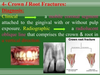
  • 1. 4- Crown I Root Fractures: Diagnosis: Clinical a mobile coronal segment attached to the gingival with or without pulp exposure. Radiographic a radiolucent oblique line that comprises the crown & root in a vertical direction.
  • 2. Treatment objectives 1- To maintain pulp vitality. 2- To restore normal esthetics & function. 3-To induce apical closure of incomplete roots of permanent teeth.
  • 3. Treatment: 1- Primary teeth: If the tooth cannot be or should not be restored, Extraction is the treatment of choice unless removal of the apical fragment would result in damage to the successor tooth bud. II- Permanent teeth: •Emergency treatment: stabilize (reattach) the coronal fragment. •Definite treatment: removal of the coronal fragment followed by a supra-gingival restoration.
  • 4. a) If the fracture extends deeper sub- gingivally, gingivectomy, osteotomy or surgical or orthodontic extrusion is done to prepare for restoration. b) If the pulp is exposed, alternatives treatment include; direct pulp capping, pulpotomy or root canal treatment. Prognosis: Fractures extending deeply below the gingival margin may not be restorable.
  • 5. Closed apex Open apex RCT Apexification Class IV complicated fracture
  • 6.
  • 7. 5-Root Fractures: Diagnosis: Clinical : mobile coronal fragment attached to the gingival that may be displaced. Radiographic : 1 or more, radiolucent lines that separate the tooth fragment in horizontal fractures. Multiple radiographic exposures may be required for diagnosis of other types of fractures.
  • 8. Treatment objectives: 1. Reposition as soon as possible 2. Stabilize the coronal fragment in its anatomically correct position to optimize healing of the periodontal ligament & neurovascular supply while maintaining esthetic & functional integrity.
  • 9. Treatment: I- Primary teeth: 1. Extraction of the coronal fragment without insisting on removing apical fragment. 2. Wait & See is considered as alternative treatment.
  • 10. II- Permanent teeth: A) Apical root fractures  No treatment is required, just follow-up by x-rays up to 6 months.  Instruct the patient not to overload the tooth
  • 11. •1 month later, union may occur by a calcific or fibrous tissue. •If the fracture line increases in width, this indicates failure of union RCT followed by surgical removal of the apical fragment.
  • 12. B) Middle-third root fractures: •Splinting is the treatment because the root part attached to the crown isn't enough to stabilize the tooth. *Types of splints: 1- Composite splint: it is used when the adjacent teeth are structurally healthy & well-aligned. 2- Light stainless steel wire splint. 3- Arch bar splint. 4- acrylic resin splint. Teeth with root fractures should be splinted for 4-6 weeks.
  • 13.
  • 14. * Ideal requirements of a splint: 1- Quick & easy construction. 2- Stability throughout the healing period. 3- Should allow physiologic response of the teeth. 4- Should allow easy access for Endodontic treatment. 5- Should be passive & atraumatic. 6- Should be esthetically pleasant. 7- Shouldn't enhance plaque accumulation.
  • 15. C) Cervical-third root fractures: •If the fracture occurs at the bone level, the coronal fragment is removed, RCT is performed. •If the fracture occurs 1-2 mm infrabony, localized osteoplasty may be performed to enable treatment followed by RCT. •If the fracture is too far infrabony, then extraction is the treatment of choice.
  • 16. •Requisites of successful treatment of root fractures: 1. The fragments must be in close contact. 2. The fragments must be immobilized. 3. Absence of infection. *Patterns of healing of root fractures: 1. Healing by calcified tissue. 2. Healing by connective tissue. 3. Healing by bone & connective tissue. 4. Healing by granulation tissue.
  • 17. Prognosis: The best prognosis occurs in 1- Young age. 2- Immature roots. 3- Positive pulp response at the time of injury. 4- Approximating the dislocation within I mm. Generally, apical fractures have better prognosis than cervical ones.
  • 18. 6. Concussion Diagnosis: Clinical : The tooth is tender to pressure & percussion due to inflammation of the PDL without mobility or sulcular bleeding. Radiographic There may or may not be thickening of the periodontal ligament space. Treatment objectives: To optimize healing of the PDL & maintain pulp vitality.
  • 19. Treatment: I- Primary teeth: Unless associated infection exists, no pulpal therapy is indicated. II- Permanent teeth: Although there is, a minimal risk of pulp necrosis teeth with closed apices may undergo pulpal necrosis due to associated injury to blood vessels at the apex, & therefore must be carefully followed-up. Soft diet & analgesics .
  • 20. Prognosis: The younger the patient the better the chances for tooth remaining. Concussion may result in: 1- Asymptomatic pulp necrosis. 2- Discoloration of teeth. 3- Internal or external resorption. 4- No change & complete recovery.
  • 21. 7. Subluxation: Diagnosis: C. P. Mobile tooth without displacement which may or may not have sulcular bleeding. Radiographic No change. Treatment objectives: To enhance healing of the PDL & neurovascular supply. Treatment: I- Primary teeth: Followed-up .
  • 22. II- Permanent teeth: Tooth stabilization & relieve any occlusal interference. For comfort, a flexible splint can be used for less than 2 w. Prognosis: Primary tooth usually returns to normal within 2 weeks. Mature permanent teeth may undergo pulpal necrosis, so must be carefully followed-up.
  • 23. 8. Lateral Luxation Diagnosis C.P. -The tooth is displaced laterally with the crown usually in a palatal or lingual direction & may be locked firmly into this new position. - The tooth is usually not mobile nor tender to touch. Radiographic: Increase in the periodontal ligament space & displacement of the apex.
  • 24. Treatment I- Primary teeth Allow passive repositioning or actively reposition & splint for 2 weeks to allow for healing. If injury is severe or the tooth is nearing exfoliation Extraction. II- Permanent teeth •Reposition and stabilization as soon as possible in its anatomically correct position to optimize healing of the PDL & neurovascular supply, while maintaining esthetic & functional integrity.
  • 25. •Repositioning of the tooth is done with digital pressure & little force. •The tooth may extruded to free apical lock in the cortical bone. Splinting an additional 2-4 weeks may be needed with breakdown of marginal bone.
  • 26. Prognosis: Active repositioning increased risk of pulp necrosis compared to teeth that are left for passive repositioning. In mature permanent teeth with closed apices, pulp necrosis & obliteration are common healing complications while progressive root resorption is less likely to occur.
  • 27. 9. Intrusion: C.P.  The tooth appears to be shortened or missed in severe cases (more than 6 mm intrusion).  The tooth apex is usually displaced labially towards or through the labial cortical plate of bone in primary teeth or driven into the alveolar process in permanent teeth  The tooth isn't mobile or tender to touch.
  • 28. Radiographic •Tooth appears to be displaced apically & the PDL isn't continuous. •The relationship of the intruded primary tooth and the successor's tooth follicle should be determined; •If the apex is displaced labially, the apical tip can be seen radiographically with the tooth appearing shorter than its contralateral.
  • 29. •If the apex is displaced palatally towards the permanent tooth germ, the apical tip won't be seen radiographically & the tooth appears elongated. Extra-oral lateral radiograph may be used to detect displacement of the apex towards or through the labial plate of bone. •An intruded young permanent tooth may mimic an erupting tooth.
  • 30. Treatment: I- Primary teeth: Spontaneous re-eruption (within 3-4W.) unless it endangering the developing tooth germ, otherwise Extraction is indicated. II- Permanent teeth:  Passive repositioning.  Active repositioning (Traction) or surgical repositioning if intrusion is greater than 6mm.  Tooth stabilization to allow PDL healing & maintain esthetic & function Intrusion greater than 6 mm indicates surgical repositioning
  • 31. •In mature teeth, reposition the tooth via surgical or orthodontic extrusion & initiate endodontic treatment within 3 weeks of the traumatic incidence. •In teeth with immature root formation, the objective is to allow spontaneous eruption.
  • 32. Prognosis: 1- Primary teeth: 90% re-erupt spontaneously (either partially or completely) in 2-6 months. Even in cases of complete intrusion & displacement of primary teeth through the labial plate, re-eruption may occurs for more than 36 months. Ankylosis may occur if the periodontal ligament was severely damaged, thereby delaying or altering the eruption of the permanent successor.
  • 33. Complications following intrusion of primary teeth: 1-Dilacerations: It occurs in permanent successors of intruded primary teeth where the calcified portion is twisted providing a new direction of growth. It may lead to a sharp angle between the crown and the root. 2-Gemination: Partial duplication of the affected teeth in that part of the tooth formed after injury.
  • 34. 3.Turner's hypoplasia: Localized small pigmented hypoplastic areas. 4. Appearance of additional cusps or denticles.
  • 35. II- Permanent teeth: • Mature teeth with closed apices have a risk for pulp necrosis, pulp canal obliteration, and progressive root resorption. • Immature teeth that are allowed to reposition spontaneously show the lowest risk for complications. • An intrusion of 7 mm or more have a negative influence on healing.
  • 36. Primary dentition 0pen apex Closed apex / less than 5 mm intrusion Allow 6 months for spontaneous re-eruption & advise the parents about the potential the damage to the tooth Extract the tooth if root tip is displaced into the permanent tooth bud Permanent dentition Closed apex/greater than 6 mm intrusion INTRUSION INJURIES Consider slight surgical Luxation & allow for spontaneous re-eruption or orthodontic repositioning. -surgical repositioning &physiological splinting for 1-2 weeks of injury -Ca(OH)2 pulpectomy within tow weeks of the injury. -surgical or orthodontic repositioning. -Ca(OH)2 pulpectomy within two weeks of the injury. Follow up is recommended to monitor signs of pathology, via radiographs. -Follow up for 4 weeks. -Advise the parents about possible.
  • 37. 10. Extrusion: (partial Avulsion) Diagnosis: C.P. The tooth appears elongated & is mobile. Radiographic findings: An increase in the width of the periodontal ligament space apically. Treatment: I- Primary teeth: Reposition and splinting the tooth for 1-2 weeks & allow for healing, except when there are indications for extraction (e.g.: sever injury or exfoliation).
  • 38. II- Permanent teeth: •Reposition the tooth as soon as possible & then stabilize the tooth in its anatomically correct position to optimize healing of the periodontal ligament & neurovascular supply while maintaining esthetic & functional integrity. •Repositioning may be done with slow & steady apical pressure to gradually displace the coagulum formed between the apex & the floor of the socket •Splint the tooth for up to 3 weeks.
  • 39. Prognosis: •There is a lack of clinical evidence evaluating repositioning of extruded primary teeth ( NO DATA). •In mature permanent teeth with closed apices, there is a considerable risk for pulp necrosis & pulp canal obliteration, therefore these teeth must be followed-up carefully.
  • 41. 11. Avulsion: Diagnosis: Clinical & radiographic features: Reveal that the tooth is not present in its socket or that the tooth already has been replanted. Treatment Objectives: I- Primary teeth: To prevent further injury to the developing successor, Avulsed primary teeth should not be replanted.
  • 42. II- Permanent teeth: Replant & stabilization the tooth as soon as possible in its anatomically correct position to optimize healing of the periodontal ligament and neurovascular supply while maintaining esthetic and functional integrity, except when replantation is contraindicated.
  • 43. Contraindications of tooth replantation: 1-The child's stage of dental development (the risk of ankyloses where considerable alveolar growth has to take place) 2-Compromising medical condition; such as: Immunocompromise, severe congenital cardiac anomalies, severe uncontrolled seizure disorder, severe mental disability & severe uncontrolled diabetes. 3-Compromised integrity of the avulsed tooth or the supporting structures. Treatment: The treatment strategies are directed towards avoiding the inflammation that may occur as a result from the tooth's attachment damage and/or pulpal infection.
  • 44. Technique of replantation: 1- Patient preparation: Inform the parents -and if possible, the patient- about: - The need for splinting, for 1-2 weeks. •The need for RCT, 1-2 weeks after replantation. •The uncertain prognosis of tooth replantation. the cementum.
  • 45. 2- Tooth preparation: •The tooth is held from the crown. •The dentist should avoid using any brush or sharp instrument on the root surface as it might remove remnants of the periodontal fibers and exposed the cementum.
  • 46. 3- Socket preparation: •The socket and the surrounding tissues should be carefully inspected and cleaned with sterile saline. •Use a soft tissue curette to remove any coagulated tissue from the socket, then carefully irrigate the socket.
  • 47. 4- Tooth replantation: The tooth is placed into the socket carefully using gentle pressure. 5- Checking occlusion: The replanted tooth should be free of contact with the opposing teeth to avoid overloading the tooth, which may interfere with proper reattachment. 6- Splinting: Flexible splinting for 1-2 weeks is indicated.
  • 48. Prognosis: depends on: A) The stage of root development: The chance for revascularization is better in case of open apices than in case of closed apices. B) The extra-oral dry time: The tooth has the best prognosis if replanted immediately. If the tooth cannot be replanted within 5 minutes, it should be stored in a medium that will help to maintain vitality of the periodontal ligament fibers.
  • 49. Transportation media for avulsed teeth: 1- Viaspan. 2-Hank's balanced salt solution-HBSS- (tissue culture medium). 3- Cold milk. 4- Saliva (buccal vestibule or floor of the mouth). 5- Physiologic saline or water.
  • 50. Limited tooth storage in a cell-compatible medium prior to replantation has produced similar healing results as compared with immediately replanted teeth. The risk of ankylosis increases with an extra-oral dry time of 15 minutes. An extra-oral dry time of 60 minutes is considered as the crtical point where survival of the periodontal cells is unlikely to occur.. In permanent anterior teeth, there is a considerable risk for pulp necrosis, root resorption, ankylosis, and subsequent infra-occlusion during adolescent growth.
  • 51. First aid for avulsed teeth 1-Keep the patient calm 2-Find the tooth & pick it up by the crown 3-Clean the tooth 4-Place the tooth in a suitable storage medium Seek emergency dental treatment immediately
  • 52. Immediate Delayed Reactions of tooth to trauma 1. Pulpal Hyperemia 2. Internal Hemorrhage 3. Pulp Calcification 4. Internal Resorption 5. External Resorption 6. Pulp Necrosis 7. Ankylosis 1. Infraction 2. Uncomplicated crown fracture 3. Complicated crown fracture 4. Crown /root fracture 5. Root fracture 6. Concussion 7. Subluxation 8. Lateral luxation 9. Extrusion 10.Intrusion 11.Avulsion
  • 53.
  • 54. Instructions to the patient on Telephone