vascular anomalies.pptx
•Download as PPTX, PDF•
3 likes•214 views

it is about vascular anomalies and hemangiomas

Recommended
More Related Content
What's hot
What's hot (20)
Similar to vascular anomalies.pptx
Similar to vascular anomalies.pptx (20)
Recently uploaded
Recently uploaded (20)
vascular anomalies.pptx
- 2. Introduction Most common congenital abnormality Most confusing and misunderstood conditions Inconsistent terminology used for its classification
- 3. • Functional disability • Cosmetic problems • Life threatening hemorrhage
- 4. History Virchow and Wegener in 1880 Vascular tumors into angiomas and lymphangiomas Simplex, Cavernosum and Racemosum
- 5. Classification Mulliken and Glowacki Biological classification Clinical behavior and endothelial cell characteristics Hemangiomas Vascular malformations
- 6. ISSVA Classification 2014 Hemangioma Vascular Malformations • Infantile hemangioma • Congenital hemangioma – RICH and NICH • Tufted angioma • Kaposiform hemangioendothelioma • Spindle cell hemangioendothelioma • Other hemangioendotheliomas • Dermatologic acquired vascular tumors Slow Flow vascular malformation - Capillary malformation - Venous malformation - Lymphatic malformation Fast Flow vascular malformation - Arterial malformation - Arteriovenous malformation - Arteriovenous fistula Complex combined vascular malformation - CVM, CLM, LVM, CLVM - AVM-LM, CM-AVM
- 7. Comparison of Previous Terminology and ISSVA Terminology Capillary or Cavernous hemangioma of any organ Infantile hemangioendothelioma of liver Hepatic hemangioma, Cavernous hemangioma Lymphangioma, Cystic hygroma Port wine stain, Capillary hemangioma Previous ISSVA • Infantile hemangioma • Hepatic or Infantile hemangioma • Venous malformation • Lymphatic malformation • Capillary malformation
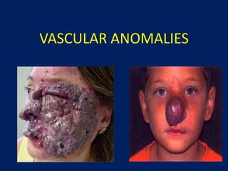
- 8. Hemangioma
- 9. Hemangioma True benign endothelial cell tumor Overgrowth of normal vascular tissue Most common tumor of the head and neck in children 7% of all benign soft tissue tumors Common in females - 3 : 1 ratio
- 10. Development Usually not present at the time of birth Appears weeks or months after birth Erythematous, macular patch
- 11. Hemangioma Stages Proliferating stage – first year Involuting stage – few years Involuted stage – most resolved by 10 years of age
- 12. Proliferative Stage Grows by endothelial proliferation Increased number of mast cells within the endothelial wall Grows faster than the growth of the child
- 13. Involutory Stage 12 months of age - signs of involution 5 to 9 years
- 14. Types of Hemangioma Infantile hemangioma – most common Congenital hemangioma – rare, present at birth, blue/gray - Rapidly involuting congenital hemangioma (RICH) - Non involuting congenital hemangioma (NICH) Intramuscular hemangioma Kaposiform hemangioendothelioma (KHE)
- 15. Sites Hemangiomas may involve superficial tissue, deep tissue or both Deep hemangiomas involve muscle or visceral organs - liver, lung, spleen, and gastrointestinal tract Intra-osseous hemangiomas are extremely rare and may deform the underlying skeleton
- 16. Clinical Presentation Superficial hemangioma - raised, reddish to purple tumor with a distinct margin Deep subcutaneous hemangiomas - deep bluish hue with normal overlying skin Both the lesions are firm on palpation Do not pulsate or exhibit any thrills or bruits
- 17. Investigations CT MRI - to document the extent of the deep hemangioma Arteriography - rarely indicated
- 18. Management Observation and parental support Intervention is necessary if functional compromise such as visual, airway or masticatory compromise, bleeding, ulceration or infection occurs
- 19. Management Corticosteroids Interferon alfa-2a Surgery - small lesions and as a secondary procedure after initial therapy and involution Sclerosing agents Cryotherapy Argon laser ablation
- 21. Vascular Malformations Malformed vessels secondary to aberrant development during the various stages of embryogenesis Between the 4th and 10th week of embryogenesis
- 22. Incidence - 15% of live births Present at birth Do not involute Grow proportionately with the patient growth
- 23. Trauma, infection and hormonal fluctuation (pregnancy/puberty) No endothelial proliferation - normal mast cells Alteration in the flow dynamics within and around the lesion Recruitment of collateral vessels Dilatation of involved vessels
- 24. Pelvis, extremities and intracranial circulation 88% are asymptomatic Wide range of presentation with an unpredictable course Male : Female = 1 : 1
- 25. Pathophysiology Normal endothelium Defect in vascular smooth muscle Progressive dilation of vascular channels
- 26. Mulliken Classification High flow lesions Low flow lesions
- 27. High Flow Vascular Malformations Arterial malformation (AM) Arteriovenous malformation (AVM) Arteriovenous fistula (AVF)
- 28. Arterial Malformation Aneurysm Coarctation Ectasia
- 29. Arteriovenous malformation Present at birth Skin shows reddish vascular hue Warm and Pulsatile Thrill and bruit present Absence of developed capillary bed Pain, ulceration, bleeding, compression / displacement of organs High output cardiac failure
- 30. Stages of AVM Stage I - pinkish-bluish stain and warmth Stage II - pulsations, thrill and bruit Stage III - dystrophic skin changes, ulceration, bleeding, pain Stage IV - high-output cardiac failure
- 31. Arteriovenous Fistula Simple arteriovenous connections Secondary to penetrating injuries after birth
- 32. Low Flow Lesions Capillary malformation Venous malformation Lymphatic malformation Combined malformation
- 33. Capillary Malformations Port wine stain Pink in infancy and darken to deep purple in childhood Do not change colour when compressed Do not disappear
- 34. Venous Malformations Present at birth Bluish, soft and easily compressible No thrills nor bruits Thin walled, dilated veins with inadequate smooth muscle layer Maneuvers that increase venous pressure (Valsalva maneuver or supine positioning) can enlarge a venous malformation Complications – Bleeding, Thrombosis, Pulmonary embolism
- 36. Phleboliths that may be noted on radiographic examination are found only in low flow lesions
- 37. Lymphatic Malformations Colourless Soft Micro cystic – irregular surface, clear or dark haemmorhagic bullae and vesicles – Salmon eggs Macro cystic – large cystic spaces containing lymphatic fluid – transilluminate - Cystic hygroma Mixed
- 38. Sturge-Weber syndrome - Port wine staining of the ophthalmic and maxillary dermatome of the Trigeminal nerve with convulsions, mental retardation and glaucoma Klippel – Trenaunay syndrome – capillary malformation with lateral megaveins and bone & soft tissue hypertrophy Blue Rubber Bleb Nevus syndrome (Bean syndrome) – multiple cutaneous and GIT venous malformation
- 39. Maffucci syndrome – Venous malformation with enchondromatosis Parkes Weber syndrome – overgrowth of the affected limb with small AV fistulas Proteus syndrome – Vascular malformation with connective tissue nevus with disproportionate overgrowth of limbs, ovarian cystadenoma, localized absence of fat Kasabach Merritt syndrome – fast growing vascular tumor hemangioendothelioma with thrombocytopenia
- 40. Investigations X-Ray - Soap bubble or honeycomb appearance MRI - differentiate low-flow from high flow lesions. Fatty deposits, venous lakes, phleboliths are all indicative of low flow lesions CT scan - extension into the surrounding soft tissue Doppler imaging - distinguish high flow lesion from low flow lesion Angiography delineates the size of the lesion and can also assess the rate of flow through the lesion
- 41. Treatment Indications Hemorrhage Risk of high-output cardiac failure Chronic venous hypertension Airway impingement Lesion threatening vital functions
- 42. Disabling pain Functional disability Cosmetic deformity Recurrent infection Persistent lymph leakage
- 44. Capillary Malformation Pulsed dye laser therapy Argon laser Surgical excision
- 45. Lymphatic Malformation Steroids Sclerotherapy Bleomycin sclerotherapy O.K 432 sclerotherapy Radiation therapy Surgery – Serial excision / Debulking
- 46. Venous Malformation Sclerotherapy for smaller malformations Sodium morrhuate, boiling water, alcohol and ethibloc are effective in fibrosing smaller lesions Treatment of choice is transcutaneous sclerotherapy 80% ethanol is the most commonly used agent
- 47. Arterial malformation & AV malformation Selective arterial embolization This procedure is usually done 24 – 72 hours before surgery in order to prevent the risk of haemorrhage during surgery The nidus needs to be embolized Goal of surgery should be total excision of the malformation
- 48. Lesion present at birth Hemangioma Vascular Malformation No Yes Proliferating Yes No Life Threatening Yes No Steroids Interferon Surgery Observation Cosmetic Resection Involuting Yes Investigations High Flow Lesion Low Flow Lesion Preop Embolisation Surgical Ablation Large vessel lakes Sclerotherapy No Yes
- 49. Take Home Message Hemangioma True tumor Endothelial proliferation Female 3 : 1 Absent at birth Rapid growth during infancy Self limited Diagnosis by history and appearance Vascular malformation • No tumor • Dysplastic vessels • 1:1 male : female • Present at birth • Growth proportional to child • Do not disappear • Diagnosis by MRI, Doppler, Angiography
- 50. QUIZ
- 51. 1 Who first classified vascular anomalies ? How was it classified ?
- 52. 2 Who classified vascular anomalies based on the biological characteristics ? How was it classified ?
- 53. 3 Types of Hemangioma ? Kasabach – Merritt syndrome ?
- 54. 4 Classify Vascular malformations ?
- 55. 5 Difference between Klippel Trenaunay syndrome and Parkes Weber syndrome ?
- 56. 6 Phleboliths are seen in which vascular anomaly ? What are Salmon eggs ?
- 57. 7 In venous malformation, which layer of the vessel wall is abnormal ? Port wine stain also known as ?
- 58. 8 In AVM, which has to be embolized ? After embolization, surgery should be done within what period ?
- 59. 9 ISSVA ?
- 60. 10 3 differences between hemangioma and vascular malformation ?
- 61. THANK YOU
Editor's Notes
- Most common congenital abnormalities observed in infants and children. Unfortunately, these lesions are also among the most confusing and misunderstood conditions, largely because of a history of inconsistent terminology used for its classification
- Vascular lesions can result in significant cosmetic problems for the patient, and some may lead to even serious life threatening hemorrhage.
- Virchow and his student Wegener, in 1880, separated all vascular tumors into angiomas and lymphangiomas characterized as "simplex", "cavernosum", and "racemosum". This continued to cause confusion and potential mismanagement.
- In 1982, Mulliken and Glowacki “biologically classified” the vascular anomalies based on their clinical behavior and endothelial cell characteristics into two groups
- ISSVA adopted and expanded this classification in 2014
- A true vascular tumor that results from a overgrowth of normal vascular tissue. Most common tumor of the head and neck in infancy and childhood, comprising approximately 7% of all benign soft tissue tumors
- Usually not present at the time of birth but are noted by the parent within the first month of life. Hemangiomas are initially noticed as an erythematous, macular patch, which progresses through a PROLIFERATIVE PHASE whereby it changes its color and grows faster than the commensurate growth of the child.
- It grows by endothelial proliferation. During the rapid growth phase, an increased number of mast cells are seen within the endothelial wall
- By 12 months of age most hemangiomas show signs of involution. The INVOLUTORY PHASE is normally slow and will not be completed until the age of 5 to 9 years.
- On examination, the superficial hemangioma usually consists of a raised, reddish to purple tumor with a distinct margin. In contrast, deep subcutaneous hemangiomas often have a deep bluish hue with normal overlying skin, making diagnosis more difficult. Both the lesions are firm to palpation and do not pulsate or exhibit any thrills or bruits. .
- Observation and parental support are the initial approaches in the management. Intervention is necessary if functional compromise such as visual change, airway or masticatory compromise, bleeding, ulceration or infection occurs. This may initially involve corticosteroids for rapidly proliferating lesions or therapy with interferon alfa-2a.
- Surgery is generally reserved for small lesions and as a secondary procedure after initial therapy and involution. Treatment modalities include routine excision, injection of sclerosing agents, cryotherapy, and ablation using an argon laser.
- Vascular malformations are thought to “result when there is interruption at a particular stage of development of a vessel”.
- Vascular malformations are present at birth and unlike hemangiomas, do not go through a “rapid proliferative phase” and they do not involute. They grow commensurately with the patient.
- Trauma, infection, and hormonal fluctuation (pregnancy or puberty) may stimulate increased growth of the vascular malformation.
- considered to be congenital vascular anomalies, but are usually first noted several years after birth or after certain triggering changes such as trauma or the hormonal changes of puberty or pregnancy.
- Capillary malformations may be smooth initially but become more “pebble – like” as the patient grows.
- , however, combined lesions take on the hue of the additional vessel type. Lymphatic-venous malformations” may appear deep purple while “capillary-lymphatic malformations” can be pink to purple.
- More difficult to treat because of the poor demarcation and the infiltrative nature of the lymphatic vessels