ICT role in 21st century education and it's challenges.
P Col III B.Ph ANS 2 12-2015
1. III B.Pharm Pharmacology I 2015-16 Dr.KPS Gowda PESCP Page 1
Chapter No-2 Drugs acting on ANS
a)Introduction-Neurohumoral transmission
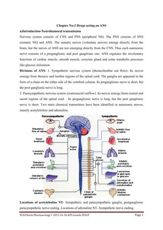
Nervous system consists of CNS and PNS (peripheral NS). The PNS consists of SNS
(somatic NS) and ANS. The somatic nerves (voluntary nerves) emerge directly from the
brain, but the nerves of ANS are not emerging directly from the CNS. Thus each autonomic
nerve consists of a preganglionic and post ganglionic one. ANS regulates the involuntary
functions of cardiac muscle, smooth muscle, exocrine gland and some metabolic processes
like glucose utilization.
Divisions of ANS- 1. Sympathetic nervous system (thoracolumbar out flow)- Its nerves
emerge from thoracic and lumbar regions of the spinal cord. The ganglia are appeared in the
form of a chain on the either side of the vertebral column. Its preganglionic nerve is short, but
the post ganglionic nerve is long.
2. Parasympathetic nervous system (craniosacral outflow). Its nerves emerge from cranial and
sacral regions of the spinal cord. . Its preganglionic nerve is long, but the post ganglionic
nerve is short. Two main chemical transmitters have been identified in autonomic nerves,
namely acetylcholine and adrenaline.
Locations of acetylcholine NT- Sympathetic and parasympathetic ganglia, postganglionic
parasympathetic nerve ending. Locations of adrenaline NT- Sympathetic nerve ending.
2. III B.Pharm Pharmacology I 2015-16 Dr.KPS Gowda PESCP Page 2
Steps involved in neurohumoral transmission-
a.Axonal conduction- The resting potential in the axon is -70mv. In the resting state more
K+ ions present in the axoplasma and more Na+ and Cl- ions present in the extracellular
fluid. This is regulated by energy dependent (ATP) Na+/K+ Atpase. If the nerve receives an
impulse, it increases the influx of Na+ ions into the axoplasma. This leads to depolarization
and it is essential for the conduction of an impulse.
b.Junctional transmission- i) Release of NT- The NTs are synthesized in the nerve ending
and they get stored in the synaptic vesicles. When an impulse is reached nerve ending, the
voltage gated calcium channels get opened causing the influx of Ca2+ into the nerve ending.
These ions causes exocytosis, this leads to the rapture of synaptic vesicles leading to the
release of NTs.
ii)Combination of the transmitter with postjunctional receptors and production of
postjunctional potential- The released NTs diffuses into synoptic cleft and may bind to the
receptors present on the presynaptic or postsynaptic membranes. This interaction leads to the
increased permeability to cations (Na+ ions). This leads to depolarization of the membrane i.e
Excitatory post synaptic potential (EPSP). The NT and receptor may also leads to the
increased permeability to anions (mainly Cl-). This leads to hyperpolarization of the
membrane i.e Inhibitory post synaptic potential (IPSP).
c.Initiation of postjunctional activity- EPSP initiates actions like muscle contraction,
vasoconstriction, tachycardia, increased secretions, while IPSP initiates actions like
relaxation, vasodilatation, bradycardia, decreased secretions, etc.
d-Destruction or dissipation of the transmitter- The NT get dissociated from the receptor
and dissipated by various enzymes. Examples-
i) Enzymatic destruction-Cholinesterase destroys acetylcholine, whereas manoamine oxidase
(MAO) and catechol-o-methyl transferase (COMT) destroys adrenaline or noradrenalin.
ii) Reuptake mechanism- NTs like noradrenaline may re-enter the synaptic vesicles by
reuptake mechanism.
b) Adrenergic and cholinergic receptors-
Adrenergic receptors- (adrenoceptors) are selective for nor adrenaline and adrenaline. There
are two types- α-adrenoceptors and β-adrenoceptors.
1). α-adrenoceptors- They are divided into α1 and α2 subclasses.
α1 adrenoceptor is a G protein coupled receptor (GPCR) associated with Gq type of G
protein. When adrenaline binds to this receptor, GDP converted to GTP, αβγ subunits of G
protein gets detached. The -GTP complex binds to the membrane bound phospho lipase-C
3. III B.Pharm Pharmacology I 2015-16 Dr.KPS Gowda PESCP Page 3
(PLC). The activated PLC converts membrane phospholipids phosphatidylinositol-4,5-
bisphosphate (PIP2) into inositol-1,4,5- triphosphate (IP3) and diacylglycerol (DAG). The
DAG remains in the membrane and activates protein kinase C (PKC).
The activated PKC phosphorylates several cellular proteins. IP3 stimulates the release of
calcium from ER into the cytosol. The released calcium ions are responsible for the action.
The calcium ions bound to the calmodulin protein (CaM). Ca2+
- CaM complex activates the
MLCK (Myosin light chain kinase). The activated MLCK causes the phosphorylation of
myosin-LC. The myosin-LC-P causes smooth muscle contraction.
Important locations of α1 adrenoceptors- Vascular smooth muscles, genitourinary smooth
muscle, radial muscle, intestinal smooth muscle, heart and liver (Except intestine all actions
are excitatory).
a)Vascular smooth muscles- Vasoconstriction (MOA as above).
b) Genitourinary smooth muscles- Contractions (MOA as above).
c)Radial muscle- Contraction –mydriasis (MOA as above).
4. III B.Pharm Pharmacology I 2015-16 Dr.KPS Gowda PESCP Page 4
d) Heart- Increases the rate and force of heart contraction. MOA- The calcium ions bind to
the troponin, this leads to the interaction of actin-myosin causing heart muscle contraction.
e) Liver- Increases glycogenolysis and gluconeogenesis. MOA- DAG causes activation of
PKC. The activated PKC causes the phosphorylation of enzymes needed for glycogenolysis
and gluconeogenesis.
f) Intestinal muscles- Relaxation. The activated protein kinase causes phosphorylation of
cellular proteins. For eg K+
channel activation leads to efflux of K+
ions leading
hyperpolarization (IPSP). The IPSP is also due to inactivation of calcium channels.
α2 adrenoceptor- is a G protein coupled receptor (GPCR) associated with Gi type of G
protein. When adrenaline binds to this receptor, GDP converted to GTP, αβγ subunits of G
protein gets detached. The α-GTP complex binds to the membrane bound adenylyl cyclase
(AC). The inhibited AC decreases the formation of cAMP. The α-GTP complex also activates
the K+
channel. This increases the removal of K+
ions from the cell to outside. This decreases
the potential inside the cell (hyperpolarization). The α-GTP complex also inactivates the Ca2+
channels. This decreases the influx of Ca2+
ions into the cell. This also decreases the potential
within the cell causing hyperpolarization. Hence the action is inhibition.
Important locations of α2 adrenoceptors-Pancreatic β cells, platelets, nerve, vascular smooth
muscles. (all actions are inhibitory except vascular smooth muscle).
a) Pancreatic β cells- Decreases insulin secretion.
b)Platelets- Aggregation.
c)Nerve- Function as auto-receptors, neuronal inhibition.
d)Vascular smooth muscle- Vasoconstriction (both α1 and α2 causes vasoconstriction).
2). β-adrenoceptors. These are divided into three types- β1, β2 and β3. All three belongs to
stimulatory (Gs) type of G protein coupled receptors.
β-adrenoceptor is a G protein coupled receptor (GPCR) associated with Gs type of G protein.
When adrenaline binds to this receptor, GDP converted to GTP, αβγ subunits of G protein
gets detached. The α-GTP complex binds to the membrane bound adenylyl cyclase (AC). The
stimulated AC increases the formation of cAMP. Increased cAMP activates protein kinases
(especially protein kinase A), which phosphorylates cellular proteins, including ion channels.
β1 receptors located on heart (SAN and myocardium), renal juxta glomerular cells. Increases
the heart rate (chronotropy) and force of heart contraction (inotropy) and increases the rennin
secretion.
β2 receptors are located in smooth muscles, liver, and skeletal muscles. Stimulation of β2
receptors in bronchial smooth muscles causes bronchodilatation. This effect is due to Gs
5. III B.Pharm Pharmacology I 2015-16 Dr.KPS Gowda PESCP Page 5
independent activation of K+
channels resulting in hyperpolarization. This relaxes the
bronchial smooth muscles. In liver stimulation of β2 receptors results in increased blood sugar
level. In skeletal muscle β2 receptors results in increased glycogenolysis.
β3 receptors are located in adipose tissue. In adipose tissue stimulation of β3 receptors results
in increased lipolysis.
Cholinergic receptors- There are two types- Muscarinic receptors and nicotinic
receptors. a)Muscarinic receptors- These are classified into 5 sub types- M1, M2, M3, M4
and M5. All muscarinic receptors belongs to GPCRs. M1, M3 and M5 belong to Gq type and
M2 and M4 belongs to Gi type.
M1 receptor- These are located in the autonomic ganglia and CNS. The activation of M1
receptors in the autonomic ganglia result in the development of excitatory postsynaptic
potential (EPSP). The activation of M1 receptors in the CNS leads to the complex actions like
arousal, attention and analgesia. M2 receptor- These are located in the heart (SAN and
myocardium). The stimulation of these receptors results in the decreased heart rate and force
of contraction (negative chronotropy and inotropy). M3 receptor- These are located in the
smooth muscles. The stimulation of these receptors results in the muscle contraction
(examples-contraction of intestinal, bronchial and circular muscles). M4 receptor- These are
located in the CNS, and the actions are inhibitory. M5 receptor- These are located in the
CNS and the actions are excitatory similar to M1.
b) Nicotinic receptors- These are ionotropic (ion channel) receptors. These are classified
into two types- NM and NN.
a)Nicotinic (NM) receptors- These are located on the sarcolemma of the skeletal muscles at
neuro muscular junction (NMJ). These receptors are sodium channels made up of 5 sub units-
2 α, β, γ/ε and δ (pentameric structure). Two of the acetyl choline binds to the two alpha
6. III B.Pharm Pharmacology I 2015-16 Dr.KPS Gowda PESCP Page 6
subunits. This interaction leads to opening of the channel causing influx of sodium ions
(cations). This increases the potential within the muscle causing depolarization (EPSP). This
leads to skeletal muscle contraction.
b).Nicotinic (NN) receptors- These are located in the autonomic ganglia, adrenal medulla
and CNS. In the autonomic ganglia, stimulation of these receptors by the acetylcholine causes
the influx of sodium ions. This depolarizes the post ganglionic neuron. The stimulation of
these receptors in the adrenal medulla results in increased secretion of catecholamines. The
stimulation of these receptors in the CNS results in arousal, attention and analgesia.
c)Adrenergic drugs (Sympathomimetics) -These are agents which produce an effect similar
to the stimulation of postganglionic sympathetic nerves. Most of these compounds have an
intact or partially substituted amino group. Hence they are also called as sympathomimetic
amines.
Classification- These are classified based on the presence or absence of catechol (ortho-
hydroxy benzene).
1.Catecholamines - Adrenaline, Noradrenaline, Dopamine, Isoprenaline.
2.Non-catecholamines-Ephedrine,Amphetamine,Methylamphetamine, Hydroxyamphetamine,
Methylphenidate, Dexmethylphenidate.
Pharmacology of adrenergic drugs (Adrenaline)-
Synthesis- The endogenous catecholamines- dopamine, noradrenaline and adrenaline are all
synthesized from tyrosine. The tyrosine enters the adrenergic nerve via aromatic L amino
acid transporter. (Na+-tyrosine symporter). Tyrosine hydroxylase oxidizes tyrosine to
7. III B.Pharm Pharmacology I 2015-16 Dr.KPS Gowda PESCP Page 7
dihydroxyphenylalanine (L-DOPA). Aromatic L-amino acid decarboxylase converts L-
DOPA into dopamine. Dopamine enters the synaptic vesicles via vesicular manoamine
transporter (VMAT) in exchange with H+ ions. Within synaptic vesicles dopamine gets
converted to noradrenaline by the enzyme dopamine-β-hydroxylase. Dopamine is the
neurotransmitter in the dopaminergic neurons. Noradrenaline is the main NT in the
adrenergic neurons. But in the adrenal medulla noradrenaline is converted into adrenaline by
the enzyme phenylethanolamine N-methyl transferase (PNMT). Hence adrenaline is the main
NT in the adrenal medulla.
Release – On arrival of an impulse at the adrenergic nerve ending, the voltage gated calcium
channels get opened. This leads to influx of calcium ions. The triggered calcium causes
rapture of synaptic vesicles by a process called exocytosis. This releases the noradrenaline
into the synaptic cleft (NEJ-neuro effector junction). The noradrenaline binds to the alpha
receptors or beta receptors present on the presynaptic or post synaptic membrane. This
initiates the pharmacological response (EPSP or IPSP).
Fate- Noradrenaline is metabolized by two main enzymes- Catechol-o-methyl transferase
(COMT) and monoamine oxidase (MAO). COMT is present in the circulating blood and this
enzyme degrades the circulating catecholamines. Whereas MAO is located in the adrenergic
8. III B.Pharm Pharmacology I 2015-16 Dr.KPS Gowda PESCP Page 8
neurons and this degrade the noradrenaline located in the adrenergic nerves (outside the
vesicles). COMT, MAO and aldehyde dehydragenase degrade the catecholamines into
multiple intermediates and finally into vanillyl mandelic acid (VMA), which is excreted
through urine.
Pharmacological actions of adrenaline-Noradrenalin mainly act on alpha receptors,
isoprenaline mainly acts on beta receptors and adrenaline acts on both alpha and beta
receptors.
Adrenergic receptors- (adrenoceptors) are selective for nor adrenaline and adrenaline. There
are two types- α-adrenoceptors and β-adrenoceptors.
1). α-adrenoceptors- They are divided into α1 and α2 subclasses.
α1 adrenoceptor is a G protein coupled receptor (GPCR) associated with Gq type of G
protein. When adrenaline binds to this receptor, GDP converted to GTP, αβγ subunits of G
protein gets detached. The -GTP complex binds to the membrane bound phospho lipase-C
(PLC). The activated PLC converts membrane phospholipids phosphatidylinositol-4,5-
bisphosphate (PIP2) into inositol-1,4,5- triphosphate (IP3) and diacylglycerol (DAG). The
(IP3) and DAG are the second messengers. Ca2+
is the third messenger. The DAG remains in
the membrane and activates protein kinase C (PKC). The activated PKC phosphorylates
several cellular proteins. IP3 stimulates the release of calcium from ER into the cytosol. The
released calcium ions are responsible for the action. The calcium ions bound to the
calmodulin protein (CaM). Ca2+
- CaM complex activates the MLCK (Myosin light chain
kinase). The activated MLCK causes the phophorylation of myosin-LC. The myosin-LC-P
causes smooth muscle contraction.
9. III B.Pharm Pharmacology I 2015-16 Dr.KPS Gowda PESCP Page 9
Important locations of α1 adrenoceptors- Vascular smooth muscles, genitourinary smooth
muscle, radial muscle, intestinal smooth muscle, heart and liver.(Except intestine all actions
are excitatory).
a)Vascular smooth muscles- Vasoconstriction (MOA as above).
b) Genitourinary smooth muscles- Contractions (MOA as above).
c) Radial muscle- Contraction –mydriasis (MOA as above).
d) Heart- Adrenaline increases the rate and force of heart contraction. MOA- The calcium
ions bind to the troponin, this leads to the interaction of actin-myosin causing heart muscle
contraction.
e) Liver- Increases glycogenolysis and gluconeogenesis. MOA- DAG causes activation of
PKC. The activated PKC causes the phophorylation of enzymes needed for glycogenolysis
and gluconeogenesis.
f) Intestinal muscles- Relaxation.
α2 adrenoceptor- is a G protein coupled receptor (GPCR) associated with Gi type of G
protein. When adrenaline binds to this receptor, GDP converted to GTP, αβγ subunits of G
protein gets detached. The α-GTP complex binds to the membrane bound adenylyl cyclase
(AC). The inhibited AC decreases the formation of cAMP. The α-GTP complex also activates
the K+
channel. This increases the removal of K+
ions from the cell to outside. This decreases
the potential inside the cell (hyperpolarization). The α-GTP complex also inactivates the Ca2+
channels. This decreases the influx of Ca2+ ions into the cell. This also decreases the potential
within the cell causing hyperpolarization. Hence the action is inhibition.
Important sites of α2 adrenoceptors -Pancreatic β cells, platelets, nerve, vascular smooth
muscles (all actions are inhibitory except vascular smooth muscle).
a) Pancreatic β cells- Decreases insulin secretion.
b)Platelets- Aggregation.
c)Nerve- Function as auto receptors, neuronal inhibition.
d)Vascular smooth muscle- Vasoconstriction (both α1 and α2 causes vasoconstriction).
2)β-adrenoceptors. These are divided into three types- β1, β2 and β3. All three belongs to
stimulatory (Gs) type of G protein coupled receptors.
β-adrenoceptor is a G protein coupled receptor (GPCR) associated with Gs type of G protein.
When adrenaline binds to this receptor, GDP converted to GTP, αβγ subunits of G protein gets
detached. The α-GTP complex binds to the membrane bound adenylyl cyclase (AC). The
stimulated AC increases the formation of cAMP. Increased cAMP activates protein kinases
(especially protein kinase A), which phosphorylates cellular proteins, including ion channels.
10. III B.Pharm Pharmacology I 2015-16 Dr.KPS Gowda PESCP Page 10
β1 receptors located on heart (SAN and myocardium), renal juxta glomerular cells. Increases the
heart rate (chronotropy) and force of heart contraction (inotropy) and increases the rennin
secretion.
β2 receptors are located in smooth muscles, liver, and skeletal muscles. Stimulation of β2
receptors in bronchial smooth muscles causes bronchodilatation. This effect is due to Gs
independent activation of K+
channels resulting in hyperpolarization. This relaxes the bronchial
smooth muscles. In liver stimulation of β2 receptors results in increased blood sugar level. In
skeletal muscle β2 receptors results in increased glycogenolysis.
β3 receptors are located in adipose tissue. In adipose tissue stimulation of β3 receptors results in
increased lipolysis.
Pharmacological actions of adrenaline- 1.Heart- Adrenaline increases heart rate (positive
chronotropy) and force of heart contraction (positive inotropy).
2.Blood vessels-Constrict the blood vessels of skin and mucous membrane (alpha effect). But
it dilates the blood vessels of skeletal muscles (beta effect).
3.Blood pressure- Dale’s vasomotor reversal- Normally adrenaline acts on both alpha and
beta receptors. Alpha actions increase the BP, where as beta actions decrease the BP. When
adrenaline is administered after the administration of an alpha blocker (ergotoxine), then
adrenaline produces only fall in BP. This effect is known as Dale’s vasomotor reversal.
4.Smooth muscles-a)GIT- Adrenaline relaxes the smooth muscles of intestine and decreases
the motility. b)Bronchi- Adrenaline relaxes the bronchial smooth muscles and causes
bronchodilation.
5.Spleen- Adrenaline contracts the spleen, hence more RBCs are released into the circulation.
6.Hair follicle- Adrenaline contracts the pili erector muscle of hair follicle, producing
erection of hair.
7.Eye- Adrenaline produces mydriasis due to contraction of radial muscles. Adrenaline also
reduces intra ocular pressure.
8.Respiration- Adrenaline produces a weak stimulation of respiration.
9.Skeletal muscles- Adrenaline stimulates glycogenolysis in the skeletal muscles. This
relieves the fatigue of the skeletal muscle.
10.Metabolic effects-Adrenaline increases blood sugar level. Adrenaline also causes
lipolysis and this increases free fatty acid level of plasma.
ADME- Adrenaline is inactivated in the GIT if administered orally. Hence it is administered
by inhalation or injection. Adrenaline is metabolized by COMT and MAO. The metabolite
VMA is excreted through urine.
11. III B.Pharm Pharmacology I 2015-16 Dr.KPS Gowda PESCP Page 11
Preparations of adrenergic drugs. Adrenaline HCl inj 1mg/ml, adrenaline spray,
noradrenaline bitartrate inj 4mg/ml, isoproterenol HCl inj (isoprenaline), ephedrine HCl tabs
and injections.
Uses of adrenergic drugs-
1.Adrenaline is used in Stokes-Adams syndrome (temporary loss of conseousness due to
insufficient blood flow to the brain).
2. Adrenaline is administered by intra cardiac route for the resuscitation of failing heart.
3. Used to treat allergy due to histamine.
4. Used in bronchial asthma, as it causes bronchodilatation.
5.As adrenaline constricts the blood vessels, it is used to prolong the effect of local
anaesthetics and also used to control blood bleeding.
6. Noradrenaline is mainly used for elevating the BP in case of shock.
7. Isoprenaline is mainly used as a bronchodilator and as a cardiac stimulant in heart block.
ADRs of adrenaline- Palpitations (abnormal heart beat), tachycardia, arrhythmia, anxiety,
tremor, hypertension, constipation, and acute pulmonary oedema.
-------------------------------------------------------------------------------------------------
d)Adrenergic receptor blockers, adrenergic neuron blockers.
Anti-adrenergic Drugs (Sympathetic or adrenergic blocking drugs)- These are drugs
which antagonize the receptor actions of adrenaline and related drugs.
12. III B.Pharm Pharmacology I 2015-16 Dr.KPS Gowda PESCP Page 12
Classification-
1.Alpha adrenergic blocking agents- 2.Beta adrenergic blocking agents-
1.Alpha adrenergic blocking agents- a)Selective alpha 1 blockers- Prazosin, terazosin,
doxazosin, tamsulosin.
b) Selective alpha 2 blockers- Yohimbine, medetomidine. Mainly used in research and
veterinary practice. Rarely used in human medicine. Yohimbine was used as aphrodiasic.
Medetomidine is given along with ketamine to induce anaesthesia in lab animals.
c)Non selective alpha blockers-) - Phentolamine, Phenoxybenzamine.
(Important locations of α1 adrenoceptors- Vascular smooth muscles, genitourinary smooth
muscle, radial muscle, intestinal smooth muscle, heart and liver.(Except intestine all actions
are excitatory). α1 adrenoceptor is a G protein coupled receptor (GPCR) associated with Gq
type of G protein. Important sites of α2 adrenoceptors-Pancreatic β cells, platelets, nerve,
vascular smooth muscles (all actions are inhibitory except vascular smooth muscle).)
Actions of alpha blockers- 1.Blockage of vasoconstrictor effect. This decreases peripheral
resistance, venous return and cardiac output. This decreases the BP. Adrenaline produces fall
in BP in presence of alpha blocker (Dale’s vasomotor reversal). In the absence of alpha
blocker adrenaline produces biphasic response (due to alpha and beta receptor activation).
2. Reflex tachycardia. This is due to the blockage of presynaptic alpha 2 receptors.
3. Nasal stiffness (due to blockage of vascular alpha receptors) and miosis (due to the
blockage of α1 receptors of radial muscles.
4. Stimulation of alpha receptors in the intestinal smooth muscles causes relaxation. The
blockage increases intestinal motility. This may leads to diarrhoea.
5. The developed hypotension by alpha blockers reduces the renal blood flow. This decreases
glomerular filtration, more fluid and salt get reabsorbed leads to salt and fluid retention. 6.
The tone of the smooth muscle of urinary bladder, trigone and urethra decreases. 7. The alpha
blockage leads to inhibition of ejaculation (impotence).
ADRs- Palpitations (abnormal heart beats), postural hypotension (positional changes of BP),
nasal stuffness, diarrhoea, fluid retention, impotence.
13. III B.Pharm Pharmacology I 2015-16 Dr.KPS Gowda PESCP Page 13
Palpitations Postural hypotension Nasal stuffness
Clinical uses of alpha blockers- 1.Used in the diagnosis and the treatment of
pheochromocytoma. 2.Used in the treatment of hypertension. 3.Used in the treatment of
congestive heart failure. 4.Used in the treatment of haemorrhagic and bacteraemia shock.
5.Used in peripheral vascular diseases. 6.Used in Benign hypertrophy of prostate (BHP).
7.Used in the treatment of migraine.
Preparations of alpha blockers-
14. III B.Pharm Pharmacology I 2015-16 Dr.KPS Gowda PESCP Page 14
2.Beta adrenergic blocking agents-
a.Selective β1 blockers (cardioselective)- Metoprolol, atenolol, acebutolol, esmolol,
betaxolol, celiprolol.
b.Selective β2 blockers- Butoxamine.
c.Nonselective (β1 and β2 blockers)- Propranolol, sotalol, timolol, pindolol, labetalol,
carvedilol.
β1 receptors located on heart (SAN and myocardium), renal juxta glomerular cells.
β2 receptors are located in smooth muscles, liver, and skeletal muscles.
β3 receptors are located in adipose tissue.
Pharmacology of Propranolol-
1.CVS-a.Heart- Propranolol blocks β1 receptors located on heart (SAN and myocardium).
This decreases the heart rate, force of contraction and cardiac output.
b.Blood pressure- They reduce blood pressure by acting on the heart and decreasing cardiac
output.
c.Bronchial smooth muscle- Propranolol block the β2 receptors located in the bronchial
smooth muscle. This prevents the relaxation caused by adrenaline. Hence this produces
bronchospasm. This is dangerous in asthmatic patients.
d.CNS- Lipid soluble beta blockers (eg propranolol) cross the BBB and produce sedative and
anticonvulsant effects.
15. III B.Pharm Pharmacology I 2015-16 Dr.KPS Gowda PESCP Page 15
e. Local anaesthetic effect- Propranalol is a potent local anaesthetic like lidnocaine. But it is
not used in clinical practice due to its irritant property.
f. Eye- The topical administration of propranolol on to the eye, decreases IOP.
g. Metabolic effect- Propranolol blocks the adrenaline induced glycogenolysis and lipolysis.
ADME-Most of the beta blockers are rapidly and completely absorbed if administered orally.
Also they are rapidly and completely metabolized in the liver. Because of rapid metabolism
only fraction of the drug enters the blood circulation.
ADRs- 1.Sudden hypotension, bradycardia leading to cardiac asystole.
2.Nausea, vomiting, constipation and broncho-spasm. 3.Cold extremities and absent pulse.
4.Prolonged use of propranolol may produce fatigue, muscle cramps, lethargy,
hallucinations and mental depression.
Lethargy muscle cramps depression
Therapeutic uses of beta blockers-
1.Hypertension (mild antihypertensives). 2.Angina pectoris. 3.Cardiac arrythmias.
4.Myocardial infarction. 5.Congestive heart failure. 6.Used to treat dissecting aneurysm
(enlargement of aorta). 7.Pheochromocytoma, 8.Thyrotoxicosis, 9.Migraine, 10.Anxiety
11.Glaucoma. 12. Hypertrophic cardiomyopathy.
16. III B.Pharm Pharmacology I 2015-16 Dr.KPS Gowda PESCP Page 16
Marketed preparations of beta blockers- Propranolol tab, inj. Atenolol tab, labetalol HCl
inj, tab, timolol maleate eye drops, carvedilol tabs.
Adrenergic neuron blockers- These are the drugs which block the release of noradrenaline
from the post ganglionic sympathetic nerve endings.
Examples- Reserpine, guanethidine.
Reserpine- It is an alkaloid from the roots of Rauwolfia serpentine. It was a popular
antihypertensive drug in between 1950- 1960, but is now used as a pharmacological tool. The
hypotensive action is due to deletion of NA from peripheral adrenergic nerve endings. It
irreversibly blocks the VMAT and also inhibits the reuptake of NA into the synoptic vesicles.
ADRs- Bradycardia, Diarrhoea, miosis, postural hypotension, nasal stiffness, impotence.
Marketed Preparation- Adelphane tabs (Reserpine and hydrochlorthiazide).
Guanethidine. It is a postganglionic adrenergic blocking agent. Uptake of guanethidine and
storage in sympathetic neurons occur via the noradrenaline transporter. Guanethidine slowly
displaces noradrenaline from its storage innerve endings and there by blocks the release of
noradrenaline. This leads to decreased vasoconstriction and decreases the BP.
Marketed Preparation- Ismelin (guanethidine monosulfate tab). It is one of the out dated
drug. But it is still licensed in some countries, e.g. UK for the rapid control of BP in a
hypertensive emergency.
-------------------------------------------------------------------
e.Cholinomimetics and anticholinesterases
17. III B.Pharm Pharmacology I 2015-16 Dr.KPS Gowda PESCP Page 17
Cholinergic drugs (Cholinomimetics / Parasympathomimetics) – These drugs act on
organs innervated by postganglionic parasympathetic nerves. They produce an effect similar
to the stimulation of parasympathetic nervous system.
Classification of cholinergic drugs-
1.Esters of choline- Acetylcholine, methacholine, carbachol, bethanechol.
2.Cholinomimetic alkaloids- Pilocarpine, arecholine, muscarine.
3.Anitcholinesterases-a)Reversible–Carbamates-Physostigmine,neostigmine,
pyriodstigmine, edrophonium, rivastigmine. Acridine- Tacrine.
b)Irriversible- Organophosphates- Di-isopropyl flurophosphate (DFP), echothiophate,
parathion, malathion, diazinon (TIK-20). Carbonyl (SEVIN), propoxur (BAYGON).
Synthesis, storage and degradation of acetylcholine.
Synthesis- Choline enters the cholinergic nerve along with Na+
through choline/Na+
symporter. Acetyl coenzyme A (derived from the mitochondria) combines with choline to
form acetylcholine. This reaction is catalyzed by choline acetyltransferase.
18. III B.Pharm Pharmacology I 2015-16 Dr.KPS Gowda PESCP Page 18
Storage- After its synthesis in the cytoplasm, Ach (acetylcholine) is transported into synaptic
vesicles for storage. Ach enter the synaptic vesicle through Ach-H+
antiporter present on the
synaptic vesicles. Each synaptic vesicle contains about 1000 to 50000 molecules of
acetylcholine.
Release of Ach. Because of arrival of an impulse the Ca2+
channel present on the membrane
gets opened. This leads to inflow of Ca2+
ions, depolarization, exocytosis, rapture of synaptic
vesicles and release of Ach molecules into the synaptic cleft. The released Ach molecules
combines with muscarinic or nicotinic receptors located on the presynaptic or post synaptic
membranes produce pharmacological effects.
Degradation of acetylcholine- Acetylcholine in the synaptic cleft is degraded by membrane
bound acetylcholinesterase (AchE) into choline and acetate. There are two types of AchE.
19. III B.Pharm Pharmacology I 2015-16 Dr.KPS Gowda PESCP Page 19
The acetylcholinesterases (true), are present on cholinergic sites and pseudocholinesterases
are present in the blood plasma, liver and intestine. The choline gets re-entered into the
cholinergic nerve ending, which may utilize for the fresh synthesis of acetylcholine.
Pharmacological actions of acetylcholine-
Cholinergic receptors- There are two types of cholinergic receptors-muscarinic receptors,
and nicotinic receptors.
a.Muscarinic receptors- These are 7 transmembrane domain G protein coupled receptors.
There are 5 types of muscarinic receptors. M1, M3 and M5 are associated with Gq type and
hence are responsible for the stimulation of phospholipase C (PLC). The remaining M2 and
M4 are coupled to Gi type and are responsible for adenylyl cyclase (AC) inhibition.
Locations and actions of muscarinic receptors:
M1 receptors- These receptors are expressed on the autonomic ganglia. If the M1 receptors
present on the autonomic ganglion acted by acetylcholine, it causes EPSP. These receptors
are also expressed in the CNS. Its actions are complex. Some of the important actions are
arousal, attention, analgesia.
M3 receptors- These receptors are present on the smooth muscles and secretary glands. The
acetylcholine combines with M3 receptors and produces smooth muscle contraction and
increased glandular secretion.
M5 receptors – These receptors are expressed in the CNS and actions are complex.
MOA (of M1 , M3 , M 5 receptors) - Gq type- activation of PLC-> PIP2 IP3 and DAG-
release of Ca2+
, activation of PKC, phosphorylation of cellular proteins pharmacological
action.
M2 receptors- These receptors are expressed on the SAN, AVN, atrial and ventricular
muscles of the heart. The receptor activation causes hyperpolarization (IPSP) decreased
conduction velocity. Hence it produces negative chronotropy (decrease in the heart rate). In
the atrial and ventricular muscle the receptor activation produces negative ionotropic effect.
M4 receptors- These are expressed in the CNS and actions are complex.
MOA- (M2 M4 receptors) Gi type - inhibition of AC and activation of K+
channels,
inactivation of Ca2+
channels-> hyperpolarization.
Nicotinic receptors- These receptors are present on the ganglia of the sympathetic and
parasympathetic nerves ( Ng, or NN). The nicotinic receptors are also present at the NMJ of
the somatic nerve ending (NM).
The nicotinic receptors are ion channel receptors. These receptors are composed of 5
subunits- two α subunits, a β subunit, a γ subunit and a δ subunit. In the absence of Ach, the
20. III B.Pharm Pharmacology I 2015-16 Dr.KPS Gowda PESCP Page 20
receptor gate is closed, and cations (like Na2+
) are not entering the channel. When two Ach
molecules bind to α subunits, the channel gets open, and the cations pass through the channel
into the cell.
Actions of acetylcholine (through muscarinic receptors)
1. Heart- Acetylcholine activates the M2 receptors present on the pacemaker cells of the
SAN/AVN causes negative chronotropy (decreased heart rate). Acetylcholine activates the
M2 receptors present on the myocardial cells causes negative ionotropy (decreased force of
heart contraction). The other effects are decreased cardiac output and bradycardia.
2.Blood vessels- Acetylcholine produces vasodilatation of the blood vessels. Hence there is
fall in B.P. This is mediated through EDRF (NO).
MOA-Acetylcholine combines with muscarinic (M3) receptor present on the endothelial
cells. Through activation of PLC, PIP2-> IP3 and DAG. IP3 triggers the Ca2+
within the
cytoplasm. The Ca2+
combines with CaM. The resultant Ca2+
-CaM complex stimulates the
endothelial nitric oxide synthase (eNOS). The activated eNOS catalyzes the formation of NO
from L.arginine. The NO diffuses to the adjacent vascular smooth muscle cells, where it
activates guanylyl cyclase. The activated guanylyl cyclase catalyzes the conversion of GTP to
cGMP, which dephosphorylates myosin- LC. This prevents actin-myosin interaction. This
leads to the relaxation of smooth muscle. The NO also acts by activating K+
channel to cause
relaxation of the smooth muscle.
3.Smooth muscles - GIT, bronchial, urinary bladder muscles get contracts. These effects are
mediated through M3 receptors (Gq type). The effects are increased intestinal motility,
bronchoconstriction, micturition.
4.Glands- On the secretary glands M3 receptors (Gq type) are present. The acetylcholine
increases sweat, saliva, gastric juice, lacrimal juice, pancreatic juice secretions.
5.Eye- Acetylcholine activates the M3 receptors located on the circular muscles, leads to
miosis.
Actions of acetylcholine through nicotinic receptors
1.Autonomic ganglia- Both sympathetic and parasympathetic ganglia are stimulated. These
effects are mediated through nicotinic receptors (NN).
2.Skeletal muscle- The skeletal muscles are innervated by somatic nerves (voluntary nerves).
When two Ach molecules combine receptor sites present on the two α unit of the nicotinic
receptor (NM). This produces skeletal muscle contraction.
21. III B.Pharm Pharmacology I 2015-16 Dr.KPS Gowda PESCP Page 21
Actions of cholinergic drugs on CNS- As acetylcholine not pass the BBB, hence no central
effects. However direct injection of acetylcholine or other cholinergic drugs enter the CNS,
produce a complex pattern of stimulation followed by depression.
Pharmacokinetics- Acetylcholine is rapidly hydrolyzed and hence ineffective orally. Some
cholinergic drugs are absorbed orally. Acetylcholine is hydrolyzed by acetyl cholinesterase
and some other cholinergic drugs are hydrolyzed by pseudocholinesterase.
ADRs- Abdominal colic, bronchoconsriction, bradycardia, hypotension, sweating, urinary
urgency, hypothermia, miosis, diarrhoea.
Contraindications- In peptic ulcer, asthma, coronary insufficiency (hypotension reduces
coronary circulation).
Therapeutical uses- 1. Acetylcholine is not used for any therapeutic purpose. But it is used
in Experimental Pharmacology. 2. Methacholine is used for the management of paroxysmal
tachycardia (a type of arrhythmia). 3. Carbachol is used in the treatment of urinary retention
and peripheral vascular diseases. 4. Bethanechol is used for the treatment of urinary retention
and paralytic ileus. It is also used in the treatment of abdominal distension following surgery.
5. Pilocarpine is used as a miotic and in glaucoma.
Preparations- Bethanechol chloride 10mg tab, Pilocarpine nitrate ophthalmic drops.
Anticholinesterases-
These are the agents which inhibit acetyl cholinesterase enzyme and protect the acetylcholine
from the hydrolysis. Hence the acetylcholine gets accumulated near the synaptic cleft to
produce its effect. These are indirectly acting cholinomimetic agents.
Classification-I-Reversible - a.Carbamates - Physostigmine, neostigmine, pyridostigmine,
edrophonium. b. Acridine- Tacrine
II Irriversible- a. Organophosphates- DFP, echothiophate, parathion, malathion,
(insecticides) diazinon (TIK20). b. Carbamates - Carbaryl (SEVIN), propoxur (BAYGON)
MOA- Each molecule of acetylcholinesterase (AChE) degrades about 25000 molecules of
acetylcholine (Ach) per second. The acetylcholinesterase has two sites anionic and esteratic.
Anionic site is negative and is formed by free carboxyl group of amino acid. The esteratic
site is located at 2.5 to 5o
A from anionic site and has two groups-hydroxyl group of serine
and an imidazole group of histidine. Initially acetylcholine binds to the active site of the
esterase. Here, the ester bond of Ach is broken, releasing acetate and choline. Edrophonium
is the main example of a reversible anticholinesterase. It binds by electrostatic forces to the
anionic site of the enzyme. It does not form covalent bonds with the esteratic site of the
enzyme and so is very short acting (2 to 10 min). The carbamate esters (eg. Neostigmine)
22. III B.Pharm Pharmacology I 2015-16 Dr.KPS Gowda PESCP Page 22
undergo hydrolysis like acetylcholine, but the breakdown of the carbamylated enzyme is
much slower (30 min to 6h). Organophosphorus compounds ( eg. Parathion) results in the
formation of phosphorous bond with esteratic site of the enzyme. This covalent phosphorus-
enzyme bond is very stable and the enzyme is inactivated for about 100 hours. Hence these
are irreversible anticholinesterases. They are extremely toxic and are used as insecticides (eg.
malathion, parathion).
Pharmacological effects- The effects are similar to cholinomimetic agents (acetylcholine).
Uses of reversible anticholinesterases-
1.Used as miotic to counteract the effect of mydriatics.
2.Neostigmine/physostgmine are used in myasthenia gravis.
3.Edrophonium is used as a diagnostic agent for myasthenia gravis.
4.Used as miotics in glaucoma.
Marketed Preparations- Neostigmine methylsulfate inj , physostigmine salicylate inj,
pyridostigmine tabs, edrophoneum chloride inj.
23. III B.Pharm Pharmacology I 2015-16 Dr.KPS Gowda PESCP Page 23
Organophophorus or insecticide poisoning- This poisoning is due to accidental or suicidal
consumption of insecticides like parathion, malathion, etc.
Signs and symptoms- Muscarinic effects- Miosis of eye, watery nasal discharge,
bronchospasm, increased bronchial secretions, tightness in the chest, bradycardia, hypotension,
involuntary defecation and urination. Nicotinic effects- Fatigability and weakness of muscles,
involuntary twitching, and fasciculation.
Central effects- ataxia (a lack of coordination while performing voluntary movements),
confusion, slurred speech, coma and respiratory paralysis.
Treatment-1.Gastric lavage - (the process of cleaning out the contents of the stomach).2
Maintenance of respiration 3.Supportive measures- maintainance of BP, hydration, control of
convulsions, etc. 4.Use of atropine- It prevents the muscarinic and central effects. 2 mg i.v.
repeated every 10 min till pupil dilates. 5. Use of cholinesterase reactivators- Oximes-
pralidoxime (2-PAM) 1-2g injected i.v. slowly.
---------------------------------------------------------------------------------------------------------------
f) Antimuscarinic agents (anticholinergics or cholinergic blocking drugs)-
Anticholinergics are the drugs that block the muscarinic actions of acetylcholine. (Nicotinic
receptor blockers include ganglionic receptor antagonists and NMJ blockers.)
Classification of anticholinergics ( muscarinic receptor blockers)-
1. Natural alkaloids- Atropine, hyoscine (scopolamine).
2. Semisynthetic atropine substitutes-
a. Used for ophthalmic purpose - Homatropine, eucotropine, cyclopentolate
b. Used as antispasmodic - Propantheline, methantheline, oxyphenonium.
c. Used in bronchial asthma- Ipratropium bromide,
d. Used in peptic ulcer- Pirenzepine
e. Used in Parkinsonism- Benztropine, benzhexol, biperiden.
24. III B.Pharm Pharmacology I 2015-16 Dr.KPS Gowda PESCP Page 24
Pharmacology of atropine- It is an alkaloid obtained from belladonna plant and is a
quaternary ammonium compound. It is a competitive antagonist of acetylcholine at
muscarinic receptors. It can block nicotinic receptors at very large doses.
1. Pharmacological actions-
a. Effects on CNS- At therapeutic dose, only fraction enter the CNS. At higher doses
atropine causes cortical stimulation. This leads to restlessness, disorientation,
hallucination, delirium, and coma. Scopolamine is a CNS depressant.
(Restlessness- lack of quiet or rest, disorientation- is the opposite of orientation,
hallucination -is a perception in the absence of stimulus. delirium -acute confusional state)
b. CVS- heart-Atropine blocks the M2 receptors located in the SAN and AVN pacemaker
cells. This blockage causes tachycardia.
Blood vessels –Atropine has little or no effect on blood pressure. At toxic doses of atropine
causes cutaneous vasodilatation. This leads to fall in BP.
c. Eyes- M3 receptors are present on the circular muscles of the iris of the eyes. The blockage
of these receptors by the atropine causes the relaxation of the circular muscles. This results in
mydriasis (dilatation of the pupil). This is associated with photophobia. When excess light
enters the eyes, miosis should occur, but due to relaxation of circular muscles, constriction of
pupil can’t occur. This causes photophobia. The relaxation of the circular muscles causes
tightening of the suspensory ligament, lens become flat and vision is fixed for the far objects.
This is cycloplegia or paralysis of accommodation. Atropine increases the IOP. This
precipitates the glaucoma.
d. Smooth muscles-M3 receptors are located on the smooth muscles. Atropine blocks these
receptors and causes relaxation.
On GIT-Atropine decreases the frequency of contractions and reduces the motility in all parts
of the GIT- from stomach to colon. It has spasmolytic action.
Urinary tract- Causes relaxation of the urinary bladder. This leads to urinary retention.
Bronchiole smooth muscle - Relaxation – bronchodilatation. Its utility in bronchial asthma is
less because in asthma spasm is due to histamine.
25. III B.Pharm Pharmacology I 2015-16 Dr.KPS Gowda PESCP Page 25
f. Effects on secretions- Atropine blocks the M3 receptors located on the secretary glands
and decreases its secretions.
Salivary secretions- Decrease. This causes dryness of mouth, difficulty in swallowing and
difficulty in speech.
Gastric secretions- Decrease in gastric acid (HCl) secretion.
Other secretions - decrease in secretions from nose, lacrimal juice, bronchial glands, sweating
(hyperthermia),
g. Local anaesthetic -Atropine has a mild anaesthetic action on the cornea.
Pharmacokinetics-Atropine can be given orally or by i.v/im injection. On local application
significant systemic absorption can occur. Atropine partly metabolized, partly excreted
unchanged. Hyoscine is more completely metabolized.
ADRs-a. Dryness of mouth b. Blurring of vision, photophobia. c.Tachycardia d. Urinary
retention. e. Constipation f. Precipitate glaucoma g. Hyperthermia h. Local allergic reactions
Acute belladonna poisoning - Fatal dose 10-20 mg in children, 20-50 mg in adults.
(Belladonna fruit contains atropine). Symptoms- Dryness of mouth, tachycardia, mydriasis,
photophobia, cycloplegia, hyperthermia, restlessness, urinary retention.
Diagnosis- Administration of edrophonium- failure to increase secretions indicates
belladonna poisoning.
Treatment- Gastric lavage with tannic acid, lowering of body temperature, administration of
IV fluids, catheterize the bladder, administration of physostigmine-1-4mg i.v slowly 1-2 hrly.
Physostigmine and neostigmine act by inhibiting the acetyl cholinesterase enzyme. This
increases the concentration of acetylcholine. In atropine poisoning there are CNS symptoms.
A drug capable of penetrating CNS is therefore required. Physostigmine is a tertiary
compound crosses the BBB, while neostigmine is a quaternary compound and cannot cross
the BBB. Hence physostigmine is preferred over neostigmine in atropine poisoning. The
patient should be kept in dark room to prevent damage to retina. Diazepam is used to control
convulsions.
Uses of atropine - a. In organophosphorus poisoning. b.Pre-anaesthetic medication- to
decrease salivary secretions, respiratory secretions, etc. c. Used as antispasmodic- Atropine is
26. III B.Pharm Pharmacology I 2015-16 Dr.KPS Gowda PESCP Page 26
used in severe intestinal, biliary colic. d. CVS- used in heart block, e. Eye- atropine is used to
produce cycloplegia in accommodative esotropia.
Contraindications-In children and elderly males, glaucoma.
Marketed preparations- Atropine sulphate amps, vials, Cyclopentolate HCl eye drops,
Ipratropium Bromide aerosol, Benztropine mesylate tabs, inj.
-----------------------------------------------------------------------------------
--------------------------------------------------------------------------------------------------------
II Sessional topics
g)Ganglionic stimulants and blockers-
Drugs acting on autonomic ganglia
The receptors present on the autonomic ganglia of sympathetic or parasympathetic nerves are
nicotinic receptors (NN). The acetylcholine released from the preganlionic nerves bind with
these receptors and stimulate the post ganglionic nerves.
These drugs are classified into
I- Ganglion stimulants II- Ganglion blockers
I- Ganglion stimulants-
These drugs stimulate the nicotinic receptors (NN) in both sympathetic and parasympathetic
ganglion. Ganglion stimulants act by depolarization therefore, excessive amounts of these
drugs will lead to sustained depolarization and transmission failure (ganglion block).
Examples of ganglion stimulants. Nicotine, TMA (tetramethylammonium), DMPP
(Dimethylphenyl piperazinium)
27. III B.Pharm Pharmacology I 2015-16 Dr.KPS Gowda PESCP Page 27
Nicotine: It is an alkaloid isolated from the leaves of tobacco, Nicotiana tabacum. Nicotine is
readily absorbed from the respiratory tract, oral membranes, and skin. Since nicotine is a
strong base it is highly ionized in the stomach and hence poorly absorbed from the stomach.
Tolerance developed after repeated use of small doses of nicotine.
Pharmacological actions:
1.Sympathetic ganglia: Its pharmacological actions are complex because its effects are on
both sympathetic and parasympathetic ganglia. It stimulates the ganglia in small doses,
however, in larger doses; the initial stimulation is followed very rapidly by depolarization
blockade. Nicotine also stimulates the nicotinic receptors of muscle (Nm), and this is
followed rapidly by depolarization blockade.
2- Cardiovascular actions: The nicotine binds to the nicotinic receptors of the sympathetic
nerve; this increases the release of adrenaline and nor-adrenaline. Hence it causes
vasoconstriction, tachycardia, hypertension and increased cardiac output.
2- Gastrointestinal tract actions: The nicotine binds to the nicotinic receptors of the
parasympathetic nerve (vagus nerve) , this increases the release of acetylcholine. Hence the
actions are increased tone and motility of the intestine, and increased gastric secretion.
3- Central nervous system effects: In the CNS, nicotine can cause tremors, which proceed
to convulsions as the dose is increased, such effects followed by depression. Small doses
stimulate respiration due to the activation of respiratory centre, and chemo receptors of
carotid body. In large doses, stimulation is followed by depression and death occurs due to
respiratory paralysis. Nicotine also stimulates the chemoreceptor trigger zone (CRT) to
induce vomiting.
Nicotine is of no therapeutic value. Investigations are going on to find any clinical use of
nicotine.
Other ganglionic stimulants- TMA and DMPP are also ganglionic stimulants. TMA is the
simplest quaternary ammonium salt and used in Pharmacological research purposes. DMPP is
used as hypertensive agent in research. They differ from nicotine primarily in the fact that
stimulation is not followed by ganglionic depolarization blockade.
II- Ganglion blockers
Ganglion blockers are classified according to the mechanism of action into:
1- Depolarizing ganglion blockers
2- Non-depolarizing ganglion blockers: a- Competitive b- Non-competitive
1- Depolarizing ganglion blockers:
Higher concentrations of nicotine causes prolonged depolarization.
28. III B.Pharm Pharmacology I 2015-16 Dr.KPS Gowda PESCP Page 28
2- Non-depolarizing ganglion blockers:
a-Competitive ganglion blockers: Tetraethylammonium (TEA), Trimethaphan,
Mecamylamines, and Pempidine
MOA-They cause non-depolarizing competitive blockage of sympathetic and
parasympathetic ganglia. They compete with acetylcholine at postsynaptic nicotinic
receptors.
Pharmacological actions- Blockage of sympathetic outflow causes vasodilatation of the
blood vessels leading to hypotension. Blockage of parasympathetic out flow causes reduction
in GIT motility, dry mouth, cycloplegia, urinary retention.
. b- Non- Competitive ganglion blockers:
Hexamethonium- It blocks the nicotinic receptors present on the sympathetic and
parasympathetic ganglia by non-competitive way. It is not a depolarizing blocker.
Therapeutic uses of ganglion blockers:
Mecamylamine- Investigations are going on for its use in patients to quit the smoking.
Trimethaphan is occasionally used in the treatment of hypertensive emergencies (short term
treatment) and used in dissecting aortic aneurysm; to produce controlled hypotension during
surgical operations. It is also used in patients undergoing electroconvulsive therapy.
Undesirable side effects:
Postural or orthostatic hypotension, constipation and paralytic ileus, dry mouth, mydriasis,
impotence in males and urinary retention
i) Drugs used in myasthenia gravis Parkinsonism-
Drugs used in myasthenia gravis.
Myasthenia gravis (Myo means “muscle” asthenia means “weakness’ and gravis means
“great”) is an autoimmune disease of skeletal muscle at neuromuscular junction causing
defective synaptic transmission. In myasthenia gravis antibodies for NN receptors are
developed. These antibodies destroy the NN receptors present on the NMJ of the skeletal
muscle. Thus in MG the number of NN receptors are decreased. This leads to muscle
weakness and easy fatigability of the muscles. MG is affecting about 1 in 10,000 population.
These diseases may be due to either congenital or acquired (autoimmune attack on an ion
channel). Up to 75% of MG patients have an abnormality of the thymus gland; 25% have a
thymoma, a tumor of the thymus. In MG, some antibodies impair the ability of acetylcholine
to bind to the NN receptors; others lead to the destruction of receptors.
29. III B.Pharm Pharmacology I 2015-16 Dr.KPS Gowda PESCP Page 29
Signs and symptoms- Disturbances in the movements of eye, eyelid, facial expression,
talking, swallowing. ptosis, diplopia (double vision), dysphagia, shortness of breath,
dysarthria (impaired speech), respiratory paralysis.
Diagnosis of MG- 1.Physical examination- Example- Looking upward and side wards for 30
secs: positive if there is development of ptosis and diplopia. 2.Blood test- It is performed to
detect certain antibodies. 3.Electromyography (EMG)- It is a diagnostic procedure to assess
the health of muscles and the nerves that control them. 4.Edrophonium test. 5.Chest X ray-
To detect thymoma. 6.Spirometry- Lung function test.
30. III B.Pharm Pharmacology I 2015-16 Dr.KPS Gowda PESCP Page 30
Drug treatment in MG- Neostigmine is the drug of choice in MG. It is administered orally
as neostigmine bromide tablet in the dose of 15 to 30g thrice daily. Its disadvantages are short
duration of action (3 to 5h), development of tolerance and waning (decrease of muscle
strength gradually) of the muscle. For long term treatment pyridostigmine bromide (60 to
240mg once daily) is used. Ephedrine sulphate and potassium chloride are used as adjuvant to
anticholinesterase in MG. Immunosuppressants like glucocorticoids (prednisolone 30 to
60mg orally once daily), azathioprine or cyclosporine have been found effective in some
patients who can’t be controlled by anticholinesterases and adjuvant drugs.