SlideShare a Scribd company logo
1 of 2
Download to read offline
Purpose: The purpose of the study was to evaluate the effects of the
rehabilitation program after simultaneously total knee replacement on
balance and functionality in patients with osteoarthritis and to compare
the results with the healthy individuals.
Methods: Two study groups of osteoarthritic patients between ages 50-
85 who underwent bilateral TKA and one control group (Group 3, n¼20)
were included in the study. Exercise program with frequent physical
therapist supervision (2 session/1 week) was performed in Group 1
(n¼20) and less supervised exercise program was performed Group 2
(n¼20) (1 session/2 week). Exercise program was continued during the
first month. All the patients were evaluated preoperatively as well as
1.and 2.month postoperatively. The patients were checked every week
by telephone interview. Functionality was evaluated with WOMAC (The
Western Ontario and McMaster Universities Arthritis) Index, dynamic
balance with Timed Up&Go (TUG) and stair climbing test (SCT), static
balance with single limb standing test (SLST).
Results: There was no statisitical significance between groups in terms
of age (p¼0,463). In preop evaluation there were significant differences
between groups in all tests in favor of the control group. WOMAC pain
scores in Groups 1 and 2 were similar in 1.month postop evaluation
while the decrease in the pain score in Group 1 was higher than the
scores of Group 2 and was close to the scores of healthy controls
(p¼0,00).
WOMAC stiffness scores were higher in postop period than preop
period in both Group 1 and 2. The stiffness scores of Group 1 were
significantly lower than Group 2 in 2. month postoperatively and were
close to healthy controls (p¼0,00).
WOMAC functionality scores of Group 1 and 2 were significantly higher
in 1.and 2.month postop compared to preop period. The functionality
scores of Group 1 were significantly higher in postop 2.month com-
pared to Group 2 and were closer to the scores of healthy controls
(p¼0,00).
The completion time of TUG test of the groups were shorter in 1.and
2.month scores and were similar to the 1.and 2.month scores of controls
(p¼0,33).
SCT time of the groups were shorter in both 1.and 2. month postop
period compared to preop period. Stair climbing time of Group 1 was
significantly shorter than Group 2 in 2.month postoperatively and was
closer to the scores of healthy controls (p¼0,00).
SLST for both legs of Group 1 were significantly higher than Group 2 in
1. and 2. month postoperatively and were closer to healthy controls
(p¼0,00).
Conclusions: The patients which have applied exercise program after
simultaneously bilateral TDR under the supervision of the physi-
otherapist 2 days a week for 1 month has obtained improvements in
balance and functional parameters and it has been shown that their
performances approach the healthy control group values.
662
INFLAMMATORY FACTORS DO NOT ENABLE BMP2-INDUCED
OSTEOPHYTE FORMATION
E.N. Blaney Davidson, M.I. Koenders, A.P. van Caam, E.L. Vitters,
M.B. Bennink, P.L. van Lent, F.A. van de Loo, P.M. van der Kraan. Radboud
Univ. Med. Ctr., Nijmegen, Netherlands
Purpose: The main hallmarks of osteoarthritis (OA) are cartilage
damage and osteophyte formation. The latter can clearly be observed in
X-rays of patients. What causes these osteophytes in OA is not entirely
clear. We have previously identified that in experimental models both
TGF-beta and BMP2 can induce de novo osteophyte formation. In
addition, when blocking TGF-beta signalling during experimental OA
we were able to prevent osteophyte formation. Recently we have shown
that chondrocyte-specific overexpression of BMP2 in a mouse model
does not induce osteophyte formation on its own, but that it greatly
enhances experimental OA-induced osteophyte formation. The latter
could be blocked by inhibition of TGF-beta activity. Apparently, TGF-
beta is involved in osteophyte formation but since there are inflam-
matory factors in (experimental) OA, it is still unclear whether they
might play a role in osteophyte formation. This led to our question: can
inflammatory factors enable BMP2 to induce osteophyte formation in a
mechanically stable joint?
Methods: We exposed Col2a1-rtTA-BMP2 mice (inducible BMP2 with
doxycycline) to a simple inflammatory trigger that has been implicated
in OA: interleukin-1, and to a more complex inflammatory trigger:
zymosan by intra-articular injection. Saline injections were used as
controls. In addition, since we have previously identified TGF-beta as a
major trigger for osteophyte formation in OA, we included a low-dose of
adenovirus overexpressing TGF-beta (ad-TGF-beta) as a trigger, which
on its own results in fibrosis but not yet osteophytes. We added ad-Luc
as a viral control. Mice were either fed standard diet (non-dox) or a diet
with doxycycline (dox) starting 7 days prior to i.a. injection for the
entire duration of the experiment and 14 days after intra-articular
injection of the adenovirus we isolated knee joints for histology.
Results: Saline injections combined with BMP2 did not lead to osteo-
phyte formation. Moreover, neither did IL-1 injection combined with
BMP2. Zymosan injection resulted in clear synovial inflammation, which
was further enhanced by BMP2 overexpression. (fig 1A) Also, there were
minor fissures in medial tibial cartilage in zymosan-injected joints, which
showed a trend to becoming slightly more severe when combined with
BMP2, but this was not significant. (fig 1B) Despite the overt inflamma-
tion induced by injection of zymosan, the combination of zymosan with
BMP2 overexpression was insufficient to induce osteophytes. (fig 1C)
Injection of adenoviral TGF-beta resulted in massive fibrosis and minor
osteophyte formation. As expected, combining TGF-beta with BMP2
resulted in clearly enhanced osteophyte formation. (fig 1D)
Conclusions: Together these data show that neither a simple inflam-
matory trigger like IL-1, nor a more complex inflammatory trigger like
zymosan enable BMP2 to induce osteophytes. In contrast, BMP-2 in
combination with TGF-beta is a results in massive osteophyte for-
mation. The latter confirmed our prior data where we showed that by
blocking TGF-beta during experimental OA we could prevent BMP2
from inducing osteophytes.
663
CARTILAGE GENE EXPRESSION CHANGES IN EARLY MOUSE DMM
ARE DETECTED IN LATE HUMAN OA
J. Soul y, S.L. Dunn y, L.H. Kung y,
z, S. Anand x, J.-M. Schwartz y,
T.E. Hardingham y, R.P. Boot-Handford y. y
The Univ. of Manchester,
Manchester, United Kingdom; z
Murdoch Childrens Res. Inst., Melbourne,
Australia; x
Stockport NHS Fndn. Trust, Stockport, United Kingdom
Purpose: The DMM mouse has become the model of choice for much
experimental OA research because of its reproducible and relatively
rapid disease onset, coupled to the mouse’s genetic tractability. Nev-
ertheless, the DMM model represents a mechanically induced form of
OA developing quickly (2e8 weeks) in a young (10e12 week) animal
whereas most human OA is in an ageing population with idiopathic and
long term disease. We therefore compared the differentially regulated
gene profiles accompanying damage in human OA cartilage with sam-
ples of cartilage progressing to damage taken from DMM mice.
Methods: Global gene expression was determined by RNAseq analysis
on i) paired intact and damaged OA cartilage from 8 patients at TKR; and
ii) mouse tibial medial compartment cartilage laser dissected from
DMM and sham operated mice 2 weeks after operation. The expression
Abstracts / Osteoarthritis and Cartilage 24 (2016) S63eS534S396
Downloaded from ClinicalKey.com.au at University of Melbourne May 05, 2016.
For personal use only. No other uses without permission. Copyright ©2016. Elsevier Inc. All rights reserved.
data was batch effect corrected with RUVSeq, normalised and the fold
changes between the paired human samples and the DMM and sham
mice calculated with DESeq2. Benjamini-Hochberg multiple testing
correction was performed. Additional expression data obtained from
mouse cartilage isolated as described above from 2 and 6 week DMM
and sham wild type mice (GSE45793) was downloaded from GEO and
analysed with limma to identify differentially expressed genes.
Genes with an absolute fold change of !1.5 and a Benjamini-Hochberg
corrected p-value of 0.1 were classified as differentially expressed. For
comparison between mouse and human gene expression data, gene IDs
were mapped to ortholog gene groups from InParanoid. Hyper-
geometric statistics were used to calculate the statistical significance of
the gene overlaps.
Results: The human and mouse gene expression datasets shared 12600
orthologue gene groups. Comparison of RNAseq data from 8 paired
samples of damaged and intact OA cartilage from patients undergoing
TKR revealed 1237 genes were significantly dysregulated in the damaged
cartilage tissue. Data from mouse DMM experiments (2 datasets obtained
at 2 weeks and 1 data set at 6 weeks post operation) were analysed
against their respective shams generating a combined list of 1756 genes
that were significantly dysregulated by DMM. Comparison of the genes
dysregulated in damaged human OA and mouse DMM cartilage revealed
a highly significant (p¼ 4.403e-05) overlap of 220 genes (Figure 1).
Reactome pathway assessment of these 220 genes revealed the most
significant terms to involve extracellular matrix (ECM) including ECM
degradation / organisation, collagen formation / degradation and
integrin cell surface interactions. These terms were populated pre-
dominantly with collagen and other genes encoding structural ECM
proteins such as FN1, ACAN and LUM. Other significant terms included
peptide ligand binding receptors / G Protein-Coupled Receptor ligand
binding (which shared the AGTR2, F2R, EDNRA, JAK3, C3, CCBP2,
ANXA1, CXCR7 and ATGR1 genes) and signal transduction.
Of the 220 dysregulated genes common to human and one, or more of
the mouse DMM datasets, 21 genes were co-ordinately dysregulated in
all of the datasets (Table 1). These core set of genes were predominantly
involved with signalling and included chordin 2 (BMP signalling), GDF6
(joint development), PTGS2 (prostaglandin synthesis), Retinoic Acid
Receptor B and a limited number of ECM associated genes included
Lumican and TGFb induced protein In addition, the dysregulation of
three of these genes (NPR3, MOXD1, PTGS2) may be linked to their
reported sensitivity to mechanical stimulation and the increased stress
on chondrocytes associated with, or causing the tissue damage.
Conclusions: Around 18% of the genes found to be dysregulated in the
damaged cartilage taken from patients at TKR are also dysregulated in
cartilage from DMM mice at either 2 and/or 6 weeks post-operation.
Many of these genes encode proteins that are part of, or associated with
the cartilage ECM. Of particular note is the core of 21 genes which are
involved in different aspects of signalling that are changed in expression
at the earliest stages of cartilage damage in the DMM mouse and also
changed in late stage human OA cartilage. These results provide sup-
porting evidence that the DMM model, although of joint destabilisation
in a young mouse, nevertheless reveals changes that suggest similar
processes are active in late human OA and these may represent
potential targets for future therapy. Supported by AR UK 19501 & 20414.
664
RAAV-MEDIATED COMBINED GENE TRANSFER AND
OVEREXPRESSION OF TGF-b AND SOX9 REMODELS HUMAN
OSTEOARTHRITIC ARTICULAR CARTILAGE
K. TAO, Sr. y,
z, A. Rey-Rico x, J. Frisch x, J. Venkatesan x, G. Schmitt x,
H. Madry x,
k, J. Lin ¶,
z, M. Cucchiarini x. y
Peking Univ. People's Hosp.,
Beijing, China; z
Peking Univ. Hlth. Sci. Ctr., Beijing, China; x
Ctr. of
Experimental Orthopaedics, Saarland Univ., Homburg, Saar, Germany;
k
Dept. of Orthopaedic Surgery, Saarland Univ., Homburg, Saar, Germany;

Inst. of Arthritis, Peking Univ. People's Hosp., Beijing, China
Purpose: Direct administration of therapeutic candidate gene sequen-
ces using the safe and effective recombinant adeno-associated virus
(rAAV) vectors is a promising strategy to stimulate the biologic activities
of articular chondrocytes as an adapted tool to treat human osteo-
arthritic (OA) cartilage [1].
Methods: In the present study, we developed a combined gene transfer
approach based on the co-delivery of the pleiotropic transformation
growth factor beta (TGF-b) with the specific transcription factor SOX9
via rAAV to human normal and OA chondrocytes in vitro and cartilage
explants in situ in light of the mitogenic and pro-anabolic properties of
these factors [2, 3].
Results: Effective, durable co-overexpression of TGF-b and sox9 sig-
nificantly enhanced the levels of cell proliferation both in human nor-
mal and OA chondrocytes and cartilage explants over an extended
period of time (21 days) (Fig. 1A), while stimulating the biosynthesis of
key matrix components (proteoglycans, type-II collagen) compared
with control conditions (reporter lacZ gene transfer, absence of vector
treatment) (Fig. 1B, C, D). Of further note, expression of hypertrophic
type-X collagen significantly decreased following co-treatment by the
candidate vectors (Fig. 1B, E).
Conclusions: Here, we examined the feasibility of combining the
transfer of two crucial factors with chondroreparative activities (TGF-
b and SOX9) via direct, dual rAAV gene transfer as a means to
enhance the remodelling of human OA chondrocytes in vitro and OA
cartilage in situ. Our results first show that concomitant expression of
TGF-b and sox9 was successfully achieved via rAAV both in human
normal and OA chondrocytes in vitro and in human normal and OA
cartilage explants in situ for at least 21 days, probably due to a good
penetration and maintenance of the vectors in the targets as noted
when using individual gene treatments. The data further indicate
that high, durable levels of rAAV-co-delivered TGF-b/sox9 were
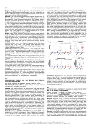
capable of activating the biological (proliferation, ECM synthesis)Figure 1. Genes dysregulated in damaged human OA and mouse DMM
cartilage and the overlap between the two populations.
Table 1. Genes dysregulated co-ordinately in damaged human OA and in all
mouse DMM cartilage datasets
Function Gene Human1
2 Wk DMM1
2 Wk DMM2
6 Wk DMM2
(log2FoldChange)
Extracellular COL8A1 1.18 1.28 2.64 2.80
Matrix LEPREL1 1.26 0.80 1.34 1.14
LUM 1.14 1.10 2.86 3.71
TGFBI 1.22 0.96 2.00 3.80
Transcription FHL2 1.14 1.95 2.35 1.61
Regulator ID2 0.74 1.19 2.37 1.65
SOX8 À0.58 À1.45 À2.41 À1.85
Signalling ARHGAP24 0.88 1.28 2.53 3.34
CHRDL2 À1.30 À1.72 À2.68 À2.75
EMP1 0.64 1.23 2.34 1.72
GDF6 0.82 2.58 2.74 2.22
MOXD1 1.06 1.55 1.62 3.08
NBL1 0.71 1.97 1.32 2.13
NFKBIZ 0.67 1.04 1.36 1.00
NPR3 1.49 1.97 3.20 2.24
NRP2 0.60 1.16 1.12 2.29
PTGS2 1.07 0.76 5.10 3.85
RARB 0.83 1.16 2.50 2.13
RNF152 0.78 0.82 1.60 1.83
TM4SF1 0.74 1.28 1.49 1.53
TSPAN2 0.79 0.79 1.53 1.13
1
¼ RNAseq data generated in-house (unpublished);
2
¼ microarray data from GSE45793
Abstracts / Osteoarthritis and Cartilage 24 (2016) S63eS534 S397
Downloaded from ClinicalKey.com.au at University of Melbourne May 05, 2016.
For personal use only. No other uses without permission. Copyright ©2016. Elsevier Inc. All rights reserved.

More Related Content

What's hot

Engineered Ligament Optimization Poster 2014 Updated
Engineered Ligament Optimization Poster 2014 UpdatedEngineered Ligament Optimization Poster 2014 Updated
Engineered Ligament Optimization Poster 2014 UpdatedMichael Selep
 
Osteoporosis : The Silent Thief Osteoporosis : The Silent Thief
Osteoporosis : The Silent Thief 	 Osteoporosis : The Silent ThiefOsteoporosis : The Silent Thief 	 Osteoporosis : The Silent Thief
Osteoporosis : The Silent Thief Osteoporosis : The Silent ThiefMedicineAndHealthCancer
 
Biomechanical properties of Anterolateral ligament of the knee compared with...
Biomechanical properties of  Anterolateral ligament of the knee compared with...Biomechanical properties of  Anterolateral ligament of the knee compared with...
Biomechanical properties of Anterolateral ligament of the knee compared with...KHALIFA ELMAJRI
 
5. Frank Buttgereit. Fin40 min rheumatic diseases and osteoporosis
5. Frank Buttgereit. Fin40 min rheumatic diseases and osteoporosis5. Frank Buttgereit. Fin40 min rheumatic diseases and osteoporosis
5. Frank Buttgereit. Fin40 min rheumatic diseases and osteoporosiscrea-autoinmunidad
 
FinalPrattPresentationPoster-1
FinalPrattPresentationPoster-1FinalPrattPresentationPoster-1
FinalPrattPresentationPoster-1Alexa Turner
 
Leah Molai Conference A0 Poster
Leah Molai Conference A0 Poster Leah Molai Conference A0 Poster
Leah Molai Conference A0 Poster Leah Molai
 
Eleven year experience ijsit 2.1.6
Eleven year experience ijsit 2.1.6Eleven year experience ijsit 2.1.6
Eleven year experience ijsit 2.1.6IJSIT Editor
 
Acsm 2015 symposium ingalls-final-slide share
Acsm 2015 symposium ingalls-final-slide shareAcsm 2015 symposium ingalls-final-slide share
Acsm 2015 symposium ingalls-final-slide sharecingalls2015
 
Physiotherapy treatment approaches for tennis elbow - Vicente Lloret Vicedo
Physiotherapy treatment approaches for tennis elbow - Vicente Lloret VicedoPhysiotherapy treatment approaches for tennis elbow - Vicente Lloret Vicedo
Physiotherapy treatment approaches for tennis elbow - Vicente Lloret Vicedohandfun
 
Age related atrophy & sarcopenia Original Hypothesis Abstract - 2015
Age related atrophy  & sarcopenia Original Hypothesis Abstract - 2015Age related atrophy  & sarcopenia Original Hypothesis Abstract - 2015
Age related atrophy & sarcopenia Original Hypothesis Abstract - 2015Kenneth Craig
 
Roughan et al. (2015) Meloxicam prevents COX-2 mediated post-surgical inflamm...
Roughan et al. (2015) Meloxicam prevents COX-2 mediated post-surgical inflamm...Roughan et al. (2015) Meloxicam prevents COX-2 mediated post-surgical inflamm...
Roughan et al. (2015) Meloxicam prevents COX-2 mediated post-surgical inflamm...Johnny Roughan
 
NCPTA 2015 POSTER- KS and KL final
NCPTA 2015 POSTER- KS and KL finalNCPTA 2015 POSTER- KS and KL final
NCPTA 2015 POSTER- KS and KL finalKaitlin Letsinger
 
2010-PLOS-reambulation
2010-PLOS-reambulation2010-PLOS-reambulation
2010-PLOS-reambulationMarodyne LivMD
 
Liu_Xuhui_Resume_Jan_2017
Liu_Xuhui_Resume_Jan_2017Liu_Xuhui_Resume_Jan_2017
Liu_Xuhui_Resume_Jan_2017Xuhui Liu
 

What's hot (20)

Colagen
ColagenColagen
Colagen
 
Jhg200074
Jhg200074Jhg200074
Jhg200074
 
Engineered Ligament Optimization Poster 2014 Updated
Engineered Ligament Optimization Poster 2014 UpdatedEngineered Ligament Optimization Poster 2014 Updated
Engineered Ligament Optimization Poster 2014 Updated
 
Toigo
ToigoToigo
Toigo
 
Osteoporosis : The Silent Thief Osteoporosis : The Silent Thief
Osteoporosis : The Silent Thief 	 Osteoporosis : The Silent ThiefOsteoporosis : The Silent Thief 	 Osteoporosis : The Silent Thief
Osteoporosis : The Silent Thief Osteoporosis : The Silent Thief
 
Biomechanical properties of Anterolateral ligament of the knee compared with...
Biomechanical properties of  Anterolateral ligament of the knee compared with...Biomechanical properties of  Anterolateral ligament of the knee compared with...
Biomechanical properties of Anterolateral ligament of the knee compared with...
 
5. Frank Buttgereit. Fin40 min rheumatic diseases and osteoporosis
5. Frank Buttgereit. Fin40 min rheumatic diseases and osteoporosis5. Frank Buttgereit. Fin40 min rheumatic diseases and osteoporosis
5. Frank Buttgereit. Fin40 min rheumatic diseases and osteoporosis
 
Cruickshank NSCI197 Paper
Cruickshank NSCI197 PaperCruickshank NSCI197 Paper
Cruickshank NSCI197 Paper
 
FinalPrattPresentationPoster-1
FinalPrattPresentationPoster-1FinalPrattPresentationPoster-1
FinalPrattPresentationPoster-1
 
Leah Molai Conference A0 Poster
Leah Molai Conference A0 Poster Leah Molai Conference A0 Poster
Leah Molai Conference A0 Poster
 
Eleven year experience ijsit 2.1.6
Eleven year experience ijsit 2.1.6Eleven year experience ijsit 2.1.6
Eleven year experience ijsit 2.1.6
 
Acsm 2015 symposium ingalls-final-slide share
Acsm 2015 symposium ingalls-final-slide shareAcsm 2015 symposium ingalls-final-slide share
Acsm 2015 symposium ingalls-final-slide share
 
UTA KINE FALL 2011.Samraj
UTA KINE FALL 2011.SamrajUTA KINE FALL 2011.Samraj
UTA KINE FALL 2011.Samraj
 
Physiotherapy treatment approaches for tennis elbow - Vicente Lloret Vicedo
Physiotherapy treatment approaches for tennis elbow - Vicente Lloret VicedoPhysiotherapy treatment approaches for tennis elbow - Vicente Lloret Vicedo
Physiotherapy treatment approaches for tennis elbow - Vicente Lloret Vicedo
 
Age related atrophy & sarcopenia Original Hypothesis Abstract - 2015
Age related atrophy  & sarcopenia Original Hypothesis Abstract - 2015Age related atrophy  & sarcopenia Original Hypothesis Abstract - 2015
Age related atrophy & sarcopenia Original Hypothesis Abstract - 2015
 
Roughan et al. (2015) Meloxicam prevents COX-2 mediated post-surgical inflamm...
Roughan et al. (2015) Meloxicam prevents COX-2 mediated post-surgical inflamm...Roughan et al. (2015) Meloxicam prevents COX-2 mediated post-surgical inflamm...
Roughan et al. (2015) Meloxicam prevents COX-2 mediated post-surgical inflamm...
 
NCPTA 2015 POSTER- KS and KL final
NCPTA 2015 POSTER- KS and KL finalNCPTA 2015 POSTER- KS and KL final
NCPTA 2015 POSTER- KS and KL final
 
Gene therapy
Gene therapyGene therapy
Gene therapy
 
2010-PLOS-reambulation
2010-PLOS-reambulation2010-PLOS-reambulation
2010-PLOS-reambulation
 
Liu_Xuhui_Resume_Jan_2017
Liu_Xuhui_Resume_Jan_2017Liu_Xuhui_Resume_Jan_2017
Liu_Xuhui_Resume_Jan_2017
 

Similar to Effects of rehabilitation programs on balance and functionality after total knee replacement

Contents lists available at ScienceDirectJournal of Orthop.docx
Contents lists available at ScienceDirectJournal of Orthop.docxContents lists available at ScienceDirectJournal of Orthop.docx
Contents lists available at ScienceDirectJournal of Orthop.docxbobbywlane695641
 
To Compare the Effect of Muscle Energy Technique and Ultrasound on Jaw Range ...
To Compare the Effect of Muscle Energy Technique and Ultrasound on Jaw Range ...To Compare the Effect of Muscle Energy Technique and Ultrasound on Jaw Range ...
To Compare the Effect of Muscle Energy Technique and Ultrasound on Jaw Range ...ijtsrd
 
The Effect of Recombinant Bone Morphogenetic Protien-2 in Revison Tibiotaloca...
The Effect of Recombinant Bone Morphogenetic Protien-2 in Revison Tibiotaloca...The Effect of Recombinant Bone Morphogenetic Protien-2 in Revison Tibiotaloca...
The Effect of Recombinant Bone Morphogenetic Protien-2 in Revison Tibiotaloca...skisnfeet
 
Psoralen Promotes Myogenic Differentiation of Muscle Cells to Repair Fracture
Psoralen Promotes Myogenic Differentiation of Muscle Cells to Repair Fracture Psoralen Promotes Myogenic Differentiation of Muscle Cells to Repair Fracture
Psoralen Promotes Myogenic Differentiation of Muscle Cells to Repair Fracture Associate Professor in VSB Coimbatore
 
Foot-Ankle-Research-Review-Issue-27
Foot-Ankle-Research-Review-Issue-27Foot-Ankle-Research-Review-Issue-27
Foot-Ankle-Research-Review-Issue-27Dr Matthew Carroll
 
The importance of matrix metalloproteases in the pathology of the tempo-mandi...
The importance of matrix metalloproteases in the pathology of the tempo-mandi...The importance of matrix metalloproteases in the pathology of the tempo-mandi...
The importance of matrix metalloproteases in the pathology of the tempo-mandi...SubmissionResearchpa
 
Union Rate of Tibiotalocalcaneal Nail with Internal or External Bone Stimulat...
Union Rate of Tibiotalocalcaneal Nail with Internal or External Bone Stimulat...Union Rate of Tibiotalocalcaneal Nail with Internal or External Bone Stimulat...
Union Rate of Tibiotalocalcaneal Nail with Internal or External Bone Stimulat...skisnfeet
 
PMR Buzz Magazine_Oct 2022.pdf
PMR Buzz Magazine_Oct 2022.pdfPMR Buzz Magazine_Oct 2022.pdf
PMR Buzz Magazine_Oct 2022.pdfmrinal joshi
 
EVALUATION OF THE EFFICIENCY OF BISPHOSPHONATES IN THE TREATMENT OF OSTEOPORO...
EVALUATION OF THE EFFICIENCY OF BISPHOSPHONATES IN THE TREATMENT OF OSTEOPORO...EVALUATION OF THE EFFICIENCY OF BISPHOSPHONATES IN THE TREATMENT OF OSTEOPORO...
EVALUATION OF THE EFFICIENCY OF BISPHOSPHONATES IN THE TREATMENT OF OSTEOPORO...indexPub
 
Stemcell Research Paper on avascular necrosis-AVN-by Dr.Pradeep Mahajan
Stemcell Research Paper on avascular necrosis-AVN-by Dr.Pradeep MahajanStemcell Research Paper on avascular necrosis-AVN-by Dr.Pradeep Mahajan
Stemcell Research Paper on avascular necrosis-AVN-by Dr.Pradeep MahajanDr Pradeep Mahajan
 
A comparative study on the clinical and functional outcome of limb salvage su...
A comparative study on the clinical and functional outcome of limb salvage su...A comparative study on the clinical and functional outcome of limb salvage su...
A comparative study on the clinical and functional outcome of limb salvage su...NAAR Journal
 
Tzekov_PLOSOne_2016 Publication Roskamp.PDF
Tzekov_PLOSOne_2016 Publication Roskamp.PDFTzekov_PLOSOne_2016 Publication Roskamp.PDF
Tzekov_PLOSOne_2016 Publication Roskamp.PDFMegan Orlando
 
1-s2.0-S1063458416006427
1-s2.0-S10634584160064271-s2.0-S1063458416006427
1-s2.0-S1063458416006427Dr. Louise Kung
 
Estudio clínico espalda, revista spine
Estudio clínico espalda, revista spineEstudio clínico espalda, revista spine
Estudio clínico espalda, revista spineDr. Manuel Concepción
 
jounal club iprf to temporomandibular joint
jounal club iprf to temporomandibular jointjounal club iprf to temporomandibular joint
jounal club iprf to temporomandibular jointMalaM67
 
Quantitative analysis of patellar tendon size and structure in asymptomatic ...
Quantitative analysis of patellar tendon size and structure  in asymptomatic ...Quantitative analysis of patellar tendon size and structure  in asymptomatic ...
Quantitative analysis of patellar tendon size and structure in asymptomatic ...Medical_Lab
 
Analytical Study of Clinicopathological Data of Saudi Patients with Osteoarth...
Analytical Study of Clinicopathological Data of Saudi Patients with Osteoarth...Analytical Study of Clinicopathological Data of Saudi Patients with Osteoarth...
Analytical Study of Clinicopathological Data of Saudi Patients with Osteoarth...Prof. Hesham N. Mustafa
 

Similar to Effects of rehabilitation programs on balance and functionality after total knee replacement (20)

Contents lists available at ScienceDirectJournal of Orthop.docx
Contents lists available at ScienceDirectJournal of Orthop.docxContents lists available at ScienceDirectJournal of Orthop.docx
Contents lists available at ScienceDirectJournal of Orthop.docx
 
To Compare the Effect of Muscle Energy Technique and Ultrasound on Jaw Range ...
To Compare the Effect of Muscle Energy Technique and Ultrasound on Jaw Range ...To Compare the Effect of Muscle Energy Technique and Ultrasound on Jaw Range ...
To Compare the Effect of Muscle Energy Technique and Ultrasound on Jaw Range ...
 
The Effect of Recombinant Bone Morphogenetic Protien-2 in Revison Tibiotaloca...
The Effect of Recombinant Bone Morphogenetic Protien-2 in Revison Tibiotaloca...The Effect of Recombinant Bone Morphogenetic Protien-2 in Revison Tibiotaloca...
The Effect of Recombinant Bone Morphogenetic Protien-2 in Revison Tibiotaloca...
 
Psoralen Promotes Myogenic Differentiation of Muscle Cells to Repair Fracture
Psoralen Promotes Myogenic Differentiation of Muscle Cells to Repair Fracture Psoralen Promotes Myogenic Differentiation of Muscle Cells to Repair Fracture
Psoralen Promotes Myogenic Differentiation of Muscle Cells to Repair Fracture
 
Foot-Ankle-Research-Review-Issue-27
Foot-Ankle-Research-Review-Issue-27Foot-Ankle-Research-Review-Issue-27
Foot-Ankle-Research-Review-Issue-27
 
The importance of matrix metalloproteases in the pathology of the tempo-mandi...
The importance of matrix metalloproteases in the pathology of the tempo-mandi...The importance of matrix metalloproteases in the pathology of the tempo-mandi...
The importance of matrix metalloproteases in the pathology of the tempo-mandi...
 
Union Rate of Tibiotalocalcaneal Nail with Internal or External Bone Stimulat...
Union Rate of Tibiotalocalcaneal Nail with Internal or External Bone Stimulat...Union Rate of Tibiotalocalcaneal Nail with Internal or External Bone Stimulat...
Union Rate of Tibiotalocalcaneal Nail with Internal or External Bone Stimulat...
 
PMR Buzz Magazine_Oct 2022.pdf
PMR Buzz Magazine_Oct 2022.pdfPMR Buzz Magazine_Oct 2022.pdf
PMR Buzz Magazine_Oct 2022.pdf
 
EVALUATION OF THE EFFICIENCY OF BISPHOSPHONATES IN THE TREATMENT OF OSTEOPORO...
EVALUATION OF THE EFFICIENCY OF BISPHOSPHONATES IN THE TREATMENT OF OSTEOPORO...EVALUATION OF THE EFFICIENCY OF BISPHOSPHONATES IN THE TREATMENT OF OSTEOPORO...
EVALUATION OF THE EFFICIENCY OF BISPHOSPHONATES IN THE TREATMENT OF OSTEOPORO...
 
Stemcell Research Paper on avascular necrosis-AVN-by Dr.Pradeep Mahajan
Stemcell Research Paper on avascular necrosis-AVN-by Dr.Pradeep MahajanStemcell Research Paper on avascular necrosis-AVN-by Dr.Pradeep Mahajan
Stemcell Research Paper on avascular necrosis-AVN-by Dr.Pradeep Mahajan
 
Relationship of Bone Marrow Density (Hip and Spine) in Cerebral Palsy: A Case...
Relationship of Bone Marrow Density (Hip and Spine) in Cerebral Palsy: A Case...Relationship of Bone Marrow Density (Hip and Spine) in Cerebral Palsy: A Case...
Relationship of Bone Marrow Density (Hip and Spine) in Cerebral Palsy: A Case...
 
A comparative study on the clinical and functional outcome of limb salvage su...
A comparative study on the clinical and functional outcome of limb salvage su...A comparative study on the clinical and functional outcome of limb salvage su...
A comparative study on the clinical and functional outcome of limb salvage su...
 
Stem cell research
Stem cell researchStem cell research
Stem cell research
 
Tzekov_PLOSOne_2016 Publication Roskamp.PDF
Tzekov_PLOSOne_2016 Publication Roskamp.PDFTzekov_PLOSOne_2016 Publication Roskamp.PDF
Tzekov_PLOSOne_2016 Publication Roskamp.PDF
 
1-s2.0-S1063458416006427
1-s2.0-S10634584160064271-s2.0-S1063458416006427
1-s2.0-S1063458416006427
 
Estudio clínico espalda, revista spine
Estudio clínico espalda, revista spineEstudio clínico espalda, revista spine
Estudio clínico espalda, revista spine
 
jounal club iprf to temporomandibular joint
jounal club iprf to temporomandibular jointjounal club iprf to temporomandibular joint
jounal club iprf to temporomandibular joint
 
Paper icchou
Paper icchouPaper icchou
Paper icchou
 
Quantitative analysis of patellar tendon size and structure in asymptomatic ...
Quantitative analysis of patellar tendon size and structure  in asymptomatic ...Quantitative analysis of patellar tendon size and structure  in asymptomatic ...
Quantitative analysis of patellar tendon size and structure in asymptomatic ...
 
Analytical Study of Clinicopathological Data of Saudi Patients with Osteoarth...
Analytical Study of Clinicopathological Data of Saudi Patients with Osteoarth...Analytical Study of Clinicopathological Data of Saudi Patients with Osteoarth...
Analytical Study of Clinicopathological Data of Saudi Patients with Osteoarth...
 

Effects of rehabilitation programs on balance and functionality after total knee replacement

  • 1. Purpose: The purpose of the study was to evaluate the effects of the rehabilitation program after simultaneously total knee replacement on balance and functionality in patients with osteoarthritis and to compare the results with the healthy individuals. Methods: Two study groups of osteoarthritic patients between ages 50- 85 who underwent bilateral TKA and one control group (Group 3, n¼20) were included in the study. Exercise program with frequent physical therapist supervision (2 session/1 week) was performed in Group 1 (n¼20) and less supervised exercise program was performed Group 2 (n¼20) (1 session/2 week). Exercise program was continued during the first month. All the patients were evaluated preoperatively as well as 1.and 2.month postoperatively. The patients were checked every week by telephone interview. Functionality was evaluated with WOMAC (The Western Ontario and McMaster Universities Arthritis) Index, dynamic balance with Timed Up&Go (TUG) and stair climbing test (SCT), static balance with single limb standing test (SLST). Results: There was no statisitical significance between groups in terms of age (p¼0,463). In preop evaluation there were significant differences between groups in all tests in favor of the control group. WOMAC pain scores in Groups 1 and 2 were similar in 1.month postop evaluation while the decrease in the pain score in Group 1 was higher than the scores of Group 2 and was close to the scores of healthy controls (p¼0,00). WOMAC stiffness scores were higher in postop period than preop period in both Group 1 and 2. The stiffness scores of Group 1 were significantly lower than Group 2 in 2. month postoperatively and were close to healthy controls (p¼0,00). WOMAC functionality scores of Group 1 and 2 were significantly higher in 1.and 2.month postop compared to preop period. The functionality scores of Group 1 were significantly higher in postop 2.month com- pared to Group 2 and were closer to the scores of healthy controls (p¼0,00). The completion time of TUG test of the groups were shorter in 1.and 2.month scores and were similar to the 1.and 2.month scores of controls (p¼0,33). SCT time of the groups were shorter in both 1.and 2. month postop period compared to preop period. Stair climbing time of Group 1 was significantly shorter than Group 2 in 2.month postoperatively and was closer to the scores of healthy controls (p¼0,00). SLST for both legs of Group 1 were significantly higher than Group 2 in 1. and 2. month postoperatively and were closer to healthy controls (p¼0,00). Conclusions: The patients which have applied exercise program after simultaneously bilateral TDR under the supervision of the physi- otherapist 2 days a week for 1 month has obtained improvements in balance and functional parameters and it has been shown that their performances approach the healthy control group values. 662 INFLAMMATORY FACTORS DO NOT ENABLE BMP2-INDUCED OSTEOPHYTE FORMATION E.N. Blaney Davidson, M.I. Koenders, A.P. van Caam, E.L. Vitters, M.B. Bennink, P.L. van Lent, F.A. van de Loo, P.M. van der Kraan. Radboud Univ. Med. Ctr., Nijmegen, Netherlands Purpose: The main hallmarks of osteoarthritis (OA) are cartilage damage and osteophyte formation. The latter can clearly be observed in X-rays of patients. What causes these osteophytes in OA is not entirely clear. We have previously identified that in experimental models both TGF-beta and BMP2 can induce de novo osteophyte formation. In addition, when blocking TGF-beta signalling during experimental OA we were able to prevent osteophyte formation. Recently we have shown that chondrocyte-specific overexpression of BMP2 in a mouse model does not induce osteophyte formation on its own, but that it greatly enhances experimental OA-induced osteophyte formation. The latter could be blocked by inhibition of TGF-beta activity. Apparently, TGF- beta is involved in osteophyte formation but since there are inflam- matory factors in (experimental) OA, it is still unclear whether they might play a role in osteophyte formation. This led to our question: can inflammatory factors enable BMP2 to induce osteophyte formation in a mechanically stable joint? Methods: We exposed Col2a1-rtTA-BMP2 mice (inducible BMP2 with doxycycline) to a simple inflammatory trigger that has been implicated in OA: interleukin-1, and to a more complex inflammatory trigger: zymosan by intra-articular injection. Saline injections were used as controls. In addition, since we have previously identified TGF-beta as a major trigger for osteophyte formation in OA, we included a low-dose of adenovirus overexpressing TGF-beta (ad-TGF-beta) as a trigger, which on its own results in fibrosis but not yet osteophytes. We added ad-Luc as a viral control. Mice were either fed standard diet (non-dox) or a diet with doxycycline (dox) starting 7 days prior to i.a. injection for the entire duration of the experiment and 14 days after intra-articular injection of the adenovirus we isolated knee joints for histology. Results: Saline injections combined with BMP2 did not lead to osteo- phyte formation. Moreover, neither did IL-1 injection combined with BMP2. Zymosan injection resulted in clear synovial inflammation, which was further enhanced by BMP2 overexpression. (fig 1A) Also, there were minor fissures in medial tibial cartilage in zymosan-injected joints, which showed a trend to becoming slightly more severe when combined with BMP2, but this was not significant. (fig 1B) Despite the overt inflamma- tion induced by injection of zymosan, the combination of zymosan with BMP2 overexpression was insufficient to induce osteophytes. (fig 1C) Injection of adenoviral TGF-beta resulted in massive fibrosis and minor osteophyte formation. As expected, combining TGF-beta with BMP2 resulted in clearly enhanced osteophyte formation. (fig 1D) Conclusions: Together these data show that neither a simple inflam- matory trigger like IL-1, nor a more complex inflammatory trigger like zymosan enable BMP2 to induce osteophytes. In contrast, BMP-2 in combination with TGF-beta is a results in massive osteophyte for- mation. The latter confirmed our prior data where we showed that by blocking TGF-beta during experimental OA we could prevent BMP2 from inducing osteophytes. 663 CARTILAGE GENE EXPRESSION CHANGES IN EARLY MOUSE DMM ARE DETECTED IN LATE HUMAN OA J. Soul y, S.L. Dunn y, L.H. Kung y, z, S. Anand x, J.-M. Schwartz y, T.E. Hardingham y, R.P. Boot-Handford y. y The Univ. of Manchester, Manchester, United Kingdom; z Murdoch Childrens Res. Inst., Melbourne, Australia; x Stockport NHS Fndn. Trust, Stockport, United Kingdom Purpose: The DMM mouse has become the model of choice for much experimental OA research because of its reproducible and relatively rapid disease onset, coupled to the mouse’s genetic tractability. Nev- ertheless, the DMM model represents a mechanically induced form of OA developing quickly (2e8 weeks) in a young (10e12 week) animal whereas most human OA is in an ageing population with idiopathic and long term disease. We therefore compared the differentially regulated gene profiles accompanying damage in human OA cartilage with sam- ples of cartilage progressing to damage taken from DMM mice. Methods: Global gene expression was determined by RNAseq analysis on i) paired intact and damaged OA cartilage from 8 patients at TKR; and ii) mouse tibial medial compartment cartilage laser dissected from DMM and sham operated mice 2 weeks after operation. The expression Abstracts / Osteoarthritis and Cartilage 24 (2016) S63eS534S396 Downloaded from ClinicalKey.com.au at University of Melbourne May 05, 2016. For personal use only. No other uses without permission. Copyright ©2016. Elsevier Inc. All rights reserved.
  • 2. data was batch effect corrected with RUVSeq, normalised and the fold changes between the paired human samples and the DMM and sham mice calculated with DESeq2. Benjamini-Hochberg multiple testing correction was performed. Additional expression data obtained from mouse cartilage isolated as described above from 2 and 6 week DMM and sham wild type mice (GSE45793) was downloaded from GEO and analysed with limma to identify differentially expressed genes. Genes with an absolute fold change of !1.5 and a Benjamini-Hochberg corrected p-value of 0.1 were classified as differentially expressed. For comparison between mouse and human gene expression data, gene IDs were mapped to ortholog gene groups from InParanoid. Hyper- geometric statistics were used to calculate the statistical significance of the gene overlaps. Results: The human and mouse gene expression datasets shared 12600 orthologue gene groups. Comparison of RNAseq data from 8 paired samples of damaged and intact OA cartilage from patients undergoing TKR revealed 1237 genes were significantly dysregulated in the damaged cartilage tissue. Data from mouse DMM experiments (2 datasets obtained at 2 weeks and 1 data set at 6 weeks post operation) were analysed against their respective shams generating a combined list of 1756 genes that were significantly dysregulated by DMM. Comparison of the genes dysregulated in damaged human OA and mouse DMM cartilage revealed a highly significant (p¼ 4.403e-05) overlap of 220 genes (Figure 1). Reactome pathway assessment of these 220 genes revealed the most significant terms to involve extracellular matrix (ECM) including ECM degradation / organisation, collagen formation / degradation and integrin cell surface interactions. These terms were populated pre- dominantly with collagen and other genes encoding structural ECM proteins such as FN1, ACAN and LUM. Other significant terms included peptide ligand binding receptors / G Protein-Coupled Receptor ligand binding (which shared the AGTR2, F2R, EDNRA, JAK3, C3, CCBP2, ANXA1, CXCR7 and ATGR1 genes) and signal transduction. Of the 220 dysregulated genes common to human and one, or more of the mouse DMM datasets, 21 genes were co-ordinately dysregulated in all of the datasets (Table 1). These core set of genes were predominantly involved with signalling and included chordin 2 (BMP signalling), GDF6 (joint development), PTGS2 (prostaglandin synthesis), Retinoic Acid Receptor B and a limited number of ECM associated genes included Lumican and TGFb induced protein In addition, the dysregulation of three of these genes (NPR3, MOXD1, PTGS2) may be linked to their reported sensitivity to mechanical stimulation and the increased stress on chondrocytes associated with, or causing the tissue damage. Conclusions: Around 18% of the genes found to be dysregulated in the damaged cartilage taken from patients at TKR are also dysregulated in cartilage from DMM mice at either 2 and/or 6 weeks post-operation. Many of these genes encode proteins that are part of, or associated with the cartilage ECM. Of particular note is the core of 21 genes which are involved in different aspects of signalling that are changed in expression at the earliest stages of cartilage damage in the DMM mouse and also changed in late stage human OA cartilage. These results provide sup- porting evidence that the DMM model, although of joint destabilisation in a young mouse, nevertheless reveals changes that suggest similar processes are active in late human OA and these may represent potential targets for future therapy. Supported by AR UK 19501 & 20414. 664 RAAV-MEDIATED COMBINED GENE TRANSFER AND OVEREXPRESSION OF TGF-b AND SOX9 REMODELS HUMAN OSTEOARTHRITIC ARTICULAR CARTILAGE K. TAO, Sr. y, z, A. Rey-Rico x, J. Frisch x, J. Venkatesan x, G. Schmitt x, H. Madry x, k, J. Lin ¶, z, M. Cucchiarini x. y Peking Univ. People's Hosp., Beijing, China; z Peking Univ. Hlth. Sci. Ctr., Beijing, China; x Ctr. of Experimental Orthopaedics, Saarland Univ., Homburg, Saar, Germany; k Dept. of Orthopaedic Surgery, Saarland Univ., Homburg, Saar, Germany; ¶ Inst. of Arthritis, Peking Univ. People's Hosp., Beijing, China Purpose: Direct administration of therapeutic candidate gene sequen- ces using the safe and effective recombinant adeno-associated virus (rAAV) vectors is a promising strategy to stimulate the biologic activities of articular chondrocytes as an adapted tool to treat human osteo- arthritic (OA) cartilage [1]. Methods: In the present study, we developed a combined gene transfer approach based on the co-delivery of the pleiotropic transformation growth factor beta (TGF-b) with the specific transcription factor SOX9 via rAAV to human normal and OA chondrocytes in vitro and cartilage explants in situ in light of the mitogenic and pro-anabolic properties of these factors [2, 3]. Results: Effective, durable co-overexpression of TGF-b and sox9 sig- nificantly enhanced the levels of cell proliferation both in human nor- mal and OA chondrocytes and cartilage explants over an extended period of time (21 days) (Fig. 1A), while stimulating the biosynthesis of key matrix components (proteoglycans, type-II collagen) compared with control conditions (reporter lacZ gene transfer, absence of vector treatment) (Fig. 1B, C, D). Of further note, expression of hypertrophic type-X collagen significantly decreased following co-treatment by the candidate vectors (Fig. 1B, E). Conclusions: Here, we examined the feasibility of combining the transfer of two crucial factors with chondroreparative activities (TGF- b and SOX9) via direct, dual rAAV gene transfer as a means to enhance the remodelling of human OA chondrocytes in vitro and OA cartilage in situ. Our results first show that concomitant expression of TGF-b and sox9 was successfully achieved via rAAV both in human normal and OA chondrocytes in vitro and in human normal and OA cartilage explants in situ for at least 21 days, probably due to a good penetration and maintenance of the vectors in the targets as noted when using individual gene treatments. The data further indicate that high, durable levels of rAAV-co-delivered TGF-b/sox9 were capable of activating the biological (proliferation, ECM synthesis)Figure 1. Genes dysregulated in damaged human OA and mouse DMM cartilage and the overlap between the two populations. Table 1. Genes dysregulated co-ordinately in damaged human OA and in all mouse DMM cartilage datasets Function Gene Human1 2 Wk DMM1 2 Wk DMM2 6 Wk DMM2 (log2FoldChange) Extracellular COL8A1 1.18 1.28 2.64 2.80 Matrix LEPREL1 1.26 0.80 1.34 1.14 LUM 1.14 1.10 2.86 3.71 TGFBI 1.22 0.96 2.00 3.80 Transcription FHL2 1.14 1.95 2.35 1.61 Regulator ID2 0.74 1.19 2.37 1.65 SOX8 À0.58 À1.45 À2.41 À1.85 Signalling ARHGAP24 0.88 1.28 2.53 3.34 CHRDL2 À1.30 À1.72 À2.68 À2.75 EMP1 0.64 1.23 2.34 1.72 GDF6 0.82 2.58 2.74 2.22 MOXD1 1.06 1.55 1.62 3.08 NBL1 0.71 1.97 1.32 2.13 NFKBIZ 0.67 1.04 1.36 1.00 NPR3 1.49 1.97 3.20 2.24 NRP2 0.60 1.16 1.12 2.29 PTGS2 1.07 0.76 5.10 3.85 RARB 0.83 1.16 2.50 2.13 RNF152 0.78 0.82 1.60 1.83 TM4SF1 0.74 1.28 1.49 1.53 TSPAN2 0.79 0.79 1.53 1.13 1 ¼ RNAseq data generated in-house (unpublished); 2 ¼ microarray data from GSE45793 Abstracts / Osteoarthritis and Cartilage 24 (2016) S63eS534 S397 Downloaded from ClinicalKey.com.au at University of Melbourne May 05, 2016. For personal use only. No other uses without permission. Copyright ©2016. Elsevier Inc. All rights reserved.