Age Related Macular Degeneration Treatment In Ghatkopar, Mumbai
•Download as PPTX, PDF•
0 likes•3 views

Human eye has various important parts like Cornea, Pupil, Iris, Lens and Retina. The macula is located in the center of the retina, the light-sensitive tissue at the back of the eye. The retina instantly converts light, or an image, into electrical impulses.

Recommended
More Related Content
Similar to Age Related Macular Degeneration Treatment In Ghatkopar, Mumbai
Similar to Age Related Macular Degeneration Treatment In Ghatkopar, Mumbai (20)
Recently uploaded
Recently uploaded (20)
Age Related Macular Degeneration Treatment In Ghatkopar, Mumbai
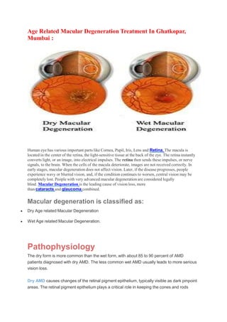
- 1. Age Related Macular Degeneration Treatment In Ghatkopar, Mumbai : Human eye has various important parts like Cornea, Pupil, Iris, Lens and Retina. The macula is located in the center of the retina, the light-sensitive tissue at the back of the eye. The retina instantly converts light, or an image, into electrical impulses. The retina then sends these impulses, or nerve signals, to the brain. When the cells of the macula deteriorate, images are not received correctly. In early stages, macular degeneration does not affect vision. Later, if the disease progresses, people experience wavy or blurred vision, and, if the condition continues to worsen, central vision may be completely lost. People with very advanced macular degeneration are considered legally blind. Macular Degeneration is the leading cause of vision loss, more than cataracts and glaucoma combined. Macular degeneration is classified as: Dry Age related Macular Degeneration Wet Age related Macular Degeneration. Pathophysiology The dry form is more common than the wet form, with about 85 to 90 percent of AMD patients diagnosed with dry AMD. The less common wet AMD usually leads to more serious vision loss. Dry AMD causes changes of the retinal pigment epithelium, typically visible as dark pinpoint areas. The retinal pigment epithelium plays a critical role in keeping the cones and rods
- 2. healthy and functioning well. Accumulation of waste products from the rods and cones can result in drusen, which appear as yellow spots. Areas of chorioretinal atrophy (referred to as geographic atrophy) occur in more advanced cases of dry AMD. There is no elevated macular scar (disciform scar), edema, hemorrhage, or exudation. Dry AMD has three stages, all of which may occur in one or both eyes: Early AMD - People with early AMD have either several small drusen or a few medium-sized drusen. At this stage, there are no symptoms and no vision loss. Intermediate AMD - People with intermediate AMD have either many medium-sized drusen or one or more large drusen. Some people see a blurred spot in the center of their vision. More light may be needed for reading and other tasks. Advanced AMD - In addition to drusen, people with advanced dry AMD have a breakdown of light-sensitive cells and supporting tissue in the central retinal area. This breakdown can cause a blurred spot in the center of your vision. Over time, the blurred spot may get bigger and darker, taking more of your central vision. You may have difficulty reading or recognizing faces until they are very close to you. Wet AMD occurs when new abnormal blood vessels develop under the retina in a process called choroidal neovascularization (abnormal new vessel formation). Localized macular edema or hemorrhage may elevate an area of the macula or cause a localized retinal pigment epithelial detachment. Eventually, untreated neovascularization causes a disciform scar under the macula. Symptoms Dry macular degeneration symptoms usually develop gradually and without pain. They may include: Visual distortions, such as straight lines seeming bent Reduced central vision in one or both eyes The need for brighter light when reading or doing close work Increased difficulty adapting to low light levels, such as when entering a dimly lit restaurant Increased blurriness of printed words Decreased intensity or brightness of colors Difficulty recognizing faces What causes macular degeneration?
- 3. Though macular degeneration is associated with aging, there is genetic component to the disease. A strong association between development of AMD and presence of a variant of a gene known as complement factor H (CFH) is observed. This gene deficiency is associated with almost half of all potentially blinding cases of macular degeneration. Other investigators have found that variants of another gene, complement factor B, may be involved in development of AMD. Specific variants of one or both of these genes, which play a role in the body's immune responses, have been found in 74 percent of AMD patients who were studied. Other complement factors also may be associated with an increased risk of macular degeneration. Oxygen-deprived cells in the retina produce a type of protein called vascular endothelial growth factor (VEGF), which triggers the growth of new blood vessels in the retina. The normal function of VEGF is to create new blood vessels during embryonic development, after an injury or to bypass blocked blood vessels. But too much VEGF in the eye causes the development of unwanted blood vessels in the retina that easily break open and bleed, damaging the macula and surrounding retina. Risk Factors The biggest risk factor for Macular Degeneration is age. Your risk increases as you age, and the disease is most likely to occur in those 55 and older. Other risk factors include: Genetics – People with a family history of AMD are at a higher risk. Race – Caucasians are more likely to develop the disease than African-Americans or Hispanics/Latinos. Smoking – Smoking doubles the risk of AMD. Diagnosis AMD is detected during a comprehensive eye exam that includes: Visual acuity test - This eye chart test measures how well you see at various distances. Dilated eye exam - Drops are placed in your eyes to widen the pupils. Your eye care professional uses a special magnifying lens to examine your retina and optic nerve for signs of AMD and other eye problems. After the exam, your close-up vision may remain blurred for several hours.
- 4. Tonometry - An instrument measures the pressure inside the eye. Numbing drops may be applied to your eye for this test. Both forms of age - related macular degeneration (AMD) are diagnosed by funduscopic examination. Visual changes can often be detected with an Amsler grid. Color photography and fluorescein angiography are done when findings suggest wet AMD. Angiography shows and characterizes subretinal choroidal neovascular membranes and can delineate areas of geographic atrophy. Optical coherence tomography (OCT) aids in identifying intraretinal and subretinal fluid and can help assess response to treatment. What Treatments Are Available for Macular Degeneration? There’s no cure for macular degeneration. Treatment may slow it down or keep you from losing too much of your vision. Your options might include: Lifestyle changes - like dieting, exercise, avoiding smoking, and protecting your eyes from ultraviolet light. Anti-angiogenesis drugs - These medications – aflibercept (Eylea), bevacizumab (Avastin), pegaptanib (Macugen), and ranibizumab (Lucentis) -- block the creation of blood vessels and leaking from the vessels in your eye that cause wet macular degeneration. Many people who’ve taken these drugs got back vision that was lost. You might need to have this treatment multiple times. Laser therapy - High-energy laser light can destroy abnormal blood vessels growing in your eye. Photodynamic laser therapy - Your doctor injects a light-sensitive drug verteporfin (Visudyne) into your bloodstream, and it’s absorbed by the abnormal blood vessels. Your doctor then shines a laser into your eye to trigger the medication to damage those blood vessels. Low vision aids - These are the devices that have special lenses or electronic systems to create larger images of nearby things. They help people who have vision loss from macular degeneration make the most of their remaining vision. Submacular surgery - This removes abnormal blood vessels or blood.
- 5. Retinal translocation - A procedure to destroy abnormal blood vessels under the center of your macula, where your doctor can’t use a laser beam safely. In this procedure, your doctor rotates the center of your macula away from the abnormal blood vessels to a healthy area of your retina. This keeps you from having scar tissue and more damage to your retina. Then, your doctor uses a laser to treat the abnormal blood vessels. Important Reminder: This information is only intended to provide guidance, not a definitive medical advice. Please consult eye doctor about your specific condition. Only a trained, experienced board certified eye doctor can determine an accurate diagnosis and proper treatment. To schedule an appointment with our experts for Age Related Macular Degeneration Management please call us at +91 8451045935, +91-8451045934 or visit our clinic at Address.