Recommended

Recommended
More Related Content
Similar to Pernas_Badrdin
Similar to Pernas_Badrdin (20)
Pernas_Badrdin
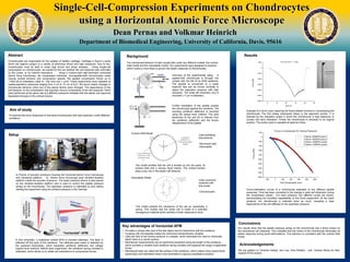
- 1. Single-Cell-Compression Experiments on Chondrocytes using a Horizontal Atomic Force Microscope Dean Pernas and Volkmar Heinrich Department of Biomedical Engineering, University of California, Davis, 95616 Chondrocytes are responsible for the upkeep of healthy cartilage. Cartilage is found in joints which are regions subject to a variety of strenuous forces and high pressures. Due to this, chondrocytes must be able to resist high forces and shear stresses. Using single-cell compression on chondrocytes, we wanted to find out whether the cell response was controlled by the cortex, or by internal mechanics. Using a custom-built high-resolution horizontal atomic force microscope, we compressed individual, micropipette-held chondrocytes under varying cortical tensions and compression speeds. We applied compression forces up to ~1500 pN at indentation rates of 100 nm/s and 1 µm/s. These experiments were repeated at pipette-aspiration pressures ranging from 2 cm to 12 cm of H2O. We found drastic changes in chondrocyte behavior when any of the above factors were changed. The dependence of the cell behavior on the compression rate exposed viscous components of the cell response. Other tests performed at the same rate but different pressures revealed that the elastic cell response depended strongly on the cortical tension. Abstract Aim of study To examine the force responses of individual chondrocytes with high resolution under different conditions. a) Picture of acoustic enclosure housing the horizontal atomic force microscope and vibrational platform. b) Atomic force microscope atop vibration-isolation platform inside the acoustic enclosure. The water pressure device is also placed on the vibration-isolation platform and is used to control the pipette pressure acting on the chondrocyte. The aspiration pressure is calibrated to zero before starting the experiment using the ambient pressure in the chamber. Results Example of a force cycle capturing the three phases involved in compressing the chondrocyte. The first phase (downward slope) is the approach portion. It is followed by the relaxation phase in which the chondrocyte is kept stationary to monitor the cell’s relaxation. Finally the chondrocyte is retracted to its original position. This entire cycle is repeated at least ten times. Force-indentation curves of a chondrocyte aspirated at two different pipette pressures. Time has been converted to the change in axial cell dimension during the compression phase. For each pressure, two different cycles are shown, demonstrating the excellent repeatability of this force experiment. At the lower pressure, the chondrocyte is indented twice as much, revealing a clear dependence of the cell stiffness on the aspiration pressure. In this schematic, a traditional vertical AFM is oriented sideways. The laser is reflected off the back of the cantilever. The reflected laser beam is detected by the quadrant photodiode, which translates cantilever deflection into voltage (optical lever method). Before each experiment, the cantilever spring constant is calibrated, which allows us to relate cell indentations to compression forces. Setup Background The mechanical behavior of cells usually falls under two different models, the cortical shell model and the viscoelastic model. Our experiments were designed to examine which model is more likely to govern the elastic response of chondrocytes. Further translation of the pipette pushes the chondrocyte against the cantilever. The resulting cantilever deflection is recorded using the optical lever method. The axial dimension of the cell (D) is inferred from the cantilever deflection and the known displacement of the pipette. Overview of the experimental setup. A pipette-held chondrocyte is brought into contact with the flat of an AFM cantilever. The pipette is connected to a water reservoir that can be moved vertically to adjust the aspiration pressure with high resolution. The initial cell dimension (D0) is recorded (~7 µm in diameter). Viscoelastic Model This model dictates that the cell is divided up into two parts, an cortical shell and a viscous liquid interior. The cortical tension plays a key role in the elastic cell behavior. This model predicts the mechanics of the cell as, essentially, a spring. This implies that the entire cell is made of a solid-like homogenous material which exhibits a linear response to force. Acknowledgements Christine Hastey, Jeni Lee, Gina McBarb, Andrew Morss Cells exhibiting this behavior: -Red blood cells -Neutrophils Cells commonly analyzed with this model: -Chondrocytes Cortical Shell Model Acknowledgements Christine Hastey, Jeni Lee, Gina McBarb, Andrew Morss Conclusions Our results show that the pipette pressure acting on the chondrocyte has a direct impact on the mechanical cell response. This indicates that the cortex of the chondrocyte dominates its elastic response during small deformations. This behavior is consistent with the cortical shell model. Acknowledgements We are grateful to Christine Hastey, Jeni Lee, Gina McBarb, and Andrew Morss for their support of this project. Key advantages of horizontal AFM • Provides a unique side view of the test object and its interactions with the cantilever • Coupling with micropipette makes the instrument extraordinarily versatile • Cells are held at low suction pressure in a pipette, which eliminates the need to chemically attach them to a carrier surface. • Mechanical measurements can be performed anywhere along the length of the cantilever, which provides a variable local cantilever spring constant and expands the range of applicable forces. • Mechanical tests can utilize the flat surface of the cantilever, which makes many experiments (particularly cell-indentation tests) more amenable to rigorous quantitative analysis