Thick walled Gall Bladder.pptx
•Download as PPTX, PDF•
0 likes•24 views

Thick walled gall bladder is very common investigation findings. Approaching this problem in algorithmic manner is necessary for improving patient outcome.

Recommended
More Related Content
Similar to Thick walled Gall Bladder.pptx
Similar to Thick walled Gall Bladder.pptx (20)
More from Dr. Shashank Agrawal
Recently uploaded
Recently uploaded (20)
Thick walled Gall Bladder.pptx
- 1. Thick walled Gall Bladder Dr Shashank Agrawal
- 2. • Gallbladder wall thickening is a commonly encountered imaging finding and can be seen in a broad spectrum of pathological conditions. • The differentiation of benign and malignant gallbladder wall thickening is critical as well as challenging. • Appropriate utilization and interpretation of imaging may allow discrimination between benign and malignant GB wall thickening
- 3. Definition • A thickened gallbladder wall measures more than 3 mm
- 4. Why it is important to differentiate • Thick walled gall bladder can be caused by- Gall stone disease leading to acute cholecystitis (reversible), Chronic cholecystitis (irreversible), Gall bladder cancer or gall bladder polyps • Management of each modality is different- As benign lesions with need some form of simple cholecystectomy but malignancy require algorithmic management
- 5. Back ground • Risk factors for GB neoplasm include gallstones and a history of chronic cholecystitis. • Others risk factors include choledochal cysts, anomalous pancreaticobiliary duct junctions, and gallbladder polyps > 1 cm in size. • Gallbladder carcinoma has a peak incidence in the sixth and seventh decades of life and is three to five times more predominant in females
- 6. There could be two scenarios Diffuse • Inflammatory- Acute cholecystitis (calculous, acalculous), chronic cholecystitis including xanthogranulomatous cholecystitis • Hepatic causes-Acute hepatitis, cirrhosis, portal hypertension • Extrinsic inflammatory condition-Pancreatitis, pyelonephritis, colitis, peritonitis • Systemic causes-Hypoalbuminemia, congestive cardiac failure, renal failure, sepsis • Neoplastic- Carcinoma gallbladder, lymphoma • Adenomyomatosis • Infective –Tubercular, dengue Focal • Polyps- Cholesterol polyposis, adenomatous • Focal adenomyomatosis, focal xanthogranulomatous cholecystitis • Neoplastic-Gallbladder cancer, metastasis
- 7. Ultrasound Radiology 1987 The type 1, or “striated,” pattern consisted of irregular, discontinuous, alternating lucent and echogenic bands, while the type 2, on “three-layer,” pattern was characterized by one smooth circumferential lucent zone interposed between two relatively uniform echogenic layers. Intrinsic GB disease No intrinsic GB disease Thickness >6mm- severe inflammation
- 9. • Adenomyomatosis- Symmetrical wall thickening, intramural cystic spaces, intramural echogenic foci • CAGB- Irregular thickening of the outer wall, focal IHL discontinuity, IHL irregularity, IHL thickening greater than 1 mm, loss of the multilayer pattern in the GB wall and intralesional vascularity on colour Doppler
- 11. Gall bladder polyps • The prevalence of gallbladder polyps varies from 0.3% to 12% in healthy adults who undergo abdominal ultrasonography (US). • GB polyps are classifed into 2 groups-- neoplastic (adenomas, adenocarcinomas) and nonneoplastic (cholesterol polyps, infammatory polyps, adenomyomatosis)
- 12. • Ultrasound features to be considered in diagnosis of polyps are • number (solitary or multiple), • size (<6 mm, 6– 10 mm,>1 cm), • shape (pedunculated or sessile), • echogenicity (hypo, iso, and hyper), • surface (smooth or nodular), • internal echogenicity (homogenous or inho-mogeneous), and • hyperechoic spots (single 1–5mm, highly echogenic dot, or partial aggregates of1–3mmsized,multiple, highly echogenic spots) • EUS is considered superior to transabdominal US for imaging the biliary system, with higher ultrasound frequencies (5–12MHz versus 2– 5MHz). • Sadamoto et al. proposed EUS formula: • Maximum diameter (in millimeters) + internal echo pattern score (heterogenous = 4, homogenous = 0) + hyperechoic spot. • With this system, the sensitivity and specificity for the risk of neoplastic polyps with scores of >12 were 77.8% and 82.7%, respectively
- 13. Vijayakumar A, Vijayakumar A, Patil V, Mallikarjuna MN, Shivaswamy BS. Early diagnosis of gallbladder carcinoma: an algorithm approach. ISRN radiology. 2012 Oct 18;2013.
- 14. Computed tomography • Goshima et al. described that the presence of three out of five findings: Diffuse wall thickening, continuous mucosal layer, intramural hypoattenuating nodules in the thickened wall, absent liver invasion and lack of biliary dilatation favors XGC • The findings of two-layered enhancing pattern of GB wall with hyperenhancement of thick inner wall ≥ 2.6 mm, non or faint enhancement of thin outer wall ≤ 3.4mm and focal irregular wall thickening were seen in GBC • Presence of enlarged lymph nodes and infiltration of liver favor GBC • Local staging
- 15. Positron Emission Tomography • Increased FDG uptake is seen in malignancy. • False-positive FDG uptake can be seen in adenomyomatosis or XGC, resulting in a misdiagnosis of GBC. • Delayed PET uptake is a feature of malignancy. • Gupta et al. showed that focal thickening with FDG avidity is a feature of malignancy. • For diffuse thickening, SUV value may be helpful. With a cut off value of 5.95, sensitivity and specificity of PET-CT are 92% and 79% respectively for diagnosing malignancy • Systemic staging
- 16. TWGB with Pancreaticobiliary Mal-junction • Pancreatic and biliary confluence is outside duodenum and either sphincters are absent or there is common channel is >6-8mm– High confluence of pancreaticobiliary ducts (HCPBD) • There is substantial increase in chance of biliary CA
- 17. IDUS/PG/ERCP Vijayakumar A, Vijayakumar A, Patil V, Mallikarjuna MN, Shivaswamy BS. Early diagnosis of gallbladder carcinoma: an algorithm approach. ISRN radiology. 2012 Oct 18;2013.
- 18. High suspicion of GBC • Diffusion weighted MR imaging (DWI) may help to differentiate between benign and malignant TWGB but is not accurate • Tumor markers e.g., CEA, CA 19-9 and CA 125 have not been found to be useful to differentiate between XGC and GBC • Preoperative FNAC may identify most GBC and some XGC but a negative FNAC does not exclude GBC. • Moreover, FNAC is not recommended in resectable GBC because of fear of tumor spread along the needle tract. • EUS guided FNAC from TWGB has been reported but requires equipment and expertise which is not available easily and everywhere.
- 19. Problem with Simple Cholecystectomy • If SC is performed for TWGB harboring GBC, it will result in breach of tumor planes between GB and liver and compromise oncological principles; this will deny the possible chance of cure in an early GBC • Moreover, if SC is done laparoscopically, GB perforation and bile spill are more likely to happen and may result in peritoneal dissemination and port site recurrence in malignant TWGB (GBC)
- 20. What should be done !! • Refer the patient to higher centres • Concept of anticipatory Extended cholecystectomy can be exploited • Extended Cholecystectomy with wedge resection– frozen proceed • In case of GBC- Radical Cholecystectomy with lymphadenectomy
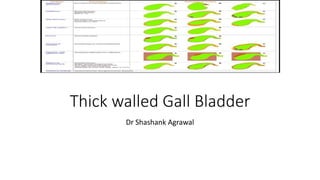
- 21. Summary of investigations Features Benign GB wall thickening Malignant GB wall thickening Degree of mural thickening Less More * Symmetry of mural thickening Symmetrical Asymmetrical, irregular, focal thickening Mural stratification Preserved Lost Enhancement pattern homogenous or stratified enhancement Presence of dotted –linear vessels (on CEUS) Delayed washout (>40 s) Inhomogeneous enhancement Branched or linear intralesional vessels (on CEUS) Early washout (<40 s) Intramural characteristics Continuous inner wall Intramural cystic spaces, echogenic foci, hypoechoic nodules, twinkling artifacts Focal discontinuity of inner and outer layer Irregularity and thickening > 1 mm of innermost layer Irregular thickening of outer layer Ancillary findings Pericholecystic fluid in absence of ascites, intraluminal membranes, sandwich sign, halo sign, visualization of Rokitansky Aschoff sinuses Diffusion restriction, direct invasion of adjacent organ, biliary obstruction, lymphadenopathy