1. Myers
1
Christopher
Myers
Augsburg
College
Genetics
Research
Project
Mode
of
Transmission
in
GloFish
Abstract
This
study
was
designed
to
study
the
mode
of
transmission
in
a
mutant
type
of
Zebrafish
known
as
GloFish.
More
specifically
the
green
fluorescent
protein
(GFP)
or
glow
transgene
was
observed
to
determine
how
it
behaves
regarding
heritability.
We
applied
a
reciprocal
cross
of
male
vs.
female,
mutant
type
vs.
wild
type
to
produce
an
F1
generation
of
both
crosses
to
a
particular
embryonic
developmental
stage
to
examine
if
the
GFP
gene
is
expressed
and
observable
if
present.
By
this
method
we
were
able
to
determine
that
the
gene
was
autosomal
dominant
and
not
sex-‐linked,
autosomal
recessive,
codominant
or
incompletely
dominant.
It
was
also
found
that
though
sex-‐linkage
was
not
present
an
apparent
maternal
effect
was.
Though
previous
studies
with
these
fish
have
been
done
at
Augsburg
College
before,
our
experiment
was
novel
in
the
way
that
it
was
done
to
determine
how,
and
if,
sex
played
a
role
in
the
mode
of
transmission.
Introduction
Over
the
past
several
decades
Zebrafish
(Danio
rerio)
have
been
extensively
studied
from
several
different
types
of
scientists
to
students
both
in
colleges
and
2. Myers
2
high
schools
around
the
globe.
This
is
due
to
the
fact
that
these
fish
are
very
inexpensive,
can
tolerate
a
reasonable
amount
of
stress,
produce
a
lot
of
eggs,
produce
these
eggs
reliably,
and
allow
observers
to
watch
embryo
development
that
is
comparable
to
other
species
(Zebrafish
FAQs).
This
embryonic
development
is
relatively
quick
and
is
easily
observable
under
a
decent
microscope.
Because
the
embryos
can
be
seen
so
easily
and
gene
expression
occurs
quickly
these
fish
have
become
ideal
research
organisms;
and
though
they
are
vastly
different
from
humans,
and
other
mammals,
their
embryonic
development
is
still
quite
similar
to
all
vertebrates
that
seem
to
follow
a
developmental
program
that
is
evolutionarily
conserved
(Kimmel
et
al.,
2012).
Recently
the
Zebrafish
have
been
genetically
altered
into
several
different
strains
of
fish
that
will
glow
different
colors
due
to
an
inserted
transgene.
These
fish
are
known
as
GloFish
and
are
readily
commercially
available
in
the
pet
market
as
well
as
offer
significant
viability
in
laboratory
experiments.
Under
normal
light
these
fish
appear
to
be
brightly
colored,
however,
when
they
absorb
certain
wavelengths
of
light
they
are
able
to
fluorescence
(glow).
Due
to
their
vast
array
to
explore
genetic
concepts
at
the
fundamental
level,
this
organism
was
chosen
for
our
experiment
in
order
to
look
at
how
this
transgene
is
inherited
(Vick
et
al.,
1995).
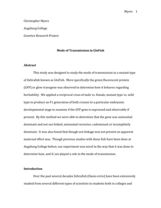
Figure
1.
Different
colors
of
GloFish
http://www.thatpetplace.com/glofish-‐danios
3. Myers
3
By
looking
at
the
main
manufacturers
and
distributor’s
of
GloFish
website
GloFish.com
several
different
types
of
GloFish
have
been
patented
and
trademarked.
The
different
types/colors
are
bright
red,
green,
orange-‐yellow,
blue,
and
purple
(Figure
1).
In
this
experiment
electric
green
was
used,
which
appear
to
be
yellow
under
normal
light.
The
source
for
the
transgene
inserted
into
the
green
GloFish
comes
from
the
Aequorea
victoria
jellyfish
(GloFish®
FAQ).
Methods
Mutant
type
GloFish
(8)
and
wild
type
Zebrafish
(6)
were
obtained
by
Professor
Beckman
from
a
pet
store
and
initially
all
placed
in
a
large
single
tank.
This
was
done
in
order
to
relieve
stress
on
the
fish
and
ease
the
acclamation
process.
The
tank
was
located
in
an
incubator
at
28.5
°C
with
a
light-‐dark
cycle.
Around
a
week
and
a
half
before
the
fish
were
mated,
both
mutant
and
wild
types
were
sexed
and
separated
accordingly:
all
males
in
one
tank
and
all
females
in
another.
The
night
before
the
intended
mating,
two
mating
tanks
were
filled
with
50/50
mix
of
water
from
the
tanks
in
which
both
the
female
and
male
fish
were
taken
from.
The
first
mating
tank
included
one
female
GloFish
and
two
male
Zebrafish.
The
second
mating
tank
included
one
female
Zebrafish
and
two
male
GloFish.
These
tanks
were
then
placed
back
into
the
incubator
and
the
fish
were
fed
again
to
make
them
comfortable.
Eggs
were
collected
the
following
morning,
roughly
4
hours
post
fertilization,
from
each
mating
tank
via
pipettes
and
microscopes
and
placed
into
two
labeled
petri
plates
containing
embryo
water
made
up
in
the
lab
(Figure
2).
Any
eggs
that
4. Myers
4
showed
abnormalities
were
discarded
from
the
samples.
The
following
day
the
eggs
were
examined
again
for
any
other
abnormalities
or
developmental
problems;
any
that
were
found
were
discarded.
Around
50
hours
post
fertilization
all
of
the
healthy
embryos
were
mounted
(five
embryos
per
slide)
and
observed
under
a
florescence
microscope
with
blue
light
settings
(Figure
2).
These
embryos
were
then
scored
on
a
positive/negative
scale
on
if
the
expression
of
GFP
was
present
by
observable
fluorescence.
Figure
2.
Example
of
egg
4
hours
post
fertilization
and
embryo
48
hours
post
fertilization.
Eggs
were
collected
4
hours
post
fertilization
and
embryos
were
examined
for
presence
of
GFP
50
hours
post
fertilization.
Photo
credit
(Kimmel
et
al.,
1995).
Results
and
discussion
The
female
Glofish
produced
a
much
larger
sample
size
of
39
viable
eggs
compared
to
the
10
viable
eggs
produced
by
the
female
Zebrafish.
The
female
Zebrafish
had
many
eggs
that
contained
abnormalities
and
many
had
to
be
discarded
of.
Out
of
the
39
embryos
produced
by
the
maternal
mutant
type
GloFish
and
paternal
wild
type
Zebrafish
all
of
them
expressed
the
GFP
gene.
For
the
maternal
wild
type
Zebrafish
and
paternal
wild
type
GloFish
4
out
of
the
10
expressed
the
GFP
gene
(Table
1).
Table
1.
GFP
presence
amongst
both
embryo
samples.
5. Myers
5
It
was
also
observed
that
in
all
of
the
maternal
Glofish
embryos
(39)
fluorescence
was
observed
throughout
most
of
the
embryo’s
tissues
(Figure
3),
however,
in
the
maternal
Zebrafish
embryos
(4)
fluorescence
was
observed
mainly
along
the
notochord
area
(Figure
4).
Also
it
should
be
noted
that
when
looking
at
the
yolk
of
the
eggs
all
of
the
maternal
GloFish
eggs
appeared
to
express
the
GFP
(Figure
5)
where
none
of
the
maternal
Zebrafish
yolks
expressed
the
GFP
even
though
embryos
later
did
express
the
transgene.
Figure
3.
Embryo
of
maternal
mutant
type
GloFish
and
paternal
wild
type
Zebrafish.
GFP
expression
in
significant
amount
of
tissue
observed.
Figure
4.
Embryo
of
maternal
wild
type
Zebrafish
and
paternal
mutant
type
GloFish.
GFP
expression
observed
around
notochord
area.
Figure
5.
Maternal
GloFish
yolk
showing
GFP
expression
before
age
where
GFP
should
be
expressed.
6. Myers
6
From
our
results
we
were
able
to
conclude
that
the
mode
of
transmission
for
the
transgene
of
GFP
expression
was
autosomal
dominant.
This
was
determined
because
if
the
transgene
was
autosomal
recessive
none
of
the
embryos
would
show
a
presence
of
GFP
because
none
of
the
wild
type
would
be
heterozygous
due
to
the
glow
gene
being
transgenic
in
nature.
But
it
was,
however,
able
for
the
GloFish
to
be
both
heterozygous
as
well
as
homozygous
dominant.
In
our
case
of
the
maternal
GloFish
it
was
almost
certain
that
she
was
homozygous
dominant
for
the
transgene.
In
the
case
of
our
paternal
GloFish,
in
order
to
produce
4
out
of
10
embryos
with
the
transgene
he
would
have
had
to
have
been
heterozygous
for
the
transgene.
However,
it
also
appears
that
there
were
some
types
of
maternal
effects
at
play
here.
This
is
due
mainly
to
two
observations.
The
first
observation
being
that
all
of
the
eggs
of
the
maternal
Glofish
contained
yolks
that
appeared
very
light
green/yellow
in
regular
light
as
well
as
fluoresced
under
blue
light
only
4
hours
after
fertilization.
None
of
the
maternal
Zebrafish
yolks
displayed
this
characteristic.
Secondly
of
all
the
embryos
that
showed
GFP
expression
the
maternal
Glofish
embryos
seemed
to
fluoresce
in
much
more
of
their
tissue
than
that
of
maternal
Zebrafish.
In
order
to
fully
examine
this
further,
next
time
I
would
have
more
than
just
one
reciprocal
cross.
If
the
results
showed
the
same
maternal
effects
across
several
matings
we
would
definitely
be
able
to
conclude
that
not
only
autosomal
dominance
is
at
play,
but
having
a
maternal
Glofish
also
significantly
alters
how
GFP
is
expressed.
Secondly,
it
would
have
been
nice
to
let
these
embryos
grow
older
to
see
if
the
tissues
in
the
4
maternal
Zebrafish
embryos
that
expressed
the
transgene
7. Myers
7
eventually
caught
up
with
the
other
39.
Lastly
looking
at
an
F2
generation
really
could
have
solidified
what
the
mode
of
transmission
is.
However,
for
the
amount
of
time
we
had
and
the
results
we
got,
I
feel
confident
in
our
autosomal
dominant
conclusion.
Effort
and
Contribution
When
dealing
with
live
animals
we
had
to
keep
them
alive,
keep
water
in
the
tank
and
make
sure
the
animals
ate
enough.
As
a
group
we
worked
well
with
feeding
them.
There
wasn’t
a
day
when
we
crossed
paths
with
each
other
and
one
of
our
group
members
was
checking
with
the
other
on
feeding
times
and
planning
on
who
was
feeding
throughout
the
week.
Even
though
not
everyone
participated
in
every
event
of
the
entire
experiment
I
do
feel
like
it
was
pretty
evenly
divided
amongst
the
group
members
and
we
each
did
a
significant
part,
from
keeping
water
healthy,
feeding,
checking
on
fish,
collecting
eggs,
examining
and
removing
bad
eggs,
setting
up
mating
tanks,
sexing
the
fish,
taking
pictures
of
eggs,
and
taking
pictures
of
a
scoring
fish.
I
did
miss
the
scoring
part,
however,
for
everything
else
I
was
present
and
put
effort
into
keeping
our
fish
alive,
our
embryos
healthy
and
preparing
for
mating.
8. Myers
8
Works
Cited
GloFish®
FAQ.
(n.d.).
Retrieved
December
13,
2014,
from
http://www.glofish.com/about/faq/
Kimmel,
C.
B.,
Ballard,
W.
W.,
Kimmel,
R.
S.,
Ullmann,
B.,
&
Schilling,
T.
F.
(1995).
Stages
of
Embryonic
Development
of
the
Zebrafish.
Developmental
Dynamics,
203:253-‐310.
Vick,
B.
M.,
Pollak,
A.,
Welsh,
C.,
&
Liang,
J.
O.
(2012).
Learning
the
Scientific
Method
Using
GloFish.
Zebrafish,
9(4),
226-‐241.
Doi:10.1089/zeb.2012.0758
Zebrafish
FAQs.
(n.d.).
Retrieved
December
13,
2014,
from
http://www.neuro.uoregon.edu/k12/FAQs.html#high
school