SlideShare a Scribd company logo
1 of 1
Developing an Engineered Synthetic Microenvironment
for 3D Vasculogenesis
Catherine E. Oliver1, Jorge Valdez2, Linda Griffith2
1Department of Biomedical Engineering, University of Connecticut
2Department Bioengineering, Massachusetts Institute of Technology
Research completed as part of the MIT Emergent Behaviors of Integrated Cellular Systems REU program.
Introduction
2D Assay:
o Performed to learn how the individual peptides and peptide combinations affect
endothelial cell attachment
Chemistry of Bioactive Gel Solution:
o Synthetic PEG hydrogels covalently crosslinked via Michael-type addition chemistry
Preparation:
o Fixed using 4% PFA
o Stained with DAPI and phalloidin to visualize nuclei and actin, respectively
o Imaged using epi-fluorescent microscopy and quantified using Image J software
3D Assay:
o Performed to assess the overall matrix remodeling within a 3D microenvironment due to
vasculogenesis through proteolysis with encapsulated endothelial cells
Chemistry of Bioactive Gel Solution:
o Synthetic PEG hydrogels crosslinked using thiol-norbornene photochemistry
The continual enhancement of standard experimental methods to expedite the
evaluation process of new drug agents is critical to the progression of cancer therapy and
disease treatment. The scope of this project, therefore, involves developing an engineered
synthetic microenvironment for 3D vasculogenesis with an overall long-term goal of
developing physiologically relevant in vitro systems incorporating endothelial networks to
be used for drug screenings. Synthetic PEG hydrogels were used in this study due to their
advantage of modularity, a characteristic which allows for the independent control of
variables such as adhesion and stiffness. The PEG hydrogels were decorated with the
widely used peptide ligand, Arginine-Glycine-Aspartic RGD, and the novel peptide ligands,
fibronectin-derived Synergy-RGD and collagen-mimetic GFOGER, which are believed to
target α5β1 and α2β1, respectively—two integrins implicated in angiogenesis. By engaging
these specific integrins, which RGD has very low affinity towards, we hypothesize that
Synergy-RGD and GFOGER will demonstrate greater adhesive qualities leading to increased
endothelial cell attachment and vasculogenesis. Thus, the following experimentation
involved the use of Synergy-RGD and GFOGER as well as potentially synergistic peptide
combinations to study how their effects in 2D and 3D compare to RGD.
Methods
Results and Conclusions
Acknowledgements
This research was supported by the National Science Foundation Science and Technology Center
Emergent Behaviors of Integrated Cellular Systems (EBICS) Grant Number CBET-0939511.
HS-
PEG-Acrylate
HS-
HS-
-SH
-SH
Thiol cross-linker Thiol ligand
Preparation:
o Fixed using 4% PFA
o Stained with DAPI and phalloidin to visualize nuclei and actin, respectively
o Imaged using confocal microscopy and quantified using Image J software
PEG-Norbornene
HS-
-SH
Thiol cross-linker Thiol ligand
HS-
2D Results:
Figure 1. Plot of number of attached cells per cm2 versus ligand concentration (μM) comparing individual effect of RGD, Syn-RGD and
GFOGER on IPS-endothelial cell attachment.
o GFOGER has greatest effect on IPS-endo cell attachment compared to RGD and Syn-RGD
Figure 2. Plot of number of attached cells per cm2 versus ligand concentration (μM) comparing individual effect of RGD and GFOGER
to its combined effect on IPS-endothelial cell attachment.
Figure 3. Plot of number of attached cells per cm2 versus ligand concentration (μM) comparing individual effect of RGD, Syn-RGD and
GFOGER to their combined effect with a heparin-binding ligand on IPS-endothelial cell attachment.
o Combining RGD and GFOGER showed increased cell attachment compared to when used
independently
o Heparin-binding peptide shows potential to increase cell attachment when used
synergistically with RGD, Syn-RGD and GFOGER
3D Results:
Figure 4, 5 and 6. Brightfield images of RGD, Syn-RGD and GFOGER, respectively, on Day 2 of incubation within a 3D synthetic PEG-
hydrogel microenvironment at a concentration of 6M cells/ml.
o GFOGER shows highly developed vascular structure formation by Day 2 compared
to RGD and Syn-RGD
Figure 7, 8, 9 and 10. Confocal images of GFOGER on Day 2 of incubation within a 3D synthetic PEG-hydrogel microenvironment at
concentrations of 1, 4, 6 and 9M cells/ml.
o GFOGER shows most prominent vascular structure formation at 6 and 9M cells/ml
Future Work
o Development of quantitative metrics to assess vasculogenesis in 3D assays
o Study synergistic peptide combinations in 3D
o Create functional assays using microfluidic devices to show perfusability of the
networks
UV
5-7 hours
2&4. 3.1.
Inert Gel Precursor
Solution + Cells
Well Plate
5. Fix
6. Stain
7. Image
5 hours
1. 2.
Precursor
Solution
Cells
Well Plate
3. Fix
4. Stain
5. Image

More Related Content

Viewers also liked (7)

Challenges with vascular injuries in peripheral setting
Challenges with vascular injuries in peripheral settingChallenges with vascular injuries in peripheral setting
Challenges with vascular injuries in peripheral setting
 
Arterial trauma
Arterial traumaArterial trauma
Arterial trauma
 
extremity Vascular trauma / injury
extremity Vascular trauma / injuryextremity Vascular trauma / injury
extremity Vascular trauma / injury
 
Acute limb ischaemia
Acute limb ischaemiaAcute limb ischaemia
Acute limb ischaemia
 
Lecture11 development of the heart and blood vessels
Lecture11   development  of the heart and blood vesselsLecture11   development  of the heart and blood vessels
Lecture11 development of the heart and blood vessels
 
1º control 2ª eval. química 2º bac 2015 2016
1º control 2ª eval. química 2º bac 2015 20161º control 2ª eval. química 2º bac 2015 2016
1º control 2ª eval. química 2º bac 2015 2016
 
Peripheral Vascular Diseases
Peripheral Vascular DiseasesPeripheral Vascular Diseases
Peripheral Vascular Diseases
 

Similar to EBICSposter_CEO_2

Study of the Effect of Gedunin against Protein GTP Binding Protein RAD to Tre...
Study of the Effect of Gedunin against Protein GTP Binding Protein RAD to Tre...Study of the Effect of Gedunin against Protein GTP Binding Protein RAD to Tre...
Study of the Effect of Gedunin against Protein GTP Binding Protein RAD to Tre...YogeshIJTSRD
 
PEG for Drugs and Drug Delivery Systems.pptx
PEG for Drugs and Drug Delivery Systems.pptxPEG for Drugs and Drug Delivery Systems.pptx
PEG for Drugs and Drug Delivery Systems.pptxKrystalBeily
 
3 GPCR signaling.pdfG-coupled receptors play a significant role in cell signa...
3 GPCR signaling.pdfG-coupled receptors play a significant role in cell signa...3 GPCR signaling.pdfG-coupled receptors play a significant role in cell signa...
3 GPCR signaling.pdfG-coupled receptors play a significant role in cell signa...vincentlangat14
 
2 GPCR overview.pdfunderstanding of the the role of G coupled receptors
2 GPCR overview.pdfunderstanding of the  the role of G coupled receptors2 GPCR overview.pdfunderstanding of the  the role of G coupled receptors
2 GPCR overview.pdfunderstanding of the the role of G coupled receptorsvincentlangat14
 
Maria antonia salerno 20 6-10
Maria antonia salerno 20 6-10Maria antonia salerno 20 6-10
Maria antonia salerno 20 6-10lab13unisa
 
Combined effects of PEGylation and particle size on uptake of PLGA particles ...
Combined effects of PEGylation and particle size on uptake of PLGA particles ...Combined effects of PEGylation and particle size on uptake of PLGA particles ...
Combined effects of PEGylation and particle size on uptake of PLGA particles ...Nanomedicine Journal (NMJ)
 
Cancer Research: Effects of Insulin-like Factor -2 (IGF-2), Collagen, and Fib...
Cancer Research: Effects of Insulin-like Factor -2 (IGF-2), Collagen, and Fib...Cancer Research: Effects of Insulin-like Factor -2 (IGF-2), Collagen, and Fib...
Cancer Research: Effects of Insulin-like Factor -2 (IGF-2), Collagen, and Fib...Raul Soto
 
Final_Poster_Presentation_TD
Final_Poster_Presentation_TDFinal_Poster_Presentation_TD
Final_Poster_Presentation_TDTyler DiStefano
 
Receptor Effector coupling by G-Proteins Zarlish attique 187104
Receptor Effector coupling by G-Proteins Zarlish attique 187104 Receptor Effector coupling by G-Proteins Zarlish attique 187104
Receptor Effector coupling by G-Proteins Zarlish attique 187104 ZarlishAttique1
 
PEGylation technique
PEGylation techniquePEGylation technique
PEGylation techniqueKushal Saha
 
Genome Scineces Poster
Genome Scineces PosterGenome Scineces Poster
Genome Scineces PosterColton McDavid
 
“Bisfenolo-A nelle otturazioni in materiale composito e riflessioni sulle low...
“Bisfenolo-A nelle otturazioni in materiale composito e riflessioni sulle low...“Bisfenolo-A nelle otturazioni in materiale composito e riflessioni sulle low...
“Bisfenolo-A nelle otturazioni in materiale composito e riflessioni sulle low...Biomed Srl
 
Protein formulation by PEGylation
Protein formulation by PEGylationProtein formulation by PEGylation
Protein formulation by PEGylationPratik Parikh
 
76201928
7620192876201928
76201928IJRAT
 

Similar to EBICSposter_CEO_2 (20)

AMI_ Rahul
AMI_ RahulAMI_ Rahul
AMI_ Rahul
 
Study of the Effect of Gedunin against Protein GTP Binding Protein RAD to Tre...
Study of the Effect of Gedunin against Protein GTP Binding Protein RAD to Tre...Study of the Effect of Gedunin against Protein GTP Binding Protein RAD to Tre...
Study of the Effect of Gedunin against Protein GTP Binding Protein RAD to Tre...
 
poster CRTD 2013
poster CRTD 2013poster CRTD 2013
poster CRTD 2013
 
PEG for Drugs and Drug Delivery Systems.pptx
PEG for Drugs and Drug Delivery Systems.pptxPEG for Drugs and Drug Delivery Systems.pptx
PEG for Drugs and Drug Delivery Systems.pptx
 
3 GPCR signaling.pdfG-coupled receptors play a significant role in cell signa...
3 GPCR signaling.pdfG-coupled receptors play a significant role in cell signa...3 GPCR signaling.pdfG-coupled receptors play a significant role in cell signa...
3 GPCR signaling.pdfG-coupled receptors play a significant role in cell signa...
 
2 GPCR overview.pdfunderstanding of the the role of G coupled receptors
2 GPCR overview.pdfunderstanding of the  the role of G coupled receptors2 GPCR overview.pdfunderstanding of the  the role of G coupled receptors
2 GPCR overview.pdfunderstanding of the the role of G coupled receptors
 
Maria antonia salerno 20 6-10
Maria antonia salerno 20 6-10Maria antonia salerno 20 6-10
Maria antonia salerno 20 6-10
 
Thesis Poster (1)
Thesis Poster (1)Thesis Poster (1)
Thesis Poster (1)
 
Combined effects of PEGylation and particle size on uptake of PLGA particles ...
Combined effects of PEGylation and particle size on uptake of PLGA particles ...Combined effects of PEGylation and particle size on uptake of PLGA particles ...
Combined effects of PEGylation and particle size on uptake of PLGA particles ...
 
Cancer Research: Effects of Insulin-like Factor -2 (IGF-2), Collagen, and Fib...
Cancer Research: Effects of Insulin-like Factor -2 (IGF-2), Collagen, and Fib...Cancer Research: Effects of Insulin-like Factor -2 (IGF-2), Collagen, and Fib...
Cancer Research: Effects of Insulin-like Factor -2 (IGF-2), Collagen, and Fib...
 
CCMB document
CCMB documentCCMB document
CCMB document
 
Final_Poster_Presentation_TD
Final_Poster_Presentation_TDFinal_Poster_Presentation_TD
Final_Poster_Presentation_TD
 
Receptor Effector coupling by G-Proteins Zarlish attique 187104
Receptor Effector coupling by G-Proteins Zarlish attique 187104 Receptor Effector coupling by G-Proteins Zarlish attique 187104
Receptor Effector coupling by G-Proteins Zarlish attique 187104
 
PEGylation technique
PEGylation techniquePEGylation technique
PEGylation technique
 
Jurnal proceeding
Jurnal proceedingJurnal proceeding
Jurnal proceeding
 
Genome Scineces Poster
Genome Scineces PosterGenome Scineces Poster
Genome Scineces Poster
 
“Bisfenolo-A nelle otturazioni in materiale composito e riflessioni sulle low...
“Bisfenolo-A nelle otturazioni in materiale composito e riflessioni sulle low...“Bisfenolo-A nelle otturazioni in materiale composito e riflessioni sulle low...
“Bisfenolo-A nelle otturazioni in materiale composito e riflessioni sulle low...
 
Protein formulation by PEGylation
Protein formulation by PEGylationProtein formulation by PEGylation
Protein formulation by PEGylation
 
76201928
7620192876201928
76201928
 
MVS_Poster_brey_FINAL
MVS_Poster_brey_FINALMVS_Poster_brey_FINAL
MVS_Poster_brey_FINAL
 

EBICSposter_CEO_2

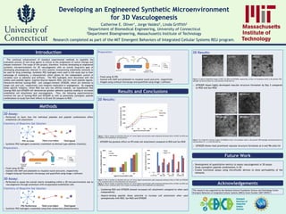
  • 1. Developing an Engineered Synthetic Microenvironment for 3D Vasculogenesis Catherine E. Oliver1, Jorge Valdez2, Linda Griffith2 1Department of Biomedical Engineering, University of Connecticut 2Department Bioengineering, Massachusetts Institute of Technology Research completed as part of the MIT Emergent Behaviors of Integrated Cellular Systems REU program. Introduction 2D Assay: o Performed to learn how the individual peptides and peptide combinations affect endothelial cell attachment Chemistry of Bioactive Gel Solution: o Synthetic PEG hydrogels covalently crosslinked via Michael-type addition chemistry Preparation: o Fixed using 4% PFA o Stained with DAPI and phalloidin to visualize nuclei and actin, respectively o Imaged using epi-fluorescent microscopy and quantified using Image J software 3D Assay: o Performed to assess the overall matrix remodeling within a 3D microenvironment due to vasculogenesis through proteolysis with encapsulated endothelial cells Chemistry of Bioactive Gel Solution: o Synthetic PEG hydrogels crosslinked using thiol-norbornene photochemistry The continual enhancement of standard experimental methods to expedite the evaluation process of new drug agents is critical to the progression of cancer therapy and disease treatment. The scope of this project, therefore, involves developing an engineered synthetic microenvironment for 3D vasculogenesis with an overall long-term goal of developing physiologically relevant in vitro systems incorporating endothelial networks to be used for drug screenings. Synthetic PEG hydrogels were used in this study due to their advantage of modularity, a characteristic which allows for the independent control of variables such as adhesion and stiffness. The PEG hydrogels were decorated with the widely used peptide ligand, Arginine-Glycine-Aspartic RGD, and the novel peptide ligands, fibronectin-derived Synergy-RGD and collagen-mimetic GFOGER, which are believed to target α5β1 and α2β1, respectively—two integrins implicated in angiogenesis. By engaging these specific integrins, which RGD has very low affinity towards, we hypothesize that Synergy-RGD and GFOGER will demonstrate greater adhesive qualities leading to increased endothelial cell attachment and vasculogenesis. Thus, the following experimentation involved the use of Synergy-RGD and GFOGER as well as potentially synergistic peptide combinations to study how their effects in 2D and 3D compare to RGD. Methods Results and Conclusions Acknowledgements This research was supported by the National Science Foundation Science and Technology Center Emergent Behaviors of Integrated Cellular Systems (EBICS) Grant Number CBET-0939511. HS- PEG-Acrylate HS- HS- -SH -SH Thiol cross-linker Thiol ligand Preparation: o Fixed using 4% PFA o Stained with DAPI and phalloidin to visualize nuclei and actin, respectively o Imaged using confocal microscopy and quantified using Image J software PEG-Norbornene HS- -SH Thiol cross-linker Thiol ligand HS- 2D Results: Figure 1. Plot of number of attached cells per cm2 versus ligand concentration (μM) comparing individual effect of RGD, Syn-RGD and GFOGER on IPS-endothelial cell attachment. o GFOGER has greatest effect on IPS-endo cell attachment compared to RGD and Syn-RGD Figure 2. Plot of number of attached cells per cm2 versus ligand concentration (μM) comparing individual effect of RGD and GFOGER to its combined effect on IPS-endothelial cell attachment. Figure 3. Plot of number of attached cells per cm2 versus ligand concentration (μM) comparing individual effect of RGD, Syn-RGD and GFOGER to their combined effect with a heparin-binding ligand on IPS-endothelial cell attachment. o Combining RGD and GFOGER showed increased cell attachment compared to when used independently o Heparin-binding peptide shows potential to increase cell attachment when used synergistically with RGD, Syn-RGD and GFOGER 3D Results: Figure 4, 5 and 6. Brightfield images of RGD, Syn-RGD and GFOGER, respectively, on Day 2 of incubation within a 3D synthetic PEG- hydrogel microenvironment at a concentration of 6M cells/ml. o GFOGER shows highly developed vascular structure formation by Day 2 compared to RGD and Syn-RGD Figure 7, 8, 9 and 10. Confocal images of GFOGER on Day 2 of incubation within a 3D synthetic PEG-hydrogel microenvironment at concentrations of 1, 4, 6 and 9M cells/ml. o GFOGER shows most prominent vascular structure formation at 6 and 9M cells/ml Future Work o Development of quantitative metrics to assess vasculogenesis in 3D assays o Study synergistic peptide combinations in 3D o Create functional assays using microfluidic devices to show perfusability of the networks UV 5-7 hours 2&4. 3.1. Inert Gel Precursor Solution + Cells Well Plate 5. Fix 6. Stain 7. Image 5 hours 1. 2. Precursor Solution Cells Well Plate 3. Fix 4. Stain 5. Image