Xoran's xCAT - high resolution low dose head CT
•
1 like•361 views

Introduction of the xCAT: Xoran's most powerful CT Scanner to date, offering sub-millimeter resolution for superior geometrical accuracy. The xCAT radiation dose is a small fraction of conventional head CT, while the entire scanner is compact and portable using a 4 wheels design.

Recommended
Recommended
More Related Content
What's hot
What's hot (20)
Similar to Xoran's xCAT - high resolution low dose head CT
Similar to Xoran's xCAT - high resolution low dose head CT (20)
Recently uploaded
Recently uploaded (20)
Xoran's xCAT - high resolution low dose head CT
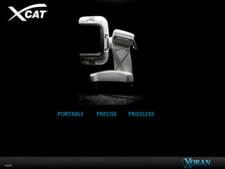
- 2. 60096 PORTABLE PRECISE PRICELESS • Agile - xCAT™ is easily positioned for intraoperative CT3 • Compact- Fits in a compact OR, small footprint with steering that pivots on a dime • Practical - A 4-wheeled solution that travels and stores well within the clinic • Real-time - Update image guidance to quickly improve accuracy intraoperatively,5,10 • Exact – confirm implant position and surgical completeness7 • Image quality - Superior geometrical accuracy with sub-millimeter resolution8,9 • Confidence – shown to alter surgical decision making1,2,10 • Safety – Radiation dose is a small fraction of conventional head CT • Quality - Aligns with the Institute of Medicine’s High Quality Care 11
- 3. 60096
- 4. 60096 ENT & Skull Base Cochlear Implants CMF & Trauma
- 5. 60096 Sinus and Skull Base Cochlear Implants CMF and Trauma • Visualize stent position • Dissection of ethmoid partitions • Visualize the need for additional resection • Detect residual bony fragments • Imaging shown to alter surgical desicions1,2 • Sub-millimeter resolution with geometric accuracy6,8 • Visualize placement of electrodes6 • Visualization of intra-cochlear trauma and Intra-scalar positioning5 • Imaging to support minimally- invasive and robotic procedures7 • Imaging supports fracture reduction • Visualize implant placement • Detect residual bony fragments • Imaging shown to alter surgical plan4 Cochlear Vanderbilt University Medical Center Nashville, TN ENT Kopf Klinic Frankfurt GmbH Germany ENT Copenhagen University Denmark ENT/CMF University Hospital of Mannheim Germany CMF Zurich Switzerland xCAT Locations with key publications: Cochlear Hannover Germany Cochlear University of Bern Switzerland CMF Dresden Germany ENT/Cochlear Ulm Germany ENT/CMF/Cochlear Dusseldorf Germany
- 6. 60096 FESS • Scan and IGS ready within minutes9 • Alters surgical plan1,2 Use of intraoperative CT scanning in endoscopic sinus surgery: a preliminary report. Jackman AH1, Palmer JN, Chiu AG, Kennedy DW. Am J Rhinol. 2008 Mar-Apr;22(2):170-4. doi: 10.2500/ajr.2008.22.3153.
- 7. 60096 Cochlear Implantation • Confirmation of electrode position5
- 8. 60096 Evaluation of portable CT scanners for otological image-guided surgery. Balachandran et al. 2012. Int J Comput Assist Radiol Surg 7(2); Design of a bone-attached parallel robot for percutaneous cochlear implantation. Kratchman et al. L.B. 2011. IEEE 58(10): Cochlear Implantation • Minimally invasive technique5
- 9. 60096 xCAT for Facial Fractures
- 10. 60096 Facial Fractures • Intraoperative confirmation of mesh placement* *Brainlab using xCAT images
- 11. 60096 C-Arm Facial Fractures Images • Alternative – 3D C-arm
- 12. 60096 xCAT ENT • Intraoperative use
- 13. 60096
- 15. 60096 Select Publications 1. Use of intraoperative CT scanning in endoscopic sinus surgery: a preliminary report. Jackman A, Palmer JN, Chiu AG, Kennedy DW, Am J Rhinol. (2008) 2. 2. Clinical utility of intraoperative volume computed tomography scanner for endoscopic sinonasal and skull base procedures. Batra PS1, Kanowitz SJ, Citardi MJ. Am J Rhinol. (2008) 3. Comparison of Intraoperative Portable CT Scanners in Skull Base and Endoscopic Sinus Surgery: Single Center Case Series. David B. Conley et.al. Skull Base. (2011) 4. Intraoperative cone beam computed tomography in the management of facial fractures. Stuck, B. A., R. Hülse, and T. J. Barth. International journal of oral and maxillofacial surgery (2012) 5. Evaluation of portable CT scanners for otologic image-guided surgery. Balachandran et al. Int J Comput Assist RadiolSurg (2012) 6. Automatic pre- to intra-operative CT registration for image-guided cochlear implant surgery Fitsum A. Reda, Jack H. Noble, Robert F. Labadie, and Benoit M. Dawant, IEEE Trans Biomed Eng. (2012) 7. 7. Minimally invasive image-guided cochlear implantation surgery: first report of clinical implementation. Labadie RF1, Balachandran R, Noble JH, Blachon GS, Mitchell JE, Reda FA, Dawant BM, Fitzpatrick JM, Laryngoscope (2014) 8. Flat-panel CT vs multislice CT to evaluate cochlear implant positioning. Zeitler DM et al. Cochlear implants International (2011) 9. The use of portable intraoperative CT for real-time image guidance: a pilot study. Das et. al. American Journal or Rhinology (2008) 10. Intraoperative IGS/CT updates for complex endoscopic frontal sinus surgery. Chennupati SK1, Woodworth BA, Palmer JN, Cohen NA, Kennedy DW, Chiu AG. (2008) 11. AAO-HNS Position Statement on Point of Care Imaging in Otolaryngology. http://www.entnet.org (2017)