7-1 ARTIFICIAL INTELLIGENCE IN PATHOLOGY semiar 2.pptx
Integrated workflow for digital pathology ina biospecimen repository enviornment
1. INTEGRATED WORKFLOW FOR DIGITAL PATHOLOGY IN A BIOSPECIMEN REPOSITORY
ENVIRONMENT
The field of Pathology requires the accurate and efficient review of
specimens for the purposes of consultation, diagnosis, research and
teaching. The increasing demand for more expedient pathology review has
led to the development of the rapidly growing and dynamic field of Digital
Pathology. This disruptive technology is allowing pathologists to overcome
many of the disadvantages of traditional glass slide review. The Biomedical
Imaging Team (BIT) in the Biopathology Center (BPC), located at The
Research Institute at Nationwide Children’s Hospital (TRINCH) in Columbus,
Ohio, has integrated digital pathology into the biorepository’s operations.
The BPC currently serves as the biorepository for the Children’s Oncology
Group (COG), the Gynecologic Oncology Group (GOG) and the pediatric
division of the Cooperative Human Tissue Network (CHTN), as well as a
Biospecimen Core Resource (BCR) for The Cancer Genome Atlas (TCGA)
project. This integration into banking operations has required the
development of specialized applications to support the utilization of this
technology. The introduction of these custom-designed applications into
BPC workflows has further increased operational efficiency.
The integration of digital pathology into the BPC’s workflow has decreased
pathology review turnaround times and eliminated shipping costs for many
projects 1,2. Simplified logistical operations have resulted in an overall
increase in operational efficiency, as the utilization of digital images
eliminates concerns about the transfer of custody of slides between
institutions. Digital pathology review has also been proven to be a reliable
means of achieving accurate results for tissue quality control and diagnostic
purposes 1,2.
1. T. Barr, D. Billiter, and S. Qualman. Formative Evaluation of Virtual
Microscopy for Tissue Quality Control Purposes; Arch Pathol Lab Med,
Vol 130, June 2006.
2. Jarzembowski J, Barr T, Prasad V, Nicol K, Suganuma R, and Shimada
H (2011, February).Virtual Microscopy as a Surrogate for Glass Slides in
Neuroblastoma, Pathology Central Review, Educational Training, and
Research; Poster session presented at the USCAP, San Antonio, TX.
3. VIPER-Virtual Imaging for Pathology, Education and Research
Retrieved from http://viper.epn.osc.edu/viper/
INTRODUCTION
The basic workflow for digital pathology begins with a glass slide and ends
with the completed pathology review of a high resolution whole slide image.
The steps involved are described in detail below.
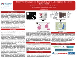
Specimen Glass Slide Preparation: The BPC acts as a central receiving
station where samples from research and healthcare institutions all over the
world are processed and banked. Depending upon customer requirements,
samples are often further processed into specimen glass slides for expert
pathology review. In this case, specimen glass slides are given to the BIT for
imaging. In preparation for scanning, the BIT is careful to first obscure any
Personal Health Information (PHI) on slide labels. In some cases barcodes
are also used to protect human-readable PHI. Slides are cleaned using lens
cleaning tissue and 70% isopropyl alcohol. This ensures the BIT obtains the
highest quality digital image possible, by removing any foreign substances
that may be adhered to the coverslip. All cleaned slides are then scanned
using specialized high-resolution imaging robots.
High Resolution Whole Slide Imaging: The BIT scanning infrastructure
includes seven Aperio ScanScope XT and one Hamamatsu NanoZoomer
2.0-HT whole slide imaging systems. These scanners can produce images of
20x or 40x optical magnification. After scanning, the high-resolution images
generated undergo a manual quality check by imaging technicians. As a
result, only pristine images representative of the original glass slide are
served to reviewers. The digital slide images are then transferred via a 1 GB
data link and stored in a digital slide repository at the Ohio Supercomputer
Center (OSC). Images are also stored locally for approximately one month to
ensure that data is fully backed up and redundant.
Digital Pathology Review: After being transferred to OSC, digital slide images
are ready to be served via the BIT’s custom-designed web-based automated
pathology review system. The VIPER application (Virtual Imaging for
Pathology, Education and Research) was designed to obtain rapid pathology
METHODS
RESULTS
REFERENCES
ACKNOWLEDGEMENTS
SPECIMEN GLASS SLIDE
DIGITAL IMAGE OF SPECIMEN SLIDE
SCANNER SYSTEM
VIPER SYSTEM
review from virtual slide images 3. OSC provides a reliable high performance
computing and communications infrastructure and has allocated 50 terabytes
of storage capacity for VIPER to facilitate the long-term storage and viewing
of digital images. BIT coordinators create cases, QC review forms, and links
to the stored images all within VIPER’s user interface. The cases and
associated slide images, pathology reports and review forms can then be
assigned to expert pathologists’ reviewer accounts. VIPER users are
automatically notified by email that they have cases pending in their review
list. After submitting finalized reviews, cases disappear from the user’s
account and the BIT receives email notification that the cases have been
completed.
DIGITAL SLIDE REPOSITORY
This disruptive technology is allowing the BPC to overcome many of the
disadvantages of traditional glass slide pathology review. Thus, growing
use of this technical tool has made pathology study more practical and
introduced new techniques such as image analysis and digital archiving.
CONCLUSION
Shital Abhange, Aaron Hobensack, Tom Barr, Dr. Nilsa Ramirez
The Research Institute at Nationwide Children’s Hospital
Columbus, Ohio