
Recommended
Recommended
More Related Content
Similar to hydroceph and spina bifida.pptx
hydroceph and spina bifida.pptx
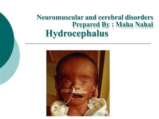
- 1. Neuromuscular and cerebral disorders Prepared By : Maha Nahal Hydrocephalus
- 2. Definition of Hydrocephalus is defined as an abnormal collection of cerebrospinal fluid with an associated dilatation of the cerebral ventricular system, a condition caused by an imbalance in the production or the absorption of the CSF through the ventricular system. An associate increase in head circumference follows. ت مع الشوكي الدماغي للسائل طبيعي غير تجميع بأنه فَّعرُي وسع بالجهاز مرتبط البطيني إنتا في خلل عن ناتجة حالة وهي ، الدماغي ج الجهاز خالل من النخاعي الدماغي السائل امتصاص أو البطيني . الرأس محيط في مرتبطة زيادة يتبع .
- 3. It could be congenital, that occurs in 3 /1000 live births or it could be acquired as in cases of hemorrhage, infection or neoplasm. , and if left untreated it may cause atrophy of the brain's white matter and severe neurological dysfunction يحدث هذا ، خلقي يكون أن يمكن في 3/1000 حي مولود األورام أو العدوى أو النزف حاالت في كما اكتسابها يمكن أو . وإذا ، الدماغ في البيضاء المادة في اًضمور تسبب فقد عالج دون تركت ًادشدي اًيعصب ً وخلال .
- 4. Any imbalance in secretion or in absorption causes increase in the accumulation of CSF in the ventricles which become dilated and compress the brain substances against the surrounding rigid bony cranium. When this occurs before fusion of cranial sutures, it provides enlargement of skull and dilation of ventricles, but if sutures were closed, it may result in diastatic or opened sutures especially the sagital. في النخاعي السائل تراكم زيادة إلى االمتصاص أو اإلفراز في خلل أي يؤدي الصلبة العظمية الجمجمة ضد الدماغ مواد على ويضغط يتوسع الذي البطينين المحيطة . الغرز اندماج قبل هذا يحدث عندما القحفية وتمد الجمجمة في اًمتضخ يوفر فإنه ، ًاد أ انبساطية خيوط حدوث إلى ذلك يؤدي فقد ، الغرز إغالق تم إذا ولكن ، للبطينين و السهمي ًةخاص مفتوحة .
- 5. Enlarged ventricles with bulging fontanels
- 6. Pathophysiology CSF circulates through out the ventricular system, CSF flows from the lateral ventricle, to the third ventricle ----then flows through (aqua duct of Sylvie's)------ -----into the fourth ventricle, where more fluids formed,---------then through foramen of luschka and the midline foramen of magendie-------- ---into the cisterna magna. Then CSF flows to the subarachnoid spaces where it is absorbed. البطين عبر النخاعي الدماغي السائل يدور الثالث البطين إلى ، الجانبي البطين من النخاعي الدماغي السائل يتدفق ، النظام - عبر يتدفق ثم ( المائية القناة لسيلفي ) ------ ----- ، السوائل من المزيد تشكل حيث ، الرابع البطين في --------- ثقبة خالل من ثم luschka من الوسط خط وثقب Magendie ----------- في سيستيرنا ماجنا . العنكبوتية تحت إلى النخاعي الدماغي السائل يتدفق ثم امتصاصه يتم حيث المساحات .
- 7. The pathway for CSF circulation is: Lateral ventricles one in each hemisphere Third ventricle Aqueduct of Sylvius Fourth Ventricle To Subarachnoid Space by (Paired Foramina of Luschka Foramen of Magendie
- 8. Types and Causes: 1. Non Communicating hydrocephaly: Obstruction to the flow of CSF through the ventricular system may result from: - Congenital abnormalities. -Structural defects, malformations, cyst, stenosis, tumors, and myelomeningocele. -Prenatal maternal infections. (Toxoplasmosis or cytomegalovirus). -neonatal infections. – (Neonatal meningo encephalitis bacterial or viral) - Perinatal hemorrhage (anoxic or traumatic).
- 9. 1 . المتصل غير الرأس استسقاء : عب النخاعي السائل تدفق إعاقة عن ينتج قد ر الجهاز البطيني : الخلقية التشوهات . - ، كيس ، تشوهات ، هيكلية عيوب السحائية النخاعية والقيلة واألورام التضيق . - الوالدة قبل األمهات التهابات . ( للخاليا المضخم الفيروس أو المقوسات داء .) - الوالدة حديثي التهابات . - ( السحايا الوليدية الفيروسي أو الجرثومي الدماغ التهاب ) بالوالدة المحيطة الفترة نزيف ( أو األكسجين نقص مؤلم .)
- 10. 2. Communicating hydrocephaly Impaired absorption of CSF within the subarachnoid space may result from -infection. -subarachnoid bleeds. -trauma. -meningeal growth, neoplasm or tumors. -Arachnoid cysts. دا النخاعي الدماغي السائل امتصاص ضعف ينتج قد العنكبوتية تحت الفضاء خل - عدوى . - العنكبوتية تحت نزيف . - صدمة . - أو ورم ، عضوي نمو األورام . - العنكبوتية الخراجات .
- 11. Hydrocephaly is associated with myelomeningocele. Arnold –chiari is One type of hydrocephaly resulted from the malformation that is characterized by herniation of small cerebellum, medulla pons and fourth ventricle into the cervical spinal canal through the enlarged foramen magnum results in obstruction in the flow of CSF causing hydrocephaly. شياري أرنولد الناتج الرأس استسقاء من واحد نوع هو هو الذي التشوه من الشوكي الرحم عنق في الرابع والبطين النخاع ، الصغير المخيخ بفتق يتميز خالل من القناة تؤدي ماغنوم إلى يؤدي مما النخاعي السائل تدفق انسداد إلى المتضخمة الثقبة الرأس استسقاء .
- 12. Clinical Manifestations -C/M various according to the onset, and the associated malformations -C / M حسب مختلفة البداية لها المصاحبة والتشوهات
- 13. Infancy: Head grows at an abnormal rate bulging fontanels + non pulsated anterior fontanel - Dilated scalp veins palpable suture with cracked sound with percussion. - Frontal protrusion with depressed eye + rotated down word. (Setting sun-sign). - Irritable – lethargic infant – feed poorly. - Changes in levels of consciousness - Opisthotonos after extreme. - Lower extremity sapasticity - منتفخ طبيعي غير بمعدل الرأس ينمو - اليافوخ + النابض غير األمامي اليافوخ - مع ملموس خياطة الرأس فروة أوردة اتساع - قرع مع متصدع صوت . - العين انخفاض مع أمامي نتوء + - كلمة أسفل استدارة ( . الشمس عالمة وضع .) - االنفعال سريع - خامل رضيع - سيء بشكل يتغذى . - الوعي مستويات في تغيرات - Opisthotonos المدقع بعد . - السفلية األطراف ترهل.
- 14. Infancy: Early infantile reflexes may persist. - Infants with ACM – may exhibit behaviors that reflect cranial nerve dysfunction as a result of brainstem compression, including swallowing difficulties, stridor, apnea, aspiration, respiratory difficulties and arm weakness. - If hydrocephalus is not treated it will disturb the development of the brainstem as manifested by poor sucking and feeding, high pitched cry – skull enlarged and cortex destroyed. - If hydrophilic progressed infant may display emesis, seizures and cardio pulmonary distress. - المبكرة الطفولية الفعل ردود تستمر قد . - بالـ المصابون الرضعACM - سلوكيات عليهم تظهر قد - انقطاع ، الصرير ، البلع صعوبات ذلك في بما ، الدماغ جذع ضغط نتيجة القحفي العصب ضعف تعكس الذراع وضعف التنفس صعوبات ، الطموح ، النفس . - يزعج فسوف الرأس استسقاء عالج يتم لم إذا - العالي والبكاء ، والتغذية الرضاعة سوء في يتجلى كما الدماغ جذع نمو - القشرة وتدمير الجمجمة تضخم . - الرئوي القلب وضيق والنوبات القيء يظهر قد للماء المحبة المتقدم الرضيع كان إذا .
- 15. Child hood S + S caused by high intracranial pressure mostly caused from posterior fossa neoplasm and aquduct stenosis. - Headache that relived after vomiting, or up right posture, papilledema strabismus, apathy, lethargic, and Ataxia. S + S ضغط ارتفاع عن ناتج في ناتج الجمجمة داخل القناة وتضيق الخلفية الحفرة ورم عن الغالب . - وذمة ، الصحيحة الوضعية أو ، القيء بعد عاد الذي الصداع الحول الحليمي والرنح ، والخمول ، والالمباالة ، .
- 16. Diagnostic evaluation: - U/S antenatal -Routine daily, measurement of HC in infancy and assessment for the presence of neurological signs. - CT + MRI are the primary diagnostic tools. -Echo en cephalography – to compare the ratio of lateral ventrice to cortex. -Sometimes isotope ventriculography. - Used to assess the flow and patency of existing shunts and to check the size of the ventricles. -U / S الوالدة قبل - قياس ، يومي روتينHC العصبية العالمات وجود وتقييم الرضع في . -CT + MRI األساسي التشخيص هي - أدوات . --Echo en cephalography - لمقارنة - القشرة إلى الجانبي البطني نسبة . - بالنظائر البطين تصوير األحيان بعض في . - الموجود التحويالت وانفتاح تدفق لتقييم تستخدم وللتحقق ة البطينين حجم من .
- 17. Therapeutic management Medical therapy is not effective; it can be used only in slowly progressed cases as to give furosemide to decrease production of CSF, or medications to lower the intra cranial pressure if surgeries are contraindicated. فعال غير الطبي العالج . في فقط استخدامه يمكن إلعطاء ببطء تتقدم التي الحاالت الفوروسيميد لتقل يل اإلنتاج الضغط لخفض األدوية أو ، النخاعي الدماغي السائل من الجراحية العمليات بطالن حالة في الجمجمة داخل .
- 18. Surgical therapy 1Direct removal of an obstruction such as resection of neoplasm, cyst, and hemotona, or in rare instances, fluid over production deceased by choroids plexus extirpation plexectomy or electric coagulation. 2Shunt: Most children require a shunt that provides drainage of CSF from the ventricles to extra cranial compartment usually the peritoneum. 3 والكيس الورم استئصال مثل لالنسداد المباشرة اإلزالة والهيموتونا حاالت في أو ، الكهرب التخثر أو المشيمية الضفيرة استئصال طريق عن الزائد السائل موت نادرة ائي . 4 التحويلة : النخ الدماغي السائل تصريف توفر تحويلة إلى األطفال معظم يحتاج اعي الحجرة إلى البطينين من القحفية الصفاق تكون ما عادة التي اإلضافية .
- 19. The preferred procedure is: 1- V.P shunts ventriculo peritoneal shunt : A shunt drains CSF from the lateral ventricles to the peritoneum (VP shunt) are used most commonly because the infection implication are moderately less severe and because the VP shunt allows the child more space to grow. Children with shunts will need numerous shunt revisions while growing, and there is a greater allowance for excess tubing, which minimizes the number of Revisions needed as the child grows. ا إلى الجانبيين البطينين من النخاعي الدماغي السائل بتصريف التحويلة تقوم لصفاق ( تحويلة VP) ملف ألن اًعشيو األكثر هي تحويلة وألن معتدل بشكل حدة أقل العدوى على المترتبة اآلثارVP للطفل تتيح للنمو أكبر مساحة . مراجعات من العديد إلى تحويالت لديهم الذين األطفال سيحتاج عدد من يقلل مما ، الزائدة لألنابيب أكبر بدل وهناك ، نموهم أثناء التحويل الطفل نمو مع الالزمة المراجعات .
- 20. 2- Ventriculo a trial shunt - V.A shunt A shunt drains CSF from the lateral ventricle to right atrium, is used for older children or for those with abdominal pathology but it is contra indicated in children with cardio pulmonary disease or elevated CSF protein. إ الجانبي البطين من النخاعي الدماغي السائل التحويلة تستنزف لى من يعانون لمن أو اًنس األكبر لألطفال وتستخدم ، األيمن األذين ولكنها ، البطن أمراض موانعة القلب بأمراض المصابين األطفال في المرتفع النخاعي السائل بروتين أو والرئة .
- 21. 3- Ventricular bypass Maybe used in older children to relieve the stenosis or masses but not used infant because spaces are poorly developed. 4- Ventriculo pleural shunts - are used sometimes for children over 5 years اًنس األكبر األطفال في تستخدم ربما الكت أو التضيق لتخفيف ولكن ل جيد بشكل متطورة غير المساحات ألن للرضع تستخدم ال . 4 - التحويالت البطينية الجنبية - لألطف األحيان بعض في تستخدم فوق ال سن 5 سنوات
- 22. Complications associated with shunt -The most common complication fro shunts is infection. This can be life threading as it involves many vital organs such as the kidneys as well as the brain. -All shunts are subject to mechanical difficulties such as kinking, plugging, separation, migration of the tubing. -Mal function caused by mechanical obstruction either within the ventricles from particular matter tissue or exudates, or from thrombosis or displacement. -The biggest indicator for shunt malfunction is the conscious level. -The child will exhibit signs and symptoms of intracranial pressure when the shunt is obstructed and should be treated as an emergency. - العدوى هي للتحويالت اًعشيو األكثر المضاعفات . من العديد يشمل ألنه الحياة خيوط بمثابة هذا يكون أن يمكن الدماغ وكذلك الكلى مثل الحيوية األعضاء . - األنبوب وترحيل ، والفصل ، والتوصيل ، االلتواء مثل الميكانيكية للصعوبات التحويالت جميع تخضع . - أو الدم تجلط من أو ، معينة إفرازات أو أنسجة من البطينين داخل إما ميكانيكي انسداد عن ناتج وظيفي خلل اإلزاحة . - - ملف هو التحويل عطل على مؤشر أكبر - واعي مستوى . -طارئة كحالة معه التعامل ويجب التحويلة إعاقة عند الجمجمة داخل الضغط وأعراض عالمات الطفل على سيظهر
- 23. Nursing considerations The goals of nursing care of the child with hydrocephalus include: - Prevent complications of hydrocephalus. -observe for signs of ICP which indicates obstruction of the shunt. - Measure head daily - Provide education and emotional supports to the family. -Neurological assessment and evaluation of pupil dilation. - Maintaining adequate nutrition -Observe signs of infection in the post operative period such as elevated temperature, poor sucking, or vomiting. مع الطفل رعاية أهداف الرأس استسقاء يشمل : الرأس استسقاء مضاعفات منع . - إعاقة إلى تشير التي الدولية المقارنات برنامج عالمات مراقبة التحويلة . يوميا الرأس قياس لـ العاطفي والدعم التعليم توفير أسرة . - التلميذ وتقويم العصبي التقييم تمدد . الكافية التغذية على الحفاظ - الجراحة بعد ما فترة في العدوى عالمات مالحظة القيء أو المص سوء أو الحرارة درجة ارتفاع مثل .
- 24. Spina Bifida
- 25. Definition: Malformation of the neural tube leading to herniation through an abnormality in the canal of the vertebral column. When the spinal cord itself is not fully formed however, the nerves do not develop as they should and the baby will have Myelomeningocele العمود قناة في خلل خالل من فتق إلى يؤدي العصبي األنبوب تشوه الفقري . تتطور ال ، التكوين مكتمل نفسه الشوكي النخاع يكون ال عندما السحائية النخاعية بالقيلة الطفل وسيصاب ينبغي كما األعصاب
- 26. Incidence In some cases it has occurred in the family before, but this is relatively uncommon, In the United States, spina bifida is the second most common birth defect and affects about one out of every one thousand pregnancies. also there is a high incidence of spina bifida in Palestine. غير هذا ولكن ، قبل من عائلة في حدث قد يكون ، الحاالت بعض في ، المتحدة الواليات في ، اًينسب شائع السنسنة ثان هي المشقوقة أكثر ي واحد كل من واحد على وتؤثر اًعشيو الخلقية العيوب . حمل حالة ألف . من عالية نسبة هناك أن كما السنسنة في المشقوقة فلسطين .
- 27. Prenatal Detection: There are several tests available to pregnant women which can be used to detect spina bifida before the baby is born, maternal serum alpha fetoprotein (MSAFP) The blood test which is done on the mother's blood around the 16th week of pregnancy. This test is not specific for spina bifida, however. An error in the dates of the pregnancy, multiple babies, as well as other birth defects can all cause an abnormal reading. If the alpha fetoprotein level is abnormal, additional testing is recommended. للكش استخدامها يمكن والتي الحوامل للنساء المتاحة االختبارات من العديد هناك ف عن السنسنة الطفل والدة قبل المشقوقة . عل االختبار هذا يقتصر ال ، ذلك ومع ى السنسنة المشقوقة . خلقي عيوب وكذلك ، األطفال وتعدد ، الحمل مواعيد في خطأ ة طبيعية غير قراءة جميعها تسبب أن يمكن أخرى . البروتين مستوى كان إذا إضافي اختبار بإجراء يوصى ، طبيعي غير ألفا الجنيني .
- 28. Causes of spina bifida Causes are unknown, but in a small number of women it appears to be caused by certain medications most often anti seizure drugs. In the majority of cases the cause of the spina bifida is never determined. However, it is now known that taking folic acid a B vitamin, before a woman becomes pregnant will reduce the chances that the baby will have spina bifida and related conditions of the brain and spinal cord. األد بعض هو سببها أن يبدو النساء من قليل عدد في ولكن ، معروفة غير األسباب وية للتشنج المضادة األدوية األحيان أغلب في . سبب تحديد يتم ال ، الحاالت معظم في السنسنة اًقمطل المشقوقة . من ب فيتامين تناول أن اآلن المعروف فمن ، ذلك ومع الطفل إصابة فرص من سيقلل الحمل قبل الفوليك حمض بالسنسنة وال المشقوقة حاالت الشوكي والحبل الدماغ في الصلة ذات .
- 29. Pathophysiology This problem occurs very early in pregnancy, Normally at the end of the fourth week of pregnancy, the neural tube formation is complete, if there is failure for the fusion of the lamina of the vertebra or splitting closed tube will result in spina bifida that can adversely affect many body systems including the nervous system, the bones and muscles as well as the kidneys and bladder. The point along the spinal cord where the undeveloped area occurs is called the "level" of the spina bifida من الرابع األسبوع نهاية في ًةعاد ، الحمل من ًادج مبكر وقت في المشكلة هذه تحدث اندماج في فشل هناك كان إذا ، اكتمل قد العصبي األنبوب تكوين يكون ، الحمل إلى سيؤدي المغلق األنبوب انقسام أو الفقرة صفيحة السنسنة أن يمكن المشقوقة يؤثر والعضالت والعظام العصبي الجهاز ذلك في بما الجسم أجهزة من العديد على اًبسل والمثانة الكلى وكذلك . المنطقة تحدث حيث الشوكي الحبل طول على النقطة تسمى النامية غير " مستوى " السنسنة المشقوقة
- 30. pathophysilogy The higher up the spinal column the "level" occurs, the greater the effect on normal nerve functions. Some individuals with low levels of spina bifida can walk with little or no assistance whereas those with higher levels will require braces and in cases of very high levels, wheelchairs, to get around. األعص وظائف على التأثير زاد ، الفقري العمود في الفقري العمود مستوى ارتفع كلما اب الطبيعية . من منخفضة مستويات من يعانون الذين األفراد لبعض يمكن السنسنة المشي المشقوقة سيحتاجو أعلى مستويات لديهم الذين أولئك أن حين في مساعدة بدون أو قليلة بمساعدة ن للتجول ، المتحركة الكراسي ، ًادج العالية المستويات حاالت وفي دعامات إلى .
- 31. Types of spina bifida 1. Spina bifida occulta: In this type the posterior vertebral arch fails to fuse, that only the bones of the spinal column will be incompletely developed, but the nerves beneath will be normal, the spinal cord and meninges does not herniated and the abnormality is not visualized externally. Does not cause neurologic problems such as paralysis or weakness and is not medically significant. 1 . السنسنة الخفية المشقوقة : في الخلفي الفقري القوس يفشل ، النوع هذا في الفقري العمود عظام سوى تندمج ال بحيث ، االندماج . وال ، طبيعية ستكون تحتها الموجودة األعصاب ولكن ، النمو مكتملة غير تكون اًيخارج الشذوذ تصور يتم وال والسحايا الشوكي الحبل ينفتق . مشاك يسبب ال ل طبية اهمية له وليس الضعف او الشلل مثل عصبية
- 32. 2. Spina bifida cystica the most severe form of spina bifida, the opening is larger, and the abnormality is shown as sac protruding in the external surface of the back and it is two types: أشكال أشد السنسنة الشذو ويظهر ، أكبر الفتحة ، المشقوقة ذ نوعان وهو للظهر الخارجي السطح في بارز ككيس :
- 33. A.Meningocele: a sac like cyst of meninges, filled with CSF, Protrude through defects in the bones of the spine B.meningomyelocele: a herniation allows for meninges, CSF, and part of spinal cord with nerves, through the bony defect. C. السحائية القيلة : كيس يشبه كيس بـ مليء ، السحائي CSF ، الفقري العمود عظام في عيوب خالل من يبرز D. السحائية النخاعية القيلة : فتق يسمح و للسحايا CSF من وجزء العظمي الخلل خالل من باألعصاب الشوكي الحبل .
- 34. Clinical manifestations a sac covered by tiny membrane likely to tear and to leak, can be located any where along spinal cord mostly lumber or lumbosacral region, and can be found at the cervical region. ويسرب يتمزق أن المحتمل من صغير بغشاء مغطى كيس الشوكي الحبل طول على مكان أي في يوجد أن يمكن ، ، العجزية القطنية المنطقة أو الخشب من الغالب في الرحم عنق منطقة في عليه العثور ويمكن .
- 35. Clinical manifestations If the defect is below the second lumber vertebra: Incontinence which means bladder nerve supply is affected. * *Partial paralysis of the lower limbs. *Sensory disturbances. *Poor anal sphincter control. If the defect is below the third sacral vertebra No motor involvement. الثاني من أقل الخلل كان إذا الخشب فقرة : المثانة عصب يعني الذي البول سلس يتأثر العرض * . * السفلية األطراف في جزئي شلل . * الحسية االضطرابات . * الشرجية العاصرة العضلة في التحكم ضعف . الثالثة العظمة تحت الخلل كان إذا فقرة حركي تورط ال .
- 36. Treatments Available for Children and Adults with Spina Bifida Because spina bifida affects so many body systems it is important that professionals from many areas be consulted to provide up-to-date, comprehensive medical, psychological and social evaluation, support and treatment. ألن اًنظر السنسنة أجهز من العديد على تؤثر المشقوقة ة من العديد من المتخصصين استشارة المهم فمن ، الجسم ودعم واجتماعي ونفسي طبي تقييم لتقديم المجاالت وشامل محدث وعالج .
- 37. What is the initial management of patients with myelomenigocele On admission the size of the defect need to be measured. The lesion needs to be covered with moist dressing, and needs to be kept continuously moist. Surgical closure needs to be performed within 24 hours. Neurological assessment: Watch for spontaneous movement of the lower extremity (good spontaneous movement correlates with better later functional outcome) The lowest level of function needs to be assessed by checking the response of the lower extremities to painful stimulation. Hydrocephalus needs to be watched for.
- 38. العيب حجم يكون أن يجب القبول عند تقاس . باستمرار رطبة تبقى أن ويجب ، رطبة بضمادة اآلفة تغطية يجب . غضون في الجراحي اإلغالق إجراء يجب 24 درجة ساعات . العصبي التقييم : السفلي للطرف التلقائية الحركة راقب ( ب الجيدة التلقائية الحركة ترتبط نتائج الحقة وظيفية ) للوظيفة مستوى أدنى تقييم يجب المؤلمة للمنبهات السفلية األطراف استجابة من التحقق طريق عن . الرأس استسقاء مراقبة يجب .
- 39. What are the surgical considerations? Closure of the defect is not associated with improvement of neurological function, but is associated with a lower infection rate. Myelomeningoceles need to be closed with 24 hours whether or not the membranes are intact. During the operative procedure extreme care needs to be taken in protecting the exposed neural tissue. This tissue needs to be dissected and placed back into as normal of a location as possible أقل إصابة بمعدل يرتبط ولكنه ، العصبية الوظيفة بتحسين يرتبط ال العيب إغالق . ت القيلة حتاج خالل اإلغالق إلى السحائية النخاعية 24 ال أم سليمة األغشية كانت سواء ساعة . اإلجراء أثناء المكشوفة العصبية األنسجة لحماية الشديد الحذر توخي يجب ، الجراحي . هذا تشريح يجب اإلمكان قدر الطبيعي مكانه إلى وإعادته النسيج
- 40. After the overlying layers need to be closed in a watertight fashion. The assistance of a plastic surgeon may be necessary to close large defects. الماء لتسرب مانعة بطريقة تعلوها التي الطبقات إغالق يتم أن بعد . تكون قد الكبيرة العيوب لسد ضرورية التجميل جراح مساعدة .