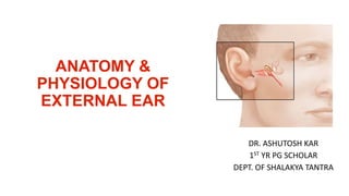
The external ear consists of the pinna, external auditory canal, and tympanic membrane. The pinna is made of elastic cartilage covered in skin and collects sound. The external auditory canal is S-shaped and carries the sound waves to the tympanic membrane. The tympanic membrane, or eardrum, separates the external ear from the middle ear and transmits vibrations to the ossicles. When viewed through an otoscope, the normal tympanic membrane appears pearly grey and concave laterally.
3. AURICLE / PINNA
• Pinna made up of a single yellow elastic cartilage covers with skin.
• 2 surface : lateral & medial
• Lateral surface : adhered to perichondrium
: presents various elevation & depression
• Medial surface : loose
5. HELIX
The prominent rim
of the auricle is
called the helix
ANTI HELIX
Another curved prominence
parallel with and in front of
the helix is called the
antihelix
CONCHA
The anti helix describes a curve
around a deep ,spacious cavity
concha’
Upper part : cymba concha
Lower part : cavum concha
9. EXTERNAL AUDITORY CANAL
Extends from conchato T
M
Measures 24mm
S shaped
2 parts outer part : cartilaginous directed UBM
inner part : bony DFM
For viewing TM : upwards, backwards& laterally
11. Cartilaginous part
Continuation of cartilage which forms the frame work of pinna
2 deficiencies
a) Fissures of Santorini : Parotid /Superficial mastoid infection appears
b) Skin covering the cartilaginous canal is thick and contains-
CERUMINOUS GLANDS which secrets cerumen (wax)
PILOSEBACEOUS GLANDS
Hair :outer canal furunculosis
12. BONY PART
SKIN :Thin continues over TM
It contains inner 2/3 part.(16mm)
Sl.no PART LOCATION IMPORTANCE
1 Isthmus 6mm lateral to TM narrow FB medial gets impacted
difficult to remove
2 Anterior Recess Antero-inferior part of deep meatus Cesspool for discharge &
debris in ext & middle ear
infection
3 Foramen of Huschke Deficiency in antero –inferior part Permitting to & from
infection from parotid
13. Blood supply of EAC
Post auricular artery
Superficial temporal
artery
14. Nerve supply of EAC
The auriculotemporal nerve : (from the mandibular branch of the trigeminal nerve)
provides sensory information from the anterior wall and roof
The auricular branch of vagus (Arnold nerve) The posterior wall and floor
sensibility is carried in the nerve fibres)
17. Partition between external and middle ear
Height : 9 to10mm
Width : 8 to 9 mm
Thickness - 0.1mm
Position - oblique postero-superior part is more lateral
Parts – Parstensa and parsflaccida
18.
19.
20.
21.
22. PARSTENSA
Formsmost of TympanicMembrane (Lower 2/3)
ANNULUS TYMPANICUS : Fibro cartilaginousring which fits in TympanicSulcus.
UMBO : Central part of parstensaistented inwardsat the levelof tip of malleus
CONE OF LIGHT- Bright part of light radiating from tip of malleus to periphery in
anteroinferior quadrant.
seen in autoscopy
23. PARS FLACIDA / SHRAPNEL’S MEMBRANE
Upper 1/3 of TM
Less tense than pars tensa
Situated abovelateral processof malleus between the notch of rivinus & anterior &
posterior mallealfold
Appears pinkish in otoscopy
24. LAYERS OF TYMPANIC MEMBRANE
3 layers
Outer epithelial layer – continuous with EAC skin
Middle fibrous layer – encloses handle of malleus
have 3 types fibres- radial ,circular ,parabolic
Inner mucosal layer - continuous with middle ear mucosa
28. Otoscopy
• Normal TM is shiny and pearly grey Concave laterally
• Transparency of TM varies from person to person
• Some middle ear structures are usually seen through TM
29. PHYSIOLOGY OF EXTERNAL EAR
• Role in hearing
pinna- collects the sound signal
passes through EAC & vibrates TM
transmitted to foot plate of stapes through chain of ossicles
EAC- along with pinna it can increase sound pressure level at the TM by
15-22db at 4000Hz