Premium Bangalore Call Girls Jigani Dail 6378878445 Escort Service For Hot Ma...
ELECTRODIAGNOSTIC STUDIES.pptx
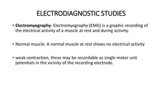
1. ELECTRODIAGNOSTIC STUDIES
• Electromyography: Electromyography (EMG) is a graphic recording of
the electrical activity of a muscle at rest and during activity.
• Normal muscle: A normal muscle at rest shows no electrical activity
• weak contraction, these may be recordable as single motor unit
potentials in the vicinity of the recording electrode.
2. • In a strong contraction, impulses of a number of motor units firing
simultaneously are superimposed, giving rise to an interference
pattern
• Denervated muscle. The denervated muscle has spontaneous
electrical activity at rest. This is called denervation potentials.
• These appear at around 15-20 days after the muscle denervation. As
nerve degeneration progresses, more and more denervation
potentials appear.
3.
4. Electromyography is useful in deciding the
following
• a) Whether or not a nerve injury is present
• b) Whether it is a complete or incomplete nerve injury
• c)Whether any regeneration occurring
• d) Level of nerve injury
5. Strength-duration curve:
• This is a graphic representation of the excitability of muscle and
nerve tissue under test .
• A small strength of current can excite a normal muscle through
neuromuscular junction, which needs a weaker current.
• In a denervated muscle, the excitation is possible on direct
stimulation of the muscle fibres with higher strength of current.
6. • A very low-strength current given for of 300 milliseconds and response
noted.
• The strength of the current is gradually increased until a minimal visible
contraction of the muscle is observed.
• This minimal current strength, required to elicit muscle contraction, is
called the Rheobase, and is measured in milliamperes.
• The Chronaxie is the duration of current required to excite a muscle with
a current-strength of double the rheobase. It is measured in milliseconds.
• graph is plotted between current-duration and corresponding
current-strength. This is called a strength-duration curve.
7.
8. Interpretation
• The pattern of the strength-duration curve of an innervated muscle is different
from that of a denervated muscle or regenerating muscle:
• Normal strength-duration curve: A normal muscle will respond to stimuli
varying in duration(300 milliseconds - 3 or even 1 millisecond) without any
increase in the strength of the current.
If the duration of current is decreased beyond it, a progressive increase in the
strength of current is required in order to produce a contraction.
A strength-duration curve plotted from such a muscle is termed a nerve curve,
because the muscle contraction is caused by stimulation of the motor nerve
entering the muscle.
9. • Denervated muscle: A totally denervated muscle will need current
either of more strength or for a longer duration. A curve from such a
muscle is termed a muscle curve.
• Partially denervated muscle: muscle recovering after nerve injury lies
between the normal and the curve of denervation, and is
characterised by an upward kink.
• The kink denotes the superimposition of the two basic types of
curves.
10. • Assessment of recovery by the strength-duration curve:
If progressive recovery is occurring the curve will, on serial
examination, become flatter with a shift to the left.
On the other hand, if the process of denervation is progressive, the
curve will become steeper and will shift to the right.
11. NERVE CONDUCTION STUDIES:
• It is a measure of the velocity of conduction of impulse in a nerve.
• A stimulating electrode is applied over a point on the nerve trunk
and the response is picked up by an electrode at a distance or directly
over the muscle.
• The velocity of the conduction of the impulse between any two points
of the nerve can be calculated.
• The normal nerve conduction velocity of motor nerve is 70
metres/second. This conduction study helps in the following:
12. Nerve Conduction studies
• This helps in following:
• Whether a nerve injury is present: If a nerve injury is present there
will be no conduction of the impulse across the suspected level
• b) Whether it is a complete or partial nerve injury: Absence of any
transmitted impulse across the suspected site is an indicator of a
complete nerve injury.
• c) Compressive lesion: The conduction velocity may simply be delayed
in compressive nerve lesions such as carpal tunnel syndrome etc.