1. DENTIN
Q1. Write a note on Dentinogenesis?
Definition:
The process of formation of dentin by odontoblasts is known as dentinogenesis. Odontoblasts
are a special type of biological cell on the outer wall of dental pulp, and it begins at the late
bell stage of a tooth development.
The different stages of dentin formation after differentiation of the cell result in different
types of dentin: mantle dentin, primary dentin, secondary dentin, and tertiary dentin.
Odontoblasts differentiate from cells of the dental papilla. This is an expression of signaling
molecules and growth factors of the inner enamel epithelium (IEE). This is known as
reciprocal induction.
They begin secreting an organic matrix around the area directly adjacent to the IEE, closest to
the area of the future cusp of a tooth. The organic matrix contains collagen fibers with large
diameters (0.1-0.2 μm in diameter). The odontoblasts begin to move toward the center of the
tooth, forming an extension called the odontoblast process. Thus, dentin formation proceeds
toward the inside of the tooth. The odontoblast process causes the secretion of hydroxyapatite
crystals and mineralization of the matrix (mineralization occurs due to matrix vesicles). This
area of mineralization is known as mantle dentin and is a layer usually about 20-150 μm
thick.
Secondary dentin is formed after root formation is finished and occurs at a much slower rate.
It is not formed at a uniform rate along the tooth, but instead forms faster along sections
closer to the crown of a tooth. This development continues throughout life and accounts for
the smaller areas of pulp found in older individuals.
Tertiary dentin is deposited at specific sites in response to injury by odontoblasts or
replacement odontoblasts from the pulp depending on the severity of the injury.
Q2. Elaborate the regional distribution of dentin according to its structure./ Types of dentin
Dentin is distributed throughout the tooth. Following are the different types of dentin
according to their structure which are found in different parts of a tooth
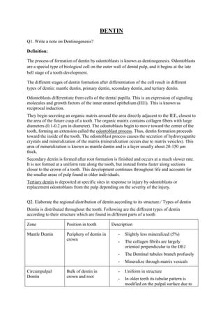
Zone Position in tooth Description
Mantle Dentin Periphery of dentin in
crown
- Slightly less mineralized (5%)
- The collagen fibrils are largely
oriented perpendicular to the DEJ
- The Dentinal tubules branch profusely
- Mineralize through matrix vesicals
Circumpulpal
Dentin
Bulk of dentin in
crown and root
- Uniform in structure
- In older teeth its tubular pattern is
modified on the pulpal surface due to
2. age related deposition of secondary
dentin
Secondary Dentin Innermost layer of
dentin between
cicumpulpal dentine
and predentin
- Deposited throughout the life
- More regular deposition
- Well-oriented dentinal tubules
Tertiary Dentin Inner layer of dentin
formed mainly in
crown response to
serious insult
Inner layer of dentin formed mainly in crown
response to serious insult (severe attrition,
fracture and caries).
i. Reactionary
Formed as a result of acute
trauma via already existing
odontoblastic cells
Haphazard dentin formation
Cells entrapped within the
calcified mass
No or irregular dentinal
tubules
ii. Reparative
Formed as result of chronic
low-grade trauma
Via newly differentiated
ectomesenchymal cells of
dental papilla
Homogenous deposition
with regular arrangement of
dentinal tubules
Predentin Unmineralized
innermost dentine in
crown and root
- Freshly laid down matrix of dentin
- Mineralizes by globular or linear
pattern
- Width is 10-40 µm
Interglobular
dentin
Typically in outer part
of coronal dentin also
seen beneath the
tome’s granular layer
in the root
- Formed as a result of failure of mineral
globules leaving behind hypo
mineralized areas
- Dentinal tubules passes without
deviation
- The peritubular dentin is absent from
the tubules that passes the interglobular
3. dentin
Grabular Layer of
Tomes
In outer part of root
dentin beneath the
hyaline layer
- Granular zone at the outer periphery of
root dentin.
- Reasons,
i. The dentinal tubules loop back
on themselves creating air
spaces in ground section.
ii. Accumulation of dentinal
proteins at the CDJ
iii. Incomplete fusion of
calcospherite
- Hypominealized
Hyaline layer Outermost part of root
dentin
- Located outside the granular layer is a
clear hyaline layer
- Inculded as a component of dentin
- Upto 20µm thick
- Nontubular and relatively structureless
- Serve to bond cementum to dentin
Q3. How Peritubular/Intratubular dentin does differ from the intertubular dentin?
Intratubular Dentin Intertubular Dentin
Surrounds the dentinal tubules Present between 2 dentinal tubules
Also known as pertubular dentine ---
Hypermineralized Less Mineralized
Less collagen More Collagen
Q4. Write notes on
1. Sclerotic dentin
- Detinal tubules becomes occluded with calcified materials.
- Give a glassy homogenous appearance in that area
- Amount increased with age
4. - Most commonly found in apical third of the tooth root and crown midway between
the DEJ and the surface of pulp
- Its is a physiological phenomenon the occurs due the continued deposition of the
peritubular dentin
- Three possible ways of its formation
i. Deposition of the mineral within the tubule WITHOUT any dentin formation
ii. Diffuse mineralization occurs with a viable odontoblastic process still present
iii. Mineralization of the process itself and tubular contents including intratubular
collagen fibrils
iv. Significance:
Reduces the permeability of dentin
Prolongs the pulp vitality
2. Dead Tracts
Trauma will cause the odontoblastic processes within the dentinal tubules to "die
back" toward the cell body. In severe cases the cells themselves may die.
Such regions of dentin (with empty dentinal tubules) are called dead tracts (B) and
appear dark in ground sections.
3. Incremental Lines of Von Ebner
- The lines of von Ebner represent cyclic activity of the odontoblasts during dentin
formation.
- These incremental lines illustrate the daily pattern of dentin deposition that progresses
at about 4 µm per day in the crown and about 2 µm per day in the root.
- Short period lines
4. Andersen Lines
5. - Long period lines
- Approx.. 16-20µ
- Depicts 5 day pattern
- Related to stria of retzius in enamel
5. Lines of Primary Curvature (Shreger Lines)
- Bands of Sigmoidal Primary curvature of dentinal tubules
- Seen in longitudinal sections
6. Lines of Secondary Curvature (Contour Lines of Owen)
- At the course of the tubules they are not a straight, smooth structure. Instead, they
exhibit minor spiral-like kinks or curvatures along the length of their primary
curvature. These minor irregularities are referred to as secondary curvatures (A)
Q5. What are dentinal Tubules? Give their contents
Dentinal Tubules:
- Dentin is permeated by dentinal tubules that run from the pulpal surface to DEJ and
CDJ.
- Dentinal tubules are the means by which the pulp and mineralized tissues surrounding
the dentine (enamel and cementum) communicate.
- Through these tubules external agents can damage the pulp and subsequently the
periodontal ligament.
- These are curved and have a sigmoid course.
- The diameter and density of the tubules are greatest near the pulp
- Tapering from the inner to the outermost surface, they have a diameter of 2.5 μm near
the pulp, 1.2 μm in the middle of the dentin, and 0.9 μm at the dentino-enamel
junction.
- Their density is 59,000 to 76,000 per square millimeter near the pulp, whereas the
density is only half as much near the enamel
Contents
- Odontoblastic process
- Afferent nerve terminals at some parts
6. - Dentinal fluid (High concentration of K+, Low
Na+, albumin, transferrin, tenascin and proteoglycans