
Recommended
More Related Content
Similar to Pneumonia.
Similar to Pneumonia. (20)
Recently uploaded
Recently uploaded (20)
Pneumonia.
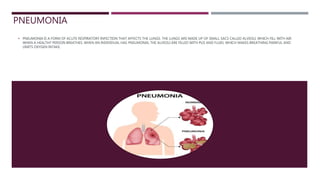
- 1. PNEUMONIA PNEUMONIA IS A FORM OF ACUTE RESPIRATORY INFECTION THAT AFFECTS THE LUNGS. THE LUNGS ARE MADE UP OF SMALL SACS CALLED ALVEOLI, WHICH FILL WITH AIR WHEN A HEALTHY PERSON BREATHES. WHEN AN INDIVIDUAL HAS PNEUMONIA, THE ALVEOLI ARE FILLED WITH PUS AND FLUID, WHICH MAKES BREATHING PAINFUL AND LIMITS OXYGEN INTAKE.
- 2. Definition: Pathological: •Inflammation of lung parenchyma distal to the terminal bronchioles, which includes the respiratory bronchioles, alveolar ducts, and alveoli. Clinical: •Lower respiratory tract symptoms AND... •{Focal chest signs (e.g. crackles) plus systemic signs (e.g. fever)} OR {unexplained CXR shadowing}. Chest infection: •A non-specific term for lower respiratory tract infections, though tends to be used for milder presentations – i.e. acute bronchitis – rather than pneumonia. •Acute bronchitis may or may not feature productive cough and fever, but has no focal chest signs and CXR is clear. Unlike pneumonia, antibiotics are not routinely indicated and only provide minimal symptomatic relief.
- 3. Causes Pneumonia is caused by a number of infectious agents, including viruses, bacteria and fungi. The most common are : •Streptococcus pneumoniae – the most common cause of bacterial pneumonia in children; •Haemophilus influenzae type b (Hib) – the second most common cause of bacterial pneumonia; •respiratory syncytial virus is the most common viral cause of pneumonia; •in infants infected with HIV, Pneumocystis jiroveci is one of the most common causes of pneumonia, responsible for at least one quarter of all pneumonia deaths in HIV-infected infants.
- 4. Types: Community-acquired pneumonia (CAP) Causes (% of all cases): •Common bacteria: Strep. pneumoniae (50%) and H. influenzae (7%). •Other bacteria: Staph. aureus (2%), Moraxella catarrhalis (2%), and the 'atypicals', Chlamydophila pneumoniae (10%), Mycoplasma pneumoniae (5-15%, depending if epidemic), and Legionella pneumophila (3%). •Viral: influenza A+B, RSV. •Pathogens in the immunosuppressed: Pneumocystis jirovecii (causing Pneumocystis pneumonia, PCP), CMV. Hospital-acquired pneumonia (HAP) •Develops >48h post-admission. •Pathogens are similar to CAP in the first 4 days. •Later, Gram -ve enterobacteria (Klebsiella, E. coli), Staph. aureus (inc. MRSA), Legionella, and Pseudomonas become increasingly common.
- 5. Aspiration pneumonia •Causes: neuromuscular problem (stroke, myasthenia gravis), ↓consciousness (anesthesia, alcohol intoxication). •Pathogens: similar to HAP, including Klebsiella in alcoholism, and anaerobes like Peptostreptococcus, Fusobacterium, and Prevotella. •Commonly affects right lower lobe. Atypical pneumonia •Broadly speaking, this refers to any pneumonia not caused by the 'typical' pathogens Strep. pneumo, H. influenzae, or Moraxella. Sometimes it more specifically refers to pneumonia caused by bacteria which cannot be easily identified by Gram stain, namely Mycoplasma, Chlamydophila, and Legionella. •Also known as walking pneumonia, as patients are often fairly well. In practice, this mild clinical picture is seen in Mycoplasma and Chlamydophila, the two commonest causes, while Legionella can cause severe disease. •Commonly results from close person to person contact, which is the case for Mycoplasma and Chlamydophila. May occur in outbreaks in schools, universities, or workplaces. Mycoplasma tends to occur in 4-yearly epidemics. •Legionella occurs in location-specific outbreaks linked to stagnant water e.g. in air-conditioning systems. Types:
- 6. Comparison of Normal lungs, Bacterial pneumonia and Viral pneumonia.
- 7. Signs and symptoms: Symptoms: •SOB •Cough with purulent sputum, possibly blood stained. •Pleuritic pain. •Systemic symptoms: fever, malaise. May be the only symptoms in the old or young. Signs: •Cyanosis, confusion. •↑RR, ↑HR, AF. •Consolidation leads to dull percussion, ↑vocal resonance (VR), and bronchial breathing (i.e. ↑breath sounds, BS). Both pneumonia and effusion cause dull percussion, but pneumonia is noisy (↑VR, ↑BS) while effusion goes shhh (↓VR, ↓BS).
- 8. Pathogen-specific features: •Strep. pneumo: lobar pneumonia, rusty sputum. Oral herpes may be present, though this is uncommon (10%) and non- specific. •H. influenzae and Moraxella: more common in COPD patients. •Staph. aureus: may be bilateral, and can occur post-influenza. •Mycoplasma pneumo: persistent dry cough, mild fever, malaise, headache, and myalgia. Usually self-resolves over weeks. Peaks in 4-yearly epidemics. Immunological complications include erythema multiforme and haemolytic anaemia. •Chlamydophila pneumo: gradual onset, initial pharyngitis/URTI symptoms, and headache. Usually self-resolves, but may take months. •Legionella: flu-like prodrome followed by cough (dry then productive or bloody), SOB, and diarrhoea and vomiting. Bilateral in severe cases. Pontiac fever is Legionella infection without lung involvement. •PCP: dry cough, bilateral pneumonia, desaturation on exertion. •Klebsiella: currant jelly sputum. •Coxiella burnetii: acquired from sheep and other farm animals. Part of Q fever, which includes flu-like symptoms, hepatitis, and endocarditis. •Chlamydophila psittaci: acquired from birds. Causes fever, dry cough, headache, splenomegaly, and arthritis.
- 9. Investigations: CURB-65 to score severity. 1 for each of: •Confusion: abbreviated mental test ≤8 or disoriented. •Urea >7. In the community, this is optional so use CRB-65 instead. •RR ≥30. •BP: SBP •Age ≥65. •0-1 is mild, 2 may require hospitalization, and ≥3 is severe and may required ITU. Community tests: •~70% of patients have mild CAP and are managed in the community. They require few, if any, investigations beyond basic obs. •If the diagnosis is unclear, get a CRP. Diagnose pneumonia and give antibiotics if CRP >100 mg/L. Consider diagnosis if CRP 20-100.
- 10. Inpatient tests: •CXR. Also arrange follow up CXR 6 weeks later to check if clear and rule out underlying lung disease. •Bloods: FBC, U+E, LFT, CRP, and blood culture. •Sputum culture. •O2 sats. ABG if <92% or severely unwell. Pathogen-specific investigations and results: •↑↑CRP suggests Strep. pneumo. •Strep. pneumo and Legionella urine antigen tests. 75% sensitive. •High-res CT for PCP may show ground glass opacities. •Legionella: ↑urea and ↑creatinine, ↓Na+ due to SIADH, ↑LFTs. While these changes are commoner in Legionella, they are not that specific. •Mycoplasma: ↓Na+, +ve cold agglutinin test. Investigations:
- 11. Management: Antibiotics for CAP: •Mild-moderate: amoxicillin PO. Clarithromycin or doxycycline if allergic. Clarithromycin if atypical suspected (monotherapy if mild, added to amoxicillin if moderate). •Severe: co-amoxiclav, cefuroxime, or cefotaxime IV (or levofloxacin if allergic), plus clarithromycin IV. •If hospitalized, start within 4 hours. Monitor response to treatment with CRP. Antibiotics in other situations: •HAP empirical treatment: piperacillin/tazobactam, 3rd generation cephalosporin, meropenem, or levofloxacin IV. Co-amoxiclav is a PO alternative or stepdown. Add vancomycin or teicoplanin or linezolid if MRSA suspected. •Aspiration pneumonia: clindamycin, levofloxacin, or piperacillin/tazobactam. •PCP: co-trimoxazole. Add steroids if moderate or severe: prednisolone PO or hydrocortisone IV.
- 12. Antibiotic duration: •5 days total usually sufficient. •Longer if remains febrile or unstable, or for certain pathogens e.g. Pseudomonas. •If starting IV, review after 48 hours for possible PO stepdown. Supportive care: •O2 •Fluids •Paracetamol Management:
- 13. Complications and prognosis: Recovery: •Fever should resolve within 1 week. •Cough and SOB may take up to 6 weeks to resolve, and fatigue may persist up to 3 months. Complications: •Respiratory failure. •Sepsis and septic shock. •Uncomplicated parapneumonic pleural effusion, empyema (purulent, colonized pleural effusion), or lung abscess. •Death: 1% in the community, 10% if admitted. All this information was collected from our application Medicos PDF which you can find us on android and apple s You can also visit our website : medicospdf.com Please keep supporting our medicos team. Thank you.