SCIENCE TRANS MED Therapeutic targeting of the MYC signal by inhibition of hi...
AY aha poster Final
1. What is the Best Age for Mice to Have Myocardial Infarction:
Modulating Matrix Metalloproteinase-9 to Answer the Question
Andriy Yabluchanskiy1, Yonggang Ma1, Dustin R. Bratton1,, Ying Ann Chiao2,
Andrew P. Voorhees1,3, Hai-Chao Han1,3, Yu-Fang Jin1,4, and Merry L. Lindsey1,5
1 San Antonio Cardiovascular Proteomics Center, Mississippi Center for Heart Research, UMMC, 2Department of Pathology, UW, 3Department of
Mechanical Engineering, UTSA, 4Department of Electrical and Computer Engineering, UTSA, and 5Research Service, G.V. (Sonny) Montgomery Veterans
Affairs Medical Center
INTRODUCTION
• Matrix metalloproteinase (MMP)-9 increases in the aging left
ventricle (LV)
• MMP-9 deletion in young mice attenuates LV remodeling and
improves cardiac function post-myocardial infarction (MI)
Inputs:
• C57BL/6J (WT, n=93) and
MMP-9 Null (Null, n=95) mice,
males and females, 11-36 month
old
• WT (n=12) and Null (n=11) mice,
males and females, 3-6 month old
(young mice, reference control)
• Left anterior descending coronary
artery ligation
Output measurements:
• Plasma MMP-9 protein levels
• Infarct area and survival rate
• LV function by echocardiography
• RT quantitative PCR: infarcted
LV and isolated macrophages
• Immunohistology: MAC-3
staining
200 μm
WT Null WT Null
WT Null WT Null
This study is supported by NIH/NHLBI SC2 HL101430,
HL095852, HHSN 268201000036C (N01-HV-00244),
R01HL075360,NIH HL051971, GM104357, and
5I01BX000505.
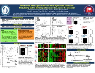
MMP-9 deletion improves LV remodeling post-MI in aged mice
Figure 1. A). MMP-9 plasma
levels at day 7 post-MI increased
with age (n=45). B). Infarct area
was similar between WT and
Null groups, confirming similar
initiating ischemic insult. C). Null
mice showed improved survival.
Infarct area (%)
WT Null
HYPOTHESIS
METHODS
RESULTS
CONCLUSION
Percent survival
Days
C
Null
75/85
WT
45/59
% stained area
p=0.041
%change from baseline
WT
ESV
r=0.38
p=0.01
Age (months) Age (months)
0 10 20 30
Figure 2. WT mice showed a linear increases in the extent of post-
MI LV dilation with age, as evidenced by increases in change of end
systolic volume (ESV; n=42), and a progressive decrease in the
ejection fraction (EF; upper panel). MMP-9 deletion abolished the
age relationship (n=75; lower panel).
WT Null
WT Null
WT
Null
Aging + MI
↑MMP-9
LV dysfunction
MMP-9
deletion
M2 macrophage
polarization
↑Inflammation
0 Age (months) 36 0 Age (months) 36
Ccl1
Ccl6
Ccl9
Ccr1
IL11
IL1r2
IL8rb
Mif
Pf4
Figure 3. WT mice showed linear age-dependent increases in 3
pro-inflammatory genes (red), while Null mice showed increases in
3 pro-inflammatory genes (red) and 7 anti-inflammatory genes
(blue). Sample sizes are n=22 for WT and n=35 for Null, all p<0.05.
Macrophages
p=0.12
WT Null
Figure 4. WT and Null
mice showed similar
numbers of
macrophages in the LV
infarct (p=0.12).
2-ΔCt
IL-1β (M1)
p=0.11
2-ΔCt
TNF-α (M1)
p=0.56
2-ΔCt
CD206 (M2)
p=0.04
2-ΔCt
TGF-β (M2)
p=0.01
Figure 5. MMP-9 deletion did not affect the expression of
M1 markers in isolated macrophages from the LV infarcts at
day 7 post-MI (upper panel, red), but promoted the M2
polarization (lower panel, blue). n=12 per group.
0 10 20 30 40
%change from baseline
Age (months)
Null
ESV
r=0.08
p=0.47
0 10 20 30
Age (months)
WT
Plasma
r=0.46
p<0.001
0 10 20 30 40
MMP-9 (ng/mL)
C3
Ccl4
Cx3cl1
Ccl5
A
p=0.33
* *
200 μm
ACKNOWLEDGEMENTS
B
Age (months)
%change from baseline
0 10 20 30 40
%change from baseline
WT
EF
r=-0.35
p=0.02
Null
EF
r=0.01
p=0.91
n=45 n=75