
Recommended
More Related Content
What's hot
What's hot (20)
Similar to History and Types of X-ray Tubes
Similar to History and Types of X-ray Tubes (20)
Recently uploaded
Recently uploaded (20)
History and Types of X-ray Tubes
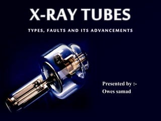
- 1. Presented by :- Owes samad
- 3. INTRODUCTION : . X-RAY TUBE:-a device which is meant for the production of x-rays. An X-ray tube is a vacuum tube. Contains a pair of electrodes i.e. a Cathode and an Anode. Cathode is a filament that releases electrons when high voltage is applied. Anode is made up of tungsten, which attracts the electrons. When the electrons released from the cathode come in contact with the tungsten , they release energy in the form of photons. These highly energized photons are channeled through a lead cylinder and series of filters creating X-rays.
- 4. X-rays were discovered by W.C. Roentgen on Friday 8th November 1895 in Germany. At the time of discovery he was experimenting about cathode rays when he passed current through crooks type tube covered with card board shield to stop any light arising from the fluorescence of glass walls in a dark room, he was surprised to note small glow coming from near placed sheet of paper painted with barium platinocynaide. He confirmed these emanation of rays coming from the tube and named them x –rays. 4 History
- 5. X-ray tube is a device based on the principle of energy conversion. When the fast moving electron comes in the vicinity of the heavy nucleus, it gets attracted towards the positively charged nucleus due to columbic force and gets deviated from its original path ,In this process kinetic energy of fast moving electron is lost and converted into heat(99%) and X-ray energy (1%). 5 Principle
- 8. In 1895: X-rays were discovered from experimental discharge tube called as Crooks tube or Cold cathode tube. In 1913 : Crooks tube was improved by William Coolidge c/as Coolidge tube or Hot cathode tube. In 1915: Hooded anode tube. In 1920: Oil cooled tube. In 1929: Rotating anode tube. In 1932: Grid controlled stationary anode tube. In 1937: Grid controlled rotating anode tube. In 1959: High speed tube. 8 History Of The Developments Of The X-ray Tubes
- 9. In 1962: Rhenium alloyed tungsten composite anode tube. In 1967: Mammography unit with Mb anode. In 1971 : Glass metal tube with Mb anode. In 1973: Three layer anode (W-Re)+ Mb ,or (W-Re)+W+(W-Zr- ………… Mb). In 1979: Metal ceramic tube. In 1981 : Three focus tube. In 1989: Direct anode cooling with noiseless rotor. In 1989: Introduction of helical CT tube ( by Siemens). In 1990: MRC tubes ( Maximus rotalix ceramic) by Philips. Cont.. 9
- 10. In 1998: Development of grid control rotating …….... anode for angiography by Toshiba. In 1999: Double focus stationary anode x-ray ……….. .tube for mobile c-arm. In 2000: Food inspection x-ray tube..................................... In 2002:High speed rotation bearing rotating ………. .anode tube for angiography..... In 2006: there is introduction of STATRON x-ray tube by Siemens. In 2003: Micro-focus X-ray tube …………… with .power supply used for …………………………………………….inspection technology . cont..
- 11. GAS TUBE (ION TUBE) OR CROOKES TUBE Historically, X-rays were discovered from experimental discharge tubes called Crookes tube. Invented by British Physicist Sir William Crookes. Also called as Cold cathode tube or Discharge tube. Early X-ray tube called as Gas tube because; its action depends upon the presence of small residual amount of gas present in it and the radiographer kept a selection of tubes of various mAs values on a rack, usually in the darkroom. When the change of mAs was required the radiographer disconnected the tube and replaced it with a different one.
- 12. Crookes X-ray tube consists of: Cathode is on the right the anode is in the center with attached ‘heat sink’ at left. The device at top is a Softener used to regulate the gas pressure. Glass bulb with around 10-6 to (5 x 10⁻⁸) atm.pressure An Al. cathode plate having curve shape to created the beam of electrons. A platinum anode target – (generate X-rays).
- 13. During operation, the residual gas in the tube would be adsorbed by the glass and the metal electrodes leading to a reduction in the x-ray output. The side tube contained a few mica sheets which would release small amount of gas when heated by an electric heater wire. A high voltage was made between the electrodes; this induces an ionization of the residual air, and thus an electron flow or "discharge" from the cathode to the anode. When these electrons hit the target, they are slowed down, producing the X-rays (Bremsstrahlung). 13 How X-rays Produced In Crookes Tube
- 14. Relatively low intensity of x-ray (not more than 5mA) was unreliable and unstable as x-ray production depended upon the gas content which was a very variable factor. This tube can not produce X-rays continuously. We can not operate the kvp and the mAs independently as there is presence of gases. 14 Limitations:
- 15. The Coolidge tube was improved by William Coolidge in 1913. The Coolidge tube, also called hot cathode tube, is the widely used. It works with a very good quality vacuum (about 10-4 Pa, or 10-6 Torr). In the coolidge tube electrons are produced by Thermionic effect i,e (on heating metal element emit electrons). 15 The Coolidge Tube (Hot Cathode X-Ray Tube)
- 16. The filament is the cathode of the tube , which is negatively charge. When high voltage potential is applied between the cathode and the anode, the electrons are thus accelerated towards positively charge anode in completely evacuated glass tube. Resulted into production of X-rays after striking the target. Coolidge tube is the prototype for modern X-ray tubes being used today. 16 Cont..
- 18. Coolidge Tube Modified Coolidge Tube With radiator ANODE ASSEMBLY RADIATOR CATHODE ASSEMBLY CATHODE ASSEMBLY X-RAY EXIT PORT 18
- 19. A source of electrons; Cathode assembly i.e.(filament and focusing cup.) A means to high voltage across anode and cathode. A means to slow down the electrons Anode assembly with target (Tungsten). Glass envelope (Borosilicate) to enclose two electrodes with vacuum level – 10-7 to 10-8 mm of Hg. 19 Basic Features Of An X-ray Tube Cathode assembly anode target Cathode& anode insulator Rotor, stator coil Focusing cup and filament Mo stem With few technical modification modern x-ray tubes are similar to coolidge tube
- 20. X-Ray Tube Components Housing Visible part of tube Glass Enclosure (insert) Vacuum Electrodes • Cathode Filament • Anode Target *
- 21. . 21 Diagram of X-ray tube:
- 22. Shields against leakage radiation It is lead lined. leakage limit:-100 mR / hour when tube operated at maximum continuous current for its maximum rated kilovoltage. Shields against high voltage • electrically grounded. • high voltage cable receptacles (wells) Housing filled with oil • Cools, • electrical insulation, • bellows on the end of tube allows oil to expand when hot. 22 TUBE HOUSING …
- 23. 23 Component of a Tube Unit: The Housing
- 24. The electrons of the X-ray tube must be sealed into an Glass enclosure. Since, it is necessary for them to operate in vacuum. It serves to support the electrons and maintain the vacuum. Special alloys having same coefficient of linear expansion as that of Pyrex glass are used. It has following features; It can withstand heat and mechanical stress. It is an electrical insulator capable to withstand high voltage. It should be capable of being sealed to the electrodes with a vacuum-tight, heat proof seal. 24 Glass Enclosure And Its Contents
- 25. It is the negative electrode of the X-ray tube. It consist of a metal structure to support the filament. It consists of a) Filament –source of electron. b) Connecting wires which supply the voltage (about 10 V) and current(3 to 5 A) c) Metallic focusing cup.. 25 Cathode
- 26. FILAMENT: Cathode filament made of thin tungsten wire which is a source of electrons . It works on the phenomenon of Thermionic emission i.e.. Tungsten spiral wire [dia : 0.2mm]. Length :1.0 cm Diameters of spiral : 0.2 cm 26 Filament
- 27. Tungsten is used as filament material because of following Reasons: It has high melting point (33700C). Less tendency to vaporize High tensile strength means it can be drawn into wires. High thermal conductivity and specific heat. Appropriate Threshold or Work- function. . 27
- 28. 28 Cont..
- 29. Used to supply voltage and current to the filament which are attached with the filament heating circuit and also H.T. Transformer. Cathode filament is mounted on two supporting or connecting wire. One wire is connected to low voltage for filament current and another is connected to high voltage current to produce high voltage b/w anode and cathode. Current 3-5 amperes and 10-12 volts are used in filament supply by filament heating transformer . 29 Connecting Wires
- 30. It is the device surrounding the cathode filament in an X-ray tube. This is actually a third electrode in the tube, called a Wehnelt electrode. Because of the forces of the mutual repulsion of large no. of electrons this electrons stream would tend to spread itself out and result in bombardment of large area on the anode of the X-ray tube. This is prevented by a focusing cup which surround the filament. If this electrode is not present, the electrons would hit the anode over a very large area. 30 Focusing Cup
- 31. The focusing is achieved by keeping the focusing cup at the same, or slightly higher, negative potential as that of the cathode filament. It is usually made up of Ni because of: a) Light wt. b) Poor thermal conductivity c) High melting point. cont 31 …
- 32. It is inability to operate the current and voltage of the x-ray tube independently . When filament is heated emission of electrons takes place. In the absence of high voltage these electrons remain around the filament . This cloud repels new electrons from the filament this tendency is known as Space charge effect. Since, Space charge is negatively charged so its presence around filament prevent further emission of electrons. When high voltage is applied electrons moved towards anode therefore, space charge effect is diminished. 32 Space Charge + - - - *
- 33. Most tubes have 2 filaments & thus 2 focal spots, only one used at a time Small focus – improved resolution Large focus – improved heat ratings – Electron beam strikes larger portion of target thus spreading heat produced to a large area 33 Focal Spots
- 34. Filament current is not tube current. It is the rate of electron flow from filament to target.Electrons / second Measured in milliamperes (mA) Limited by • filament emission (temperature / filament current) • space charge. 34 Tube Current (mA)
- 35. Kilovoltage & Space Charge Raising kvp gradually overcomes space charge • Higher fraction of electrons make it to anode as kvp increases • Below 40 kvp the current flowing is limited by space charge effect • Above 40 kvp the space charge has no influence on current flowing in the x ray tube With time, as the tube is used tungsten from hot filament vaporizes on glass insert • thins the filament • filters the x-ray beam • increases possibility of arcing • electrons attracted to glass instead of target. Saturation Voltage kVp
- 36. Positive electrode . It consist of target (focal spot) and cylindrical cu block or tungsten Rhenium disk. Functions: • Electrical conductor • Mechanical support for the Target. • Good thermal radiator. Target: The area of the anode struck by the electrons from the cathode. 36 Anode
- 37. Made up of a small plate of tungsten 2 or 3mm thick that is embedded in a large mass of cu. Square or rectangular in shape with each dimension usually greater than 1cm.Anode angle is usually 15-20 deg. We use TUNGSTEN as a target material because of following reasons: • High atomic no.(74) • High melting point (3370 °C) • High thermal conductivity. • It doesn’t vaporize easily. 37 Cont..
- 38. Target material is based on three characteristics: Atomic number:- must be high so that it results in high efficiency in x-ray production. Thermal conductivity:- must be able to conduct heat away from the target. • High melting point:- must be able to withstand high temperatures. 38 Target Material
- 39. It is the area actually bombarded by the electron stream on the target. It can be larger or smaller in size. A small focal spot is required for producing good radiographic detail but it may also lead to overheating of target. Whereas large focal spot allows greater heat loading but doesn't produce sharp image. This problem was solved in 1918 by the development of line focus principle . 39 Focal Spot
- 40. It states that as the anode angle is made small, the apparent focal spot also becomes small but with increased heat loading. Focal spot steeply slanted. 7-15 degrees typical. Adv: This slope allows x-rays produced at focal spot to leave the tube sideways In such a way that the x- ray beam emerges at right angle to the long axis of the x-ray tube. 40 Line Focus Principle
- 41. It permits large area for electron bombardment and a small x-ray source. more heat loading with good radiographic detail. Sin 6⁰= 0.104 Sin 21⁰=0.358 Target Angle Angle between target & perpendicular to tube axis Typically 7 – 15 degrees 41 Cont.. + Target Angle,Q Apparent FS = Actual FS X sin ѳ
- 42. Large • better heat ratings • better field coverage • Large fs • Reduced image quality Small • optimizes heat ratings • limits field coverage • Small fs • Better resolution 42 Target Angle (cont.) + Large Target Angle (large Actual Focal Spot) + Small Target Angle (small Actual Focal Spot)
- 43. 43 Types Of X-ray Tubes X-ray Tubes Stationary Anode x-ray tubes Rotating Anode x-ray tubes.
- 44. Advantages Compact Unit Less cost Limitations Since area covered by electrons beam on the target i.e. x-ray source and the area over which heat is spread are the same so we cannot use higher electrical loads or high mAs Applications Dental x-ray sets, small portable and mobile x-ray units with limited output Stationary Anode X-ray Tube 44
- 45. 1. A source of electrons: Cathode assembly i.e.(filament and focusing cup.) 2. A means to accelerate the electrons to high velocity voltage across anode and cathode. 3. A means to slow down the electrons Anode assembly with target (Tungsten). 4. Glass envelope (Borosilicate) to enclose two electrodes with vacuum level – with pressure of 10-7 to 10-8 mm of Hg. 45 Components Of A Stationary Anode X-ray Tubes
- 46. 46 Dental x-ray tube that showing stationary anode Fig : A fixed anode X-ray tube
- 47. Tungsten Target Plate Copper Block A Stationary Anode The above photo shows the tungsten (W) target plate mounted on its copper block.
- 48. Area of electrons bombardment should be larger enough to accommodate large amount of heat. Area of x-ray production should be as small as possible to get sharp images and good radiographic detail. Cylindrical cu-block • The small tungsten target is bounded to larger cu-block to facilitate heat dissipation. The cu prevent excessive rise in temp, because of high thermal conductivity. 48 Two important consideration which govern target design are:
- 49. Tube Housing :It is the lead lined aluminum alloy casing in which x-ray insert in contained. The metal case provides protection against 1. Radiation risk 2. Electric risk 3. Serves as a mechanical support to x-ray tube. Cont: 49
- 50. Construction of H.T. cable is: 1. Electrical conductor :- Inner most are electrical conductors which supply current to X-ray tube. 2. Insulating layer :- It is made up of special rubber which is put around the Cu core in a thick layer. 3. Metallic Sheet:- Around the rubber there is a flexible metallic sheath braided together. This metal sheath is connected to the earth and constitutes and the earthed metal screen. 4. Plastic Sheath :- Metal sheath is further covered with plastic sheath. It is designed to protect the flexible metal braiding from damage.. 50 High Tension Cables
- 52. Process by which heat is dissipated 1. Conduction 2. Convection.& 3. Radiation 52 Methods Of Heat Dissipation In Modern X-ray Tube
- 53. The first step is heat removed from the target area, passing most of the heat, by conduction into cu block of anode. The cu block conducts heat along its length and thus to out side of the tube. The target also conveys a small part of its heat by radiation across the x-ray tube to glass envelope. Through the anode and glass envelop the heat thus reaches the oil and heat is conveyed to metal casing by convection process. It is then conducted through the casing to its outer surface, which is dissipated in the room by radiation process. Cont.. 53
- 54. Fenestration: In this anode is connected to a large metal of considerable heat capacity . It is capable of absorbing many calories & becomes hot itself & radiate heat to the surroundings. USE: in fluoroscopy tube which is cooled by outside air. Water cooling:Used in therapy tubes where anode is cooled by oil which is cooled by water. Oil cooling: Used in both therapy and diagnostic tubes. 54 Methods of Heat Dissipation Fan cooling: used for heat dissipation from anode by means of fans used in low & medium therapy tubes.
- 55. Limitations of stationary anode tube were overcome by rotating anode tube which were introduced in 1936. Based on the principle of removal of target from the selection beam before it reaches too, high a temperature by replacing it by another cooler target I.e. the target from the face of the rotating disc or the end of rotating cylinder. 55 Rotating Anode X-rayTubes
- 56. The anode disc is between 55mm and 100mm in diameter and 7 mm thick machined to high tolerance to prevent in balance and wobble. Anode disc is made of Tungsten has its periphery beveled at an angle 10⁰-20°. The disc has a tungsten rhenium target area as tungsten has a high melting point 3370 ⁰C and atomic number of 74. The addition of a small quantity (5-10%) of rhenium prevents crazing of the anode surface, the tungsten is faced onto a molybdenum disc as molybdenum whilst not having as high a melting point as tungsten has twice the heat capacity 56 Main Features of Rotating Anode
- 57. The target disc is mounted on a molybdenum stem attached to the copper rotor. Rotor rotates with speed of 3000 RPM on ball bearings (made of steel) with dry lubricant( silver or lead coating). The target is cooled by radiation to the glass then oil. 57 Cont..
- 58. Rotating anode x-ray tube: The lead shield cut open 58
- 59. The anode disc needs to be rotate at high speed and this is achieved by attaching the stem to a large copper rotor, which forms the armature of a motor. The target disc, with rotor, is mounted on a shaft extending from a rotor body, which can spin on internal bearings on the rotor shaft. This rotor shaft extends through the end of the insert to the outside of the insert vacuum for connection to the anode wire, and also is the mounting point for the insert inside the housing. The rotor bearing are special as they need to operate in a vacuum, conduct a high voltage and reach high temperatures (500oC). Using steel bearings lubricated by silver powder solves the problem. 59 The Rotor and Bearing System
- 60. 60 Component of the insert: the rotor
- 61. The anode stator motor must accelerate the anode to working speed rapidly, to ready it for an x-ray exposure and then must bring it back to stop equally rapidly to prevent wear and wobble on slowing down. The “motor” consists of electromagnet coils round the glass to provide a rotating magnetic field to be induced by the currents and produce the forces needed to rotate the copper rotor 61 The Anode Stator Motor Stator coil Xray Exit port Electric supply
- 62. Advantages It permits selection of higher electrical load (exposure factor without risk of overheating) Applications Almost universal use in radiography Advances Reduced target angle New anode materials Increased speed of anode rotation Grid Controlled x-ray tube Metal / ceramic x-ray tube 62 cont..
- 63. The information in details of the electrical loads which may be applied to the X- Ray Tube without damaging it & the safe duration of such loads. It is usually provided by the manufacturer The amount of heat that can safely be used in any given tube is of course determined by the physical characteristics of the tube itself. These characteristic would include: Focal area Anode diameter Material of which anode is made Speed of anode rotation Target angle. Heat dissipations i.e volume of oil within the casing. 63 HEAT RATING: indicates load limits that a tube can safely accept
- 64. Ability of X-Ray tube to withstand a single exposure. Ability of X-Ray tube to function despite rapid exposure. Ability of X-ray tube to withstand multiple exposure during several hours of heavy use. Three types of x-ray tube rating charts are particularly important: 1.Radiographic rating charts 2.Thermal rating i.e. Anode cooling chart. 3.Angiogrphic rating chart 64 In considering the tube rating three characteristics are encountered:
- 65. Radiographic rating chart is most important because it states which radiographic technical factor are safe & which are unsafe for tube operation. Each chart contain a family of curves representing the various tube voltage in KVp. The x axis & Y axis show scale of two radiographic parameter- time & mA .For a given KVp any combination of mA & time that lies below KVp curve is safe. Any combination of mA & time that lies above the curve is unsafe. If unsafe exposure was made the tube might fail abruptly. 65 1. Radiographic rating chart
- 66. 66
- 67. Thermal rating i.e. Anode cooling chart provides information on how rapidly and how many times a single exposure can be repeated without putting too much heat into the x-ray tube. 67 2. Thermal rating Chart
- 68. Heat units HU. It is an arbitrary unit that is used to plot heating and cooling curve charts. For a single phase unit, HU = kVp x mA x s For a 3 phase 6 pulse unit, HU = 1.35 x kVp x mA x s For a 3 phase 12 pulse unit, HU = 1.41 x kVp x mA x s For a high frequency unit, HU = 1.44 x kVp x mA x s 68 Calculating Heat Units
- 69. It tells us of the total no. of exposures which can be made at various exposure rates per sec. Fluoroscopic rating chart. During fluoroscopy the heat produced is relatively small and is maintained over a relatively long period of time. It is defined as the no. of HU /sec that can be applied indefinitely with continuous running 69 3.Angiogrphic rating chart
- 70. 70 . Application Heat storage capacity Target angle Focal spot size 1.Nueroradiography 0.3 MH.U. 70 0.3mm 2.Angiography 0.4 - 1.35 MHU 12o 0.3,0.6,1.2mm 3. Mammography 0.3 MHU 100 0.6mm 4. General radiography 0.3 - 0.6 MHU 120 0.3,0.6,1.2mm 5. CT 3.5 - 7.6 MHU 120 0.6mm
- 71. The intensity of the x ray beam as it leaves the x ray tube is not uniform throughout all portions of the beam. The intensity of the beam depends on the angle at which the x rays are emitted from the focal spot . The intensity of the film exposure on the anode side of the x ray tube is less than that on the cathode side of the tube. 71 Heel effect
- 72. The decreased intensity of the x ray beam that is emitted more nearly parallel to the surface of the angled target is caused by the absorption of some of the x ray photons by the target itself. 72 Cont:
- 73. Intensity of x- rays on anode side of x-ray tube is significantly less than that on cathode side. This factor is useful in obtaining balanced densities in radiographs of body parts of different thickness. Heel effect is less noticeable when longer focus film distances are used. For equal FFD heel effect will be less for smaller films. 73 Three Clinically Important Aspects Of Heel Effect:
- 74. 74 Common faults in x-ray tube Any type of problem that deteriorate or block the output of x-rays from the x- ray unit is termed as common faults of x-ray unit…….. Faults related with the TARGET Faults related with the GLASS ENVELOPE Faults related with ROTOR STATOR ASSEMBLY Faults related with CATHODE Faults related with HOUSING SYSTEM
- 75. Fault: melted spots on anode/crazy paving of anode/ erosion of target track Cause :-due to over use specially (heat capacity exceeded).Incase of the stationary anode x-ray tube Effect :- increased heel effect ,heterogeneous x-ray output. Remedy :-use of rhenium that increase the target life by providing mechanical stability due to pitting/operate the tube in safe rating zone. 75 Common faults related with target
- 76. Fault:-no exposure/thermal shock Cause: heavy radiography exposure made on a cold target can cause (high mA on cold anode) Effect: anode disc splits radically./ can cause cracks in anode (tube death)/noisy rotation Remedy: do warm up exposure. Or use of the stress relieved anodes. As described 76 Cont:
- 77. Eliminates thermal shock from high mA exposures on cold anode. Warm-up needed whenever tube cold. Once in the morning not sufficient if tube not used for several hours. 77 Preventions
- 78. Indication : No exposure Reason Due to evaporation of filament Due to continuous exposure the filament become thinner and thinner an finally breaks. Remedy Replacement of tube insert Filament boosting time should be kept as short as possible. 78 Faults In Filament
- 79. Fault:-Formation of mirror due to heat, vaporized tungsten is deposited as a thin layer on inside of glass envelope, producing mirror like reflecting surface./ arcing of the glass. Causes – can be caused by filament evaporation deposition of filament on glass envelope as result of • high filament currents • long filament boost time Effect :-results off focus radiation. /electrons move from filament to tube housing instead of to anode • Remedy :-reduce by not holding first trigger longer than needed/use of metal ceramic tube. 79 Faults in glass envelope
- 80. Fault:-cracking of glass tube Causes – mishandling of x-ray unit specially in the case of mobile x-ray tube Effect : increased leakage radiation dose. Remedy :-Use of metal ceramic tube . Cont.. 80
- 81. electrons move from filament to tube housing instead of to anode very short exposure with instantaneously very high mA Generator often drops off line 81 High Voltage Arcs + arcing
- 82. prevents proper rotation of anode • anode can run too slow • anode can stop results in thermal damage to anode (melted spots) Filament break renders one focal spot completely inoperative 82 Tube Insert Damage
- 83. May be accompanied by air bubble in housing. Eventually causes high voltage arcing. Requires immediate service attention. 83 Oil Leaks
- 84. How To Maximize X-ray Tube Life: Minimize filament boost time. Use lower tube current (mA). Follow Tube rating chart. Do not exceed anode thermal capacity or dissipation rate of the target. Do not make high mA exposure on a cold target. Anode should not be run unnecessarily.
- 85. Tube Warm-up Procedures By warming the anode through a series of exposures and increasing kvp settings, the anode will build up heat that is needed to avoid fracture of the anode. This process takes a little over one minute put will add to the life of the tube. Close shutters of collimator.
- 86. 86 Advancement in rotating x-ray tube Advancement in target angle Advancement in target material Advancement in target diameter Advancement in speed of target Advancement with target Grid controlled x-ray tube Metal ceramic x-ray tube Stereoscopic x-ray tube Other advancement
- 87. Main feature of rotating X-ray tube: The shape of the glass envelope has been modified to accommodate the different types of the electrode and the rotating assembly. Dry lubricants are used for rotation like silver , lead Rotation of anode is achieve by the process of electro-magnetic induction in rotor and stator assembly. Cathode cup and filament are offset the target track near the periphery of the beveled anode track( angled at 6*-20*) Disc is connected to anode stem made up of Mb. 87 Advancements in rotating x-ray tube
- 88. Reduction In target angle:-as the cooling capacity is increases so now a days we can go on reduced target angle to get sharp image with small target by minimizes the ordertoobtainsharpimageas small focal spot decreases the geometric unsharpness. Now adayswehaveswitchingto6 degree from20degree.. 88 Advancement in target of x-ray tube
- 89. 89 Biangular Tubes The anode of a biangular tube has two focal tracks (Inner for fine focus) and outer for broad focus with cathode having two filaments arranged one above the other The surface of the anode disc is beveled at two angles Thus fine focus used for radiographic exam requiring more details, where as large for routine radiography.
- 90. Earlier it was tungsten alone . Rhenium 10% + tungsten 90% .(less roughening . Higher tube life) (Rhenium + tungsten ) over molybdenum base . (less weight . Inertia.) Rhenium + Tungsten (+titanium + Zirconium + molybdenum). (Rhenium + tungsten ) over molybdenum base . + graphite as backing layer (Rhenium + tungsten) + CVD Graphite base. Advantage • Higher heat storage capacity permitting higher tube rating with prolonged tube life 90 New Anode Materials
- 91. Gallium liquid alloy used as liquid within spiral grooves. Advantages Allow use of high mA and shorter exposure time. Allow possible use of smaller focal spot. Disadvantages Initial high cost of equipment Longer prepare time Greater wear and tear bearings Require breaking system 91 Spiral Grooves Bearing
- 92. The method is applied to those anodes having slits and grooves. Where the rear surface is blackened which results in reduction of anode temp. A blackened anode disc increases the fluoroscopic output of a tube due to the reduction of anode heating. At normal speed the anode rotates with the speed 3000 RPM whereas at high speed 9000-10,000 RPM with 3- phase supply. At high speed quicker rate of heat dissipation will be there allowing greater input load (higher rating capacity) but at short exposure. 92 Blackening of anode disc Increased Speed Of Anode Rotation
- 93. A more recent development in X-ray tube construction is the metal- ceramic tube, which is made from a steel (ferrochrome ) cylinder brazed to alumina ceramic (aluminum oxide )insulators at each end. These insulators carry the anode and cathode assemblies. The metal-ceramic tubes are smaller and more robust than their glass equivalents. They have another advantage, in that they enable more flexibility in the electrical circuitry associated with the tube. Offers greater heat dissipation results less load to the x-ray tube. Metal envelope grounded offers no chance of arcing of x-ray tube. Anode rotates on a axle with bearing at each end providing greater stability and reduced stress on shaft this permits use of massive anode. 93 Metal-ceramic tubes are now being used in X-ray equipment
- 94. Offer greater heat dissipation results less load to the x-ray tube . • Metal envelope grounded offers no chance of arcing of x-ray tube 94 Features of Metal ceramic X-ray tube
- 95. Higher Tube Loading Allows higher tube currents to be used because of larger heat storage capacity of anode Longer Tube Life Deposition of tungsten on the glass wall acts as electrode causing arcing bet. Glass and filament shortening tube life. When metal enclosure is grounded, this deposition will not alter grounding thus increasing its life Reduced off Focus Radiation Electrons back scattered from the anode may strike anode again producing x-rays from areas other than focal spot. The metal enclosure decreases off focus radiation by attracting off focus electrons to the grounded metal wall relatively Positive as compared to electrons. Low atomic no. of metal may produce few and low energy x-rays. 95 Advantages Of Metalceramic Tube
- 96. A third electrode called grid is used. Focusing cup surrounding the filament cup is used as third electrode to control the flow of electron. A negative bias voltage around 1500 V is applied to the cup relative to the filament to punch off the flow of electron. Thus focusing cup acts as exposure switch to turn the current ON or OFF when required. Application in capacitor discharged mobile radiographic equipment , pulsed-fluoroscopy and cardio-angiography and vascular angiography. 96 Grid Controlled X-ray Tube
- 97. Depending Upon Applications Mammography tube CT X-ray tube X-ray tube for angiography Radiotherapy X-ray tube Stereographic X-ray tube Field emission X-ray Tube 97 Different Type Of X-ray Tubes
- 98. Formaximum visualization of soft tissues of the breast having similar ability to absorb x-rays a beam of soft radiation (longer wavelength ) is required Longer wavelength can be produced by selecting x- ray tube which operate at low KVp (20-40) 98 Mammography X-ray Tube
- 99. Use of target made of molybdenum. Closer spacing of cathode and anode. Beryllium window : is used as it has low atomic no.(4) & lower absorption of x-rays. Use of molybdenum filter in place of aluminum filter. Focal spot size-0.3-0.35mm. Heat storage capacity- 0.3-0.5 M.H.U . 99 Features of a Mammography Tube
- 100. Introduction of the dual metal x ray tubes (having dual track of molybdenum/vanadium & rhodium).rhodium track & filter produces a slightly higher x ray spectrum for superior penetration of the dense breast tissue in the younger women and in those who have undergone radiation treatment or on hormone therapy. Mo /tungsten dual track with a high emission flat emitter cathode with different k-edge filters mo & rhodium meant for normal and dense breast. But with an increase in x-ray tube voltage from 25 to 30Kv simultaneously replacing Mo with a rhodium filter the x-ray spectrum for a tungsten anode is clearly shifted and higher energy especially advantageous for the radiography of large subject/dense breasts. 100 Latest developments in Mammography Units
- 101. These are oil cooled stationary anode tube. Tube for deep therapy have kvp in the range of 200-300 kv and usually work at 15-20 mA . Single focus tubes size( 6-8 mm) with hooded anode & target angle of about 35 degree. Hood is made up of Copper + Tungsten. To allow emission of required primary x-ray beam a hole is cut in the hood below the target. 101 Radiotherapy X-ray Tube
- 103. 103 Stereographic X-ray Tube Similar to conventional rotating anode x-ray except:- Rotating anode is bombarded simultaneously by two beams of electrons from two independent cathode assemblies. Used for stereographic and stereofluoro scopic x-ray examination. Anode target disk Two Filaments With focusing cup Anode target disk
- 104. FIELD EMISSION X-RAY TUBE • It is also known as cold cathode tubes. • Electrons are extracted from the cathode to anode by an intense electric field rather than by thermionic emission. Features • A conical anode surrounded by a cylindrical cathode containing facing rows of needle (actually more than two). • The x-ray beam passes through a window in the end of the tube with less intensity in the center of the field. • Needle tip diameter about 1μ • Since electrons which can be emitted are less in no. these tubes can be used only for neonatal radiography. • If higher voltage (up to 350 KV) is applied these tubes can be used for high KV chest radiography. But not useful for general radiography purpose. 104
- 105. Field emission x-ray tube Emission of electron takes place due to high potential difference
- 106. Transmission type x ray tube •In these tubes the x-rays are generate when electrons from the filament strike a thin sheet of target material. •The electrons produced at two filaments are focused with the help of deflecting and focusing coils on a thin target which may or may not be a cathode. •This unit was used in prototype for high beam x-ray therapy machines.
- 107. Since CT require longer continuous exposure time at higher KV and mA than needed for general radiography. These have been charged with heavy duty rotating anode tube with higher thermal capacity and smaller focal spot (up to 0.6mm). These tubes are air cooled with current value up to 600mA. It Should supply monochromatic X-ray beam for accurate reconstruction. Earlier model used were oil cooled ,Fixed anode relatively large (2x16mm)focal spot operated at 120 kVp & 30 mA & heavily filtered as those use in radiotherapy. 107 CT X-Ray Tube
- 108. One of the more interesting developments is the Siemens Straton x-ray tube, which is currently available as an option on Sensation 16 scanners (Fig ). The tube itself is a radical new design, where the entire tube body rotates, rather than just the anode, as is the case with conventional designs. This change allows all the bearings to be located outside the evacuated tube, and enables the anode to be cooled more efficiently. The Straton has a low inherent heat capacity of 0.8 MHU, but an extremely fast cooling rate of 5 MHU / min. 108 Developments in CT Tubes
- 109. This compares with typical figures of 7-8 MHU and up to 1.4 MHU / min for existing tubes. The heat capacity and cooling rate combine to produce a tube which Siemens claim is ‘0 MHU', implying that tube cooling considerations are a thing of the past. Sensation 16 scanners fitted with the Straton tube now have a fastest scan time of 0.37 seconds. Cont.. 109
- 110. 110 CT TUBE:
- 112. CHARACTE- RISTIS DIAGNO STIC TUBE THERAPY TUBE MAMOGRA- PHY TUBE CT TUBE 1 Tube Glass tube Glass tube Glass tube Metal ceramic tube 2Type of Anode Stationary or Rotating type Stationary type Rotating type Rotating type 3 Target Angle 6⁰-20° 25°- 35° 100 120 4 Focal spot size .3, 0.6,1.2 mm 6x6 mm 0.6 mm 0.6mm 5 Exposure time Shorter longer shorter longer 6 Heat Unit capacity 0.3-0.6 MHU 0.1-0.3 MHU 0.3-0.6 MHU 1-2 MHU 7 Type of focal spot Dual focus Single focus Dual focus Dual focus 8 Pt. radiation dose less more less more . e
- 113. Nano focus tubes X-ray diffraction tubes Miniature x-ray tubes ISOVOLT mobile Portable x-ray units Tire Inspection Thickness Gauzing Food Inspection 113 New Applications:
- 114. In case of nanofocus tubes, the focal spot is further minimized by means of a special setup consisting of lenses and diaphragms. The user may choose from a range of operating modes, each providing different tube voltages and image resolutions. The most potent operating mode offers detail detectabilities down to 200 nm (0.2 µm). Inspection technology. 114 Nano Focus Tubes
- 115. Radiography (X-ray) is a Nondestructive Testing (NDT) method that examines the volume of a specimen. Radiography (X- ray) uses X-rays and gamma-rays to produce a radiograph of a specimen, showing any changes in thickness, defects (internal and external), and assembly details to ensure optimum quality in your operation 115 Radiography (X-ray)
- 116. MINIATURE X-RAY TUBES UTILIZING CARBON- NANOTUBEBASED COLD CATHODES The electron field-emission properties of carbon nanotubes enable the fabrication of cold cathodes for a variety of vacuum device applications. The utilization of these cathodes is an attractive alternative for the replacement of thermionic or hot cathodes for generating X-rays. Miniature X-ray tubes uses triode-style carbon nano tube based cathodes. Driving gate voltages below 1000 volts for easy pulsing has been achieved, and the extended lifetime data suggests that a regulated power supply would be ideal for a constant AC operation mode. The 1mm focal spot size achieved so far is suitable for most applications. 116 Miniature X-ray Tubes
- 117. The ISOVOLT Mobil has been designed for operations where access to the inspection area is difficult. It is ideal for use in the energy, mineral and petro chemical industries where pipelines and container tanks require X-ray inspection. The ISOVOLT Mobil is equipped with a small X-ray tube and high voltage cable up to 20 m (32 ft.) in length to allow positioning in hard to reach places not accessible by other types of X-ray equipment. 117 ISOVOLT Mobile
- 118. Today special X-ray tubes are used to inspect the tires of trucks, cars, motor bikes & heavy earth movers. These tubes having flat target angles from 0*-20* and conical targets are available. 118 Tire Inspection
- 119. Thickness Gauzing: X-rays are used in rough industrial online processes to measure the thickness of sheets & foils of different sizes and materials. The transmission through the material is precisely measured in the cold and hot milling process. The X-ray tubes are designed for ultra stable emission output with high reliability. Food Inspection: various food articles( e.g. chocolate jars with jams and baby food) are controlled with fan beam x-ray tubes in digital inspection system. High x-ray output and small focal spots ensures that metal parts ,o-rings or broken glass are sorted out before shipment. 119 Cont..
- 120. Portable x-ray unit: Portable X-ray units are designed to be reliable in some of the world’s toughest conditions. Using modern compact electronics to minimize weight and provide a high power output with extremely low ripple, and a sturdy metal ceramic X-ray tube.
- 121. These are used for mobile inspection of heat-exchangers. 121 Special X-ray tubes
- 122. Analytical X-ray : Analytical X-ray is the industry standard for X-ray diffraction techniques. Standard systems are used for material characterization in research & development and for quality assurance in various industries. A variety of machines are designed for special applications. X-ray Diffraction Tubes Waves interact with crystalline structures whose repeat distance is about the same the wavelength. X-rays scattered from a crystalline structure constructively interferes and produces a diffracted beam. 122
- 123. .. These are special type of slim X-ray tubes Used in Nuclear plants to inspect weldings in narrow pipe to pipe space.
- 124. X-ray tube has experienced a rapid development from its beginning till now. The basic principle of X-ray production is same in all the x-ray tubes but some new advancements like new anode material, high speed of anode rotation, target angle, heat load and dissipation capacity etc are the factors which improved the X-ray tube .So we can say that “An x-ray equipment without an x-ray tube is like a human body without heart” All these advancements have proven to obtain a quality of image. 124
- 125. REFRENCES Physics for radiology by christensen. A practical approach to modern imaging equipment by Thompson. X-ray equipment for student radiographer by Muriel o. Chesney. http.//www. E radiography .com http.x ray tube wikipedia.com http. //www.mycopedia.com
- 126. 126 THANKS THANKS THANKS T H A N K S T H A N K S “Small experiments can leads you a big discovery”