Omentectomy in ovarian cancer
•
0 likes•99 views

The document discusses the role of omentectomy in early ovarian cancer surgery based on the gross appearance of the omentum. It notes that a healthy fatty omentum can act as a barrier against cancer spread, while a thin omentum with low fat content is more susceptible to early metastasis. Early signs of microscopic metastasis in a thin omentum include omental panniculitis. The document concludes that infracolic omentectomy is usually sufficient for staging early cancers when the omentum appears healthy, while supracolic procedures may be needed if the infracolic region shows signs of panniculitis. The gross appearance of the omentum and signs of invasion or inflammation can help determine the

Recommended
Recommended
More Related Content
What's hot
What's hot (20)
Similar to Omentectomy in ovarian cancer
Similar to Omentectomy in ovarian cancer (20)
More from Ahmed Elagwany
Recently uploaded
Recently uploaded (20)
Omentectomy in ovarian cancer
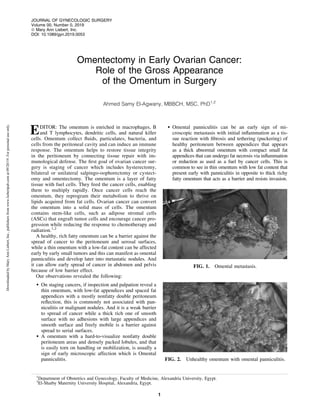
- 1. Omentectomy in Early Ovarian Cancer: Role of the Gross Appearance of the Omentum in Surgery Ahmed Samy El-Agwany, MBBCH, MSC, PhD1,2 EDITOR: The omentum is enriched in macrophages, B and T lymphocytes, dendritic cells, and natural killer cells. Omentum collect fluids, particulates, bacteria, and cells from the peritoneal cavity and can induce an immune response. The omentum helps to restore tissue integrity in the peritoneum by connecting tissue repair with im- munological defense. The first goal of ovarian cancer sur- gery is staging of cancer which includes hysterectomy, bilateral or unilateral salpingo-oophorectomy or cystect- omy and omentectomy. The omentum is a layer of fatty tissue with fuel cells. They feed the cancer cells, enabling them to multiply rapidly. Once cancer cells reach the omentum, they reprogram their metabolism to thrive on lipids acquired from fat cells. Ovarian cancer can convert the omentum into a solid mass of cells. The omentum contains stem-like cells, such as adipose stromal cells (ASCs) that engraft tumor cells and encourage cancer pro- gression while reducing the response to chemotherapy and radiation.1,2 A healthy, rich fatty omentum can be a barrier against the spread of cancer to the peritoneum and serosal surfaces, while a thin omentum with a low-fat content can be affected early by early small tumors and this can manifest as omental panniculitis and develop later into metastatic nodules. And it can allow early spread of cancer in abdomen and pelvis because of low barrier effect. Our observations revealed the following: On staging cancers, if inspection and palpation reveal a thin omentum, with low-fat appendices and spaced fat appendices with a mostly nonfatty double peritoneum reflection, this is commonly not associated with pan- niculitis or malignant nodules. And it is a weak barrier to spread of cancer while a thick rich one of smooth surface with no adhesions with large appendices and smooth surface and freely mobile is a barrier against spread to serial surfaces. A omentum with a hard-to-visualize nonfatty double peritoneum areas and densely packed lobules, and that is easily torn on handling or mobilization, is usually a sign of early microscopic affection which is Omental panniculitis. Omental panniculitis can be an early sign of mi- croscopic metastasis with initial inflammation as a tis- sue reaction with fibrosis and tethering (puckering) of healthy peritoneum between appendices that appears as a thick abnormal omentum with compact small fat appendices that can undergo fat necrosis via inflammation or reduction as used as a fuel by cancer cells. This is common to see in thin omentum with low fat content that present early with panniculitis in opposite to thick richy fatty omentum that acts as a barrier and resists invasion. FIG. 1. Omental metastasis. FIG. 2. Unhealthy omentum with omental panniculitis. 1 Department of Obstetrics and Gynecology, Faculty of Medicine, Alexandria University, Egypt. 2 El-Shatby Maternity University Hospital, Alexandria, Egypt. JOURNAL OF GYNECOLOGIC SURGERY Volume 00, Number 0, 2019 ª Mary Ann Liebert, Inc. DOI: 10.1089/gyn.2019.0053 1 DownloadedbyMaryAnnLiebert,Inc.,publishersfromwww.liebertpub.comat09/20/19.Forpersonaluseonly.
- 2. In advanced cancers, there is small omental cake mass with a low-fat content that is consumed over time or invaded with tumor cells especially with thin one. The omentum acts to engulf malignant cells before they spread to the peritoneum, especially the infracolic part, which is mobile. Thus, it is better not to perform a total omenectony in early stages, as omentum is engulfing cancer cells delays their spread to the serosal surfaces and upper abdomen leading to an omental cake. Omentectomy has no therapeutic role in addressing early cancer; so infracolic or partial omentectomy is preferable for staging early cancer. Infracolic omentecotmy is better than total one in staging of early cancers so as to leave part of omentum to localize the recurrence in the omentum and not to spread in the abdomen and pelvis (that is to engulf tumor cells).3,4 Early ovarian cancer in a patient with a thin omentum with low-fat is usually associated with early peritoneal and serosal spread. Early cancer and a thick healthy fatty omentum is usually associated with delayed spread. I can suggest that infracolic omentectomy is sufficient for early cancer staging if a patient has a healthy-looking omen- tum. I also suggest proceeding with a supracolic procedure if the infracolic is unhealthy as stated above with respect to panniculitis. Total omentectony, as it indicate invasion and so better surgery, this needs more evaluation regarding Omental gross appearance and types of surgery required. We aimed to raise awareness regarding gross appearance of omentum in ovarian tumors that can have a role in the type of surgery (Figs. 1–3) References 1. Morgan M, Boyd J, Drapkin R, Seiden MV. Cancers arising in the ovary. In: Abeloff MD, Armitage JO, Lichter AS, Niederhuber JE, Kastan MB, McKenna WG, eds. Clinical Oncology, 5th ed. Philadelphia: Elsevier; 2014:1592. 2. Rizi BS, Nagrath D. Linking omentum and ovarian cancer: NO. Oncoscience. 2015;2:797. 3. El-Agwany AS. Laparoscopy and computed tomography imaging in advanced ovarian tumors: a roadmap for pre- diction of optimal cytoreductive surgery. Gynecol Minim Invasive Ther 2018;7:66–69. 4. Meleis MH, El-Agwany AMS. Peritoneal carcinomatosis in- dex in advanced ovarian malignancy either by multislice CT verus laparotomy: A Comparative Study. Indian J Gynecol Oncolog 2015:13:24. Address correspondence to: Ahmed Samy El-Agwany, MBBCH, MSC, PhD Department of Obstetrics and Gynecology Faculty of Medicine Alexandria University Portsaid Street beside Bibliotheca Alexandrina El-Shatby, Alexandria 21526 Egypt, 21526 E-mail: Ahmedsamyagwany@gmail.com ahmed.elagwany@alexmed.edu.eg FIG. 3. Left panel shows normal mesentry; right panel shows nor- mal omentum. Both panels are showing smooth healthy fat rich omentum. 2 LETTER TO THE EDITOR DownloadedbyMaryAnnLiebert,Inc.,publishersfromwww.liebertpub.comat09/20/19.Forpersonaluseonly.