
Recommended
Recommended
More Related Content
Similar to Pathophysiology Mindmap
Similar to Pathophysiology Mindmap (20)
More from AhmedAbdElMoniem35
More from AhmedAbdElMoniem35 (20)
Recently uploaded
Recently uploaded (20)
Pathophysiology Mindmap
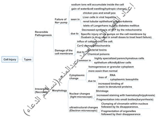
- 1. Cell Injury Types Failure of Na+ pump sodium ions will accumulate inside the cell gain of water&cell swelling(hydropic change) due to Decreased synthesis of ATP by the mitochondria Specific injury of ion pumps on the cell membrane by Ouabain (a drug used in small doses to treat heart failure). Damage of the cell membrane influx of calcium into the cell. Ca+2 damagemitochondria due to Bacterial toxins Chemical poisons Common in highly specialized parenchymatous cells epithelium ofkindly&liver cells Reversible Pathogenesis seen in chicken pox and small pox Liver cells in viral hepatitis renal tubular epithelium in hypo-kalemia islets of Langerhans in early diabetes mellitus Irreversible (Necrosis) Morphology Cytoplasmic change Nuclear changes (light microscope) ultrastructural changes (Electron microscopic) homogeneous or granular cytoplasm more eosin than normal due to loss of RNA cytoplasmic basophilia increased binding of eosin to denatured proteins Shrinkage increased staining with haematoxylin(pyknosis) Fragmentation into small bodies(karyorrhexis) Clumping of chromatin within nucleus followed by its disappearance. Fragmentation of organelles followed by their disappearance
- 2. Types of necrosis Coagulative seen in kidney, heart and spleen necrotic area pale yellow, opaque, swollen and firm Microscopically All cellular details are lost the outline of the tissue is preserved. manifestations of acute inflammation in surrounding tissues. Liquefactive seen in the central nervous system(CNS) due to its high lipid content the centers of pyogenic abscess and amoebiasis. necrotic tissue Causeous (caseation) Fat necrotic tissue necrotic tissue seen in seen in completely liquefied into a turbid fluid soon absorbed to leave an empty space tuberculous lesions. partially liquefied yellowish-grey,creamy material resembling cream cheese or casein Microscopically general architecture is preserved both the cellular details and the general architecture are totally lost homogeneous or granular, structureless,eosinophilic appearance the female breast in the omental and mesenteric fat in acute haemorrhagic pancreatitis. (traumatic fat necrosis) (enzymatic fat necrosis) the action of pancreatic lipase on the neutral fat opaque white Microscopically cloudy surrounded by chronic inflammatory cells foamy histiocytes foreign body giant cells lead to the formation of a palpable mass in the breast often clinically mistaken for cancer may undergo pathological calcification.
- 4. Irreversible(Apoptosis) Programmed cell death programmed obsolescence chromatin condensation characterized by shrinkage of cell volume an energy dependent process The chromatin is broken down in a regular fashion shed from free surfaces phagocytosed to form apoptotic bodies The dead cell breaks into fragments has a unique morphology does not elicit an inflammatory response initiation of apoptosis is regulated by growth factors physiologic endometrial cell loss during menstruation removal of the inter-digital webs during embryologic development of toes and fingers. pathologic Tissues undergoing atrophy Irradiated tissues Tissues injured by cytotoxic T lymphocytes Tumors Viral infections viral hepatitis acidophil Councilman Bodies in necrosis,groups of cells are killed, But apoptosis tends to affect single cell
- 6. Cellular disorders decrease or increases in the mass of the tissue Atrophy Hypertrophy Abnormal growth a decrease in the size of tissue increase in the size of tissue Causes Physiologic Pathologic Skeletal muscle hypertrophy in athletes. uterine muscle hypertrophy in pregnancy Left ventricular hypertrophy I hypertension Hypertrophy of one kidney due to removal of the other kidney Hyperplasia increase in the size of an organ due to increased numbers of cells, It occurs in labile and stable cells with or without decrease in the number Causes Pathologic due to effect of hormones Endometrial hyperplasia Cystic mammary hyperplasia (fibrocystic disease of the breast) Thyroid hyperplasia (Grave's disease) increase stimulation of thyroid stimulating hormone (TSH) increased estrogen stimulation particularly when it is not opposed by progesterone secretion e.g. near the menopause . Abnormal differentiation Metaplasia replacement of one normal adult cell type by a different adult (fully differentiated) cell types The alteration does not cross the histogenetic boundaries. a response to chronic inflammation. Abnormal maturation Epithelial dysplasia an abnormality of both differentiation & maturation occur in □ Cervix, vagina and vulva. □ Larynx. □ Urinary bladder. □ Large intestine in ulcerative colitis or adenomas. Physiologic breast feeding Bone marrow due to RBCs distrucyion due to decrease in the size of individual cells due to Increase in the size of individual cells Lymph node due to infection
- 7. Healing Types Composition Capillaries budding from the undamaged vessels the capillary buds are solid then become canalized develop a lumen Organization granulation(scar) tissue formation angiogenesis neovasculariztion blood flows Fibroblasts from activation of fibrocytes appear as large plump cells Initially mature collagen synthesis produce proteoglycans (ground substance of connective tissue) Macrophages (migrating monocytes) Phagocytose cell debris fibrin RBCs secretion of proteolyic enzymes collagenase elastase Clearance of dead tissue secrete fibronectin prostaglandins Fate With healing Increase Decrease collagen fibers active fibroblasts capillaries Gross Picture Granulation tissue pink soft moist bleeds in touch insensitive Resistant to bacterial infection. Microscopic Picture always present in granulation tissue. capillaries fibroblast odema inflammatory cells macrophages, lymphocytes, eosinophils, mast celles neutrophils, Macrophages. Def: the process of recovery in which the body replaces the damaged or lost tissues by new healthy tissues Resolution, Regeneration & Organization
- 10. Healing Resolution complete restoration of normal conditions after acute inflammation main features Minimal cell death and tissue damage. Rapid elimination of the causal agent Local conditions favoring removal of fluid and debris. Pathophysiology Fibrinolysin Solution of fibrin by enzyme action Removal of fluid by blood vessels and lymphatics. Removal of debris by phagocytes to regional lymph nodes hyperemia diminishes restoration to normal is complete. Ex: Resolution of lobar pneumonia bacterial inflammation of alveoli Regeneration the damaged tissue is replaced by a new one of the same type depends on type of the cell power of proliferation. Pathophysiology 2 main components Movement of surviving cells into the vacant space made available by loss of tissue due to wounding or necrosis. Proliferation of surviving cells to replace the loss. probable mechanisms Removal of contact inhibition allows movement of the cells laterally to cover wound surface cells in close contact inhibit cell migration only move vertically to replace surface loss Removal of CHALONES chemical factors inhibiting mitosis in neighboring cells of the same type by prolonging the G1 phase of the cell cycle allows proliferation of the surviving cells Release of growth stimulating factors Initiation cell division in G1&G0 due to specific growth factors Potentiation stimulate divided cells to enter S phase of cell cycle EGF PDGF due to(non-specific)growth factors(potentiators) insulin hydrocortisone growth hormone Important factors The availability of a good blood supply The survival of the supporting framework allowing the cells to grow in an organized way
- 11. Neoplasia Def: an abnormality of control of growth, differentiation and maturation of cells Recognized by the formation of amass of tissue (neoplasm or tumor) Biologic behavior Benign Neoplasm Locally Malignant Neoplasm Malignant Neoplasm neither invade nor spread Locally invasive only without distant spread Invade & spread LABORATORY DIAGNOSIS OF CANCER Histologic examination the most important method of diagnosis aided by clinical data. Fine-needle aspiration (FNA) aspiration of cells and fluids from tumors present in Palpable sites breast thyroid lymph nodes Ultrasonic guided aspiration in deep seated tumors Cytologic smears tumours of bronchi and stomach diagnose dysplasia carcinoma in situ invasive carcinoma of uterine cervix Immuno-histochemistry detection of cell products surface markers by using antibodies Tumor markers tumor associated molecules that can be detected in blood alpha-fetoprotein inhepatocellular carcinoma prostatic specific antigen in prostatic carcinoma. Chromosomal analysis diagnosis of lymphoid tumours arrangement of genes DNA ploidy analysis measurement the DNA cells to know diploid (normal DNA) aneuploid (abnormal DNA).
- 12. Carcinogenesis=Etiology of tumors Genetic Growth promoting genes (proto-oncogenes) Genes suppressor genes (anti-oncogenes). Genes that regulate apoptosis (cell death) may be converted to oncogenes by Point mutation permanent changes in DNA Chromosomal translocation Rearrangement of genetic material Burkitt s lymphoma Chromosomal deletion Deletion of one arm of a chromosome Gene amplification Reduplication of DNA production hundreds copies protoIt oncogene breast cancer proto-oncogenes normal cellular genes affect growth and Differentiation Oncogenes genes whose products are associated with neoplastic transformation
- 13. Environmental causes Factors which may activate cellular oncogenes Chemical agents Polycyclic hydrocarbons Azo and amino compounds (aniline dye and rubber industries) carcinoma of liver urinary bladder skin and bronchognic carcinoma. Asbestos malignant mesothelioma Viruses Human Papilloma Virus (HPV) benign squamous cell papilloma carcinoma of the cervix. Epstein-Barr Virus (EBV) member of herpes family nasopharyngeal carcinoma Burkitt's lymphoma Hepatitis B and C viruses (HBV and HCV) hepatocellular carcinoma. Human T cell leukaemia virus (HTLV) leukaemia lymphoma Radiation Ultraviolet rays derived from sun skin cancer malignant melanoma Ionizing irradiation myeloid leukaemia carcinoma of skin. thyroid cancer in children Hormones no evidence that hormones by themselves cause cancer the rate of growth of breast and endometrial cancer is dependent upon oestrogen prostate cancer is stimulated by testosterone prostate cancer is depressed by oestrogen. Heredity retinoblastoma breast cancer familial adenomatous polyposis diet and alcohol
- 15. HT Hypertension high blood pressure Blood pressure measurement of the force against the walls of your arteries as the heart pumps blood measured in millimeters of mercury (mmHg) usually given 120 over 80 (120/80mm Hg) One or both of these numbers can be too high The top number systolic pressure high over 140 most of the time. normal below 120 most of the time The bottom number diastolic pressure. high over 90 most of the time. normal below 80 most of the time Pre-hypertension Top number between120&139 most of the time Bottom number between80&89 most of the time most likely one to have hypertension = Cardiac output×Peripheral resistance increases the total peripheral resistance and the cardiac out Cardiac output(C.O) = H.R (heart rate) ×S.V(stroke volume). Primary=essential without apparent cause Secondary with known cause Risk Factors Increase with age Obesity Family history Tobacco use High intake of alcohol Stress High intake of sodium Sedentary life style
- 16. Pathogenesis 2-High vasomotor tone causing increases vascular resistance. 1-Thickening of the arteriolar wall which might be genetic in origin. 3-Increased blood flow resulting from renal (rennin) hormonal dysfunction aldosterone hormone corticosteroid hormone 4-Renin- angiotensin mechanism Decrease in renal blood flow Secretion of rennin The liver secretes angiotensinogen conversion of Angiotensinogen by the action of renin into angiotensin I Conversion of Angiotensin I into Angiotensin II by the action of angiotensin converting enzyme (ACE) secretion of(ACE) from the surface of pulmonary and renal epithelium. Angiotensin II causes Aldosterone secretion from the adrenal gland cortex Sympathetic activity Arteliolar vasoconstriction increase in blood pressure (passive transport) An inhibitory signal is sent to the kidney to decrease the secretion of rennin Angiotensin&aldosterone Na+ Cl reabsorption K excretion H2O retention. antidiuretic hormone H2O and salt retention increase in blood volume Perfusion of the juxtaglomerular apparatus(blood flow) increases Hypertention Theories:-
- 17. HT Complications Angina and myocardial infarction Hypertension will reduce the blood flow to the myocardium Left ventricular failure increased aortic vascular resistance backpressure Cerbro-vascular accident cerebral hemorrhage subarachnoid hemorrhage Dissecting aortic aneurysm layers of aortic wall are splitted blood passes through it severe pain bad prognosis HT crisis a severe increase in arterial blood pressure result in Pathophysiology renal, cardiac or cerebral complication death Causes Same as complications Impaired renal function Hypertensive encephalopathy Myocardial ischemia=hypoxia=cerebral infraction Eclampsia Pheochromocytoma (suprarenal medulla tumor) End organ failure
- 18. HF A state in which the heart cannot provide sufficient cardiac output to satisfy the needs of the body. The heart muscle cannot pump(eject) the blood out of the heart very well The heart muscles do not fill up with blood easily AS it become stiff the heart is no longer able to pump enough oxygen&nutrients-rich blood out to the rest of your body blood may back up other areas of the body symptoms and effects may differ from patient to patient Heart muscle has difficulty contracting. the heart's ability to contract decreases. causes pulmonary congestion(fluid in lung) Heart muscle has difficulty relaxing lead to fluid accumulation,in the feet,ankles,&legs and lung congestion. reduces organs ability to work two categories causes effects characters of patient right –sided Congestive (diastolic)HF Left –sided Congestive (systolic)HF in health the function of both ventricles is closely integrated both ventricles have the same capacity, pump virtually synchronously. the action of each ventricle is not isolated both influences and is influenced by the action of the other combined failure are common one side of the heart usually fails first, followed by other side. It is helpful in the early stages to refer as left- sided GHF or right –sided GHF
- 19. HF pathophysiology HF is secondary to systolic dysfunction diastolic dysfunction Hemodynamic Changes ex: the blood pressure&blood flow at the output of the left heart. Cellular changes affects contraction of myocardium Changes in adrenergic receptors Changes in Ca+2 ions handling found in the myocardium Slight increase in α1 receptors. β1 receptors desensitization to catecholamine Changes in contractile proteins in the muscle fibers Program cell death (Apoptosis). Increase amount of fibrous tissue β1 Receptors and α1 receptors (↑contractility). NB: Neuro hormonal changes decrease tissue perfusion(blood flow) decreasing renal perfusion salt and water retention increased blood pressure increased sympathetic output increases renin release high sympathetic & rennin angiotensin activity compensate for heart failure by increasing cardiac output through increasing peripheral resistance myocardial contractility heart rate eventually triggers myocyte hypertrophy apoptosis focal necrosis Circulating endothelin-1 (ET-1) present in high levels during heart failure causes vasoconstriction hyperplasia fibrosis