
Recommended
More Related Content
What's hot
What's hot (20)
Similar to Poriferous Patient-Specific Implants
Similar to Poriferous Patient-Specific Implants (20)
Recently uploaded
Recently uploaded (20)
Poriferous Patient-Specific Implants
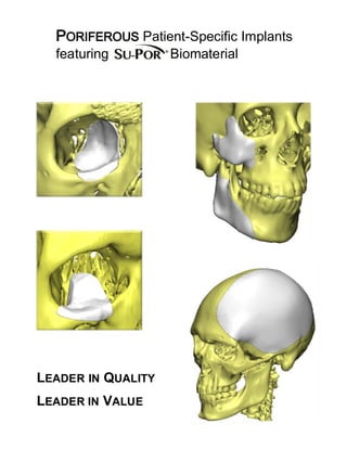
- 1. PORIFEROUS Patient-Specific Implants featuring Biomaterial LEADER IN QUALITY LEADER IN VALUE
- 2. Features and Benefits Precise Fit Each device is individually crafted to your patient's specific needs. Proven Material Poriferous polyethylene, a biocompatible material with over 30 years of proven clinical performance. Variable Thickness Variable thickness to match native bone. Modification Malleable, and allows for easy modification with a scalpel blade or burring instrument. Direct Fixation Screws may be inserted directly through the implant margins into the bone, and may eliminate the need for plate fixation. Sterile Non-pyrogenic Implant kit contains two (2) identical sterile, non-pyrogenic, implants limiting processing risk for the hospital. Virtual Approval Process Detailed electronic document allows for approval prior to manufacturing (optional) Application To meet the need of individual patients in applications of craniofacial reconstructive / cosmetic surgery and repair of craniofacial trauma. MR Safe This designation by the US FDA provides assurance of no image artifact or distortion of diagnostic MRI imaging. Tissue In- Growth The interconnecting poriferous structure may allow for fibrovascular in- growth and tissue integration. Suture/Drainage Sutures may be passed through any part of the implant. Open pores may allow drainage. Fast Turnaround Order shipment within 8 business days following design approval.
- 3. Estimated Lead Time Legend Scan data converted to virtual model Design of virtual implant template Virtual model of implant is reviewed by surgeon Implant review method Physical model produced Non-Sterile Implant template produced Non-Sterile implant and medical model shipped to client for approval Approval received Implants sterilized Request for patient-specific implant Implant design type 3 Days CT scan of patient Client makes physical design of implant on model 1 day Virtual Design kit pulled from inventory Physical Review 5 Days Virtual Review Reference CT scanning guidelines 0.5 Day 3 Days Physical Physical model produced 5 Days Model and design kit shipped to client 3 Days Model with implant design shipped to Poriferous, LLC. Ship to client 3 Days Poriferous, LLC. receives shipment 3 Days Mail CD / DVD File upload Implants produced 3 Days Process Flow Chart 7.5 business days 18.5 business days 32.5 business days Actual lead times may vary. Estimated lead time do not include external processes that are outside the control of Poriferous, LLC.
- 4. LEADER IN QUALITY. LEADER IN VALUE Poriferous, LLC. is owned and operated by professionals who were a part of the original team that introduced porous HDPE as an alloplastic material in 1985. Poriferous knows porous polyethylene and can deliver the results our surgeons demand. All processes of design, manufacturing, and sterilization are performed in our ISO 13485 Certified facility located in Newnan, Georgia. Ordering Process 1. Complete Design Input Form. 2. Utilize preferred CT scanning protocol. 3. Submit form and CT data via: Fax: 770-683-7459 Email: sales@poriferous.com Mail: Attn: Poriferous, LLC. Patient Specific Implants 535 Pine Road, Suite 206 Newnan, GA 30263 4 For upload of CT data; please contact Customer Care at (877) 631-1954, or e-mail for a secure upload request link. 5. Once your CT data has been received we will design a patient specific implant, and a graphic design proposal will be emailed for approval prior to making the actual implants. Ordering Information Cat# Description 4350 Cranial (Small, Medium, Large, X-Large) 4351 Cranial Implant Template (non-implantable) 4353 Facial (Orbital, Midface, Mandible angle, Chin) 4355 Medical Model of defect area 4356 Facial Implant Template (non-implantable)
- 5. Scanning Protocol The quality of the CT data is essential to the design and manufacture of Poriferous Patient-Specific Implants featuring Su-Por Biomaterial. Provided below is the protocol to follow: CT Scanning Guidelines The patient must be stabilized and remain completely still throughout the entire scan. If patient movement occurs, the scan must be restarted to achieve the best implant fit. The scan should include 2cm beyond the defect area or area of interest. Please provide the original DICOM slice data. Do not reformat or include viewer software with data. Important position or details should be noted as well as any asymmetrical element of the patient to indicate left and/or right. The use of a bite jib during the scanning process for the mandible or the maxilla is recommended, otherwise they will be fused in the model. SCANNING PARAMETERS: Cranial Defects Acquisition: Axial/Helical F.O.V.: Include all areas of interest. Additional 20‐25 cm above and below is preferred Gantry Tilt: 0 Spacing: Overlapping (3mm Max) Algorithm: Standard MA: 170ma/280kvp or lower Time: 2 seconds or less Facial Defects Acquisition: Axial/Helical F.O.V.: Include all areas of interest Gantry Tilt: 0 Spacing: Overlapping (1.5mm Max) Algorithm: Standard MA: 120-180ma/120kvp or lower Time: 2 Seconds or less
- 6. This document is in no way intended to be a comprehensive manual of surgical techniques. Surgeons should utilize their surgical training, clinical experience, and applicable surgical techniques to determine appropriate surgical procedures. Experienced surgical judgment should be utilized in the selection and use of SU-POR implants. Poriferous, LLC. 535 Pine Road, Suite 206 – Newnan, Georgia 30263 U.S.A T +1 770.683.3855 – F +1 770.683.7459 – info@poriferous.com – www.poriferous.com CE Certificate Number US15/842160 – ISO Certificate Number US15/842158 - 510(k)152463 © Copyright 2015. All rights reserved. SU-POR is a registered trademark of Poriferous, LLC. REV170713