1. 16
The Indian Journal of Pediatrics
March 2008; Volume 75 : Number 3
CONTENTS
Page
ORIGINAL ARTICLES
Spectral Analysis of Noise in the Neonatal Intensive Care Unit
M.D.Livera, B. Priya, A. Ramesh, P.N. Suman Rao, V. Srilakshmi, M. Nagapoornima,
A.G. Ramakrishnan, M. Dominic and Swarnarekha
..
217
..
223
..
229
..
235
..
239
..
245
..
251
..
255
..
261
..
267
..
271
..
277
..
281
Impact of Parent and Teacher Concordance on Diagnosing Attention Deficit Hyperactivity
Disorder and its Sub-types
Prahbhjot Malhi, Pratibha Singhi and Manjit Sidhu
Antithymocyte Globulin and Cyclosporin in Children with Acquired Aplastic Anemia
Jagdish Chandra, Rahul Naithani, Rakesh Ravi, Varinder Singh, Shashi Narayan, Sunita Sharma,
Harish Pemde and A.K. Dutta
Homocysteine, Vitamin B12 and Folate Status in Pediatric Acute Lymphoblastic Leukemia
M.N. Sadananda Adiga, Sunil Chandy, Girija Ramaswamy, L. Appaji and Lakshmi Krishnamoorthy
Growth and Bone Mineralization in Patients with Juvenile Idiopathic Arthritis
Ozgur Okumus, Muferet Erguven, Murat Deveci, Oznur Yilmaz and Mine Okumus
Dapsone Induced Methemoglobinemia : Intermittent vs Continuous Intravenous Methylene
Blue Therapy
Rajniti Prasad, R. Singh, O.P. Mishra and Madhukar Pandey
SPECIAL ARTICLE
Doxofylline: The Next Generation Methylxanthine
Jhuma Sankar, Rakesh Lodha and S.K. Kabra
SYMPOSIUM ON AIIMS PROTOCOLS IN NEONATOLOGY – III
Fluid and Electrolyte Management in Term and Preterm Neonates
Deepak Chawla, Ramesh Agarwal, Ashok K. Deorari and Vinod K. Paul
Sepsis in the Newborn
M. Jeeva Sankar, Ramesh Agarwal, Ashok K. Deorari and Vinod K. Paul
Minimal Enteral Nutrition
Satish Mishra, Ramesh Agarwal, M. Jeevasankar, Ashok K. Deorari and Vinod K. Paul
Approach to Inborn Errors of Metabolism Presenting in the Neonate
Suvasini Sharma, Pradeep Kumar, Ramesh Agarwal, Madhulika Kabra, Ashok K. Deorari and
Vinod K. Paul
Patent Ductus Arteriosus in Preterm Neonates
Ramesh Agarwal, Ashok K. Deorari and Vinod K. Paul
CLINICAL BRIEFS
Spinal Cord Hamartoma with Pseudopancreatic Cyst
Shalu Gupta, Ashok Kumar, A.N. Gangopadhyay and Mohan Kumar
Indian Journal of Pediatrics, Volume 75—March, 2008
210
2. 17
CONTENTS
Page
Vesicoureteric Reflux Deterioration in Monozygotic Twins
Spyridon Tsiouris, Chrissa Sioka, Anna Marinarou, Jihad Al-Bokharhli, Irene Sionti and
Andreas Fotopoulos
..
285
..
288
..
290
..
294
..
296
NOTES AND NEWS
..
248, 284
AUTHOR FOR GUIDELINES
..
298
REVIEWERS OF 2007
..
300
Tay Syndrome
S.D. Jambhekar and A.R. Dhongade
Rosai-Dorfman Disease
Madhumita Nandi, R.K. Mondal, Supratim Datta, Balai Chandra Karmakar, K. Mukherjee and
T.K. Dhibar
Langerhans Cell Histiocytosis of Mediastinal Node
Urmila N. Khadilkar, A.T.K. Rao, Kausalya Kumari Sahoo and Mukta R. Pai
Acute Eosinophilic Pneumonia Due to Round Worm Infestation
Bindu Aggarwal, Monika Sharma and Tejinder Singh
211
Indian Journal of Pediatrics, Volume 75—March, 2008
3. 77
Symposium on AIIMS Protocols in Neonatology – III
Approach to Inborn Errors of Metabolism Presenting in
the Neonate
Suvasini Sharma, Pradeep Kumar, Ramesh Agarwal, Madhulika Kabra, Ashok K. Deorari and
Vinod K. Paul
Department of Pediatrics, All India Institute of Medical Sciences, Ansari Nagar, New Delhi, India
ABSTRACT
Inborn errors of metabolism (IEM) are an important cause of acute illness in newborns. Presentation may mimic common
neonatal conditions such as sepsis. Prompt detection requires a high index of suspicion and the early measurement of
biochemical markers such as blood ammonia. Diagnosis is important not only for treatment but also for genetic counselling.
Guidelines for diagnosis and early management of IEM presenting in the neonatal period are described. [Indian J Pediatr
2008; 75 (3) : 271-276] E-mail: ashokdeorari_56@hotmail.com
Key words : Inborn errors of metabolism; Encephalopathy; Hyperammonemia
Inborn errors of metabolism (IEM) are disorders in which
there is a block at some point in the normal metabolic
pathway caused by a genetic defect of a specific enzyme.
The number of diseases in humans known to be
attributable to inherited point defects in metabolism now
exceeds 500.1 While the diseases individually are rare,
they collectively account for a significant proportion of
neonatal and childhood morbidity and mortality.
Diagnosis is important not only for treatment and
prognostication but also for genetic counselling and
antenatal diagnosis in subsequent pregnancies.
CLINICAL PRESENTATION
• Family history of neonatal deaths
• Rapidly progressive encephalopathy and seizures of
unexplained cause
• Severe metabolic acidosis
• Persistent vomiting
• Peculiar odor
• Acute fatty liver or HELLP (hemolysis, elevated liver
enzymes and low platelet counts) during pregnancy:
seen in women carrying fetuses with long-chain-3hydroxyacyl-coenzyme dehydrogenase deficiency
(LCHADD).
Table 1 describes examination findings that may
provide a clue to the underlying IEM.
TABLE 1. Clinical Pointers Towards Specific IEM’s
Severe illness in the newborn, regardless of the
underlying cause, tends to manifest with non-specific
findings, such as poor feeding, drowsiness, lethargy,
hypotonia and failure to thrive. IEM should be considered
in the differential diagnosis of any sick neonate along with
common acquired causes such as sepsis, hypoxic-ischemic
encephalopathy, duct-dependant cardiac lesions,
congenital adrenal hyperplasia and congenital infections.
Clinical pointers towards an underlying IEM include.2
• Deterioration after a period of apparent normalcy
• Parental consanguinity
Correspondence and Reprint requests : Dr Ashok K Deorari,
Professor, Department of Pediatrics, All India Institute of Medical
Sciences, Ansari Nagar, New Delhi 110029, India
[Received February 7, 2008; Accepted February 7, 2008]
Indian Journal of Pediatrics, Volume 75—March, 2008
Clinical finding
Disorder
Coarse facies
Cataract
Retinitis pigmentosa
Cherry red spot
Hepatomegaly
Renal enlargement
Eczema/alopecia
Abnormal kinky hair
Decreased pigmentation
Lysosomal disorders
Galactosemia, Zellweger syndrome
Mitochondrial disorders
Lipidosis
Storage disorders, urea cycle defects
Zellweger syndrome
Biotinidase deficiency
Menke disease
Phenylketonuria
Patterns of presentation include.2,3
(i) Encephalopathy with or without metabolic acidosis:
Encephalopathy, seizures, and tone abnormalities are
predominant presenting features of organic acidemias,
urea cycle defects and congenital lactic acidosis.
Intractable seizures are prominent in pyridoxine
dependency, non-ketotic hyperglycinemia, molybdenum
271
4. 78
S. Sharma et al
co-factor defect and folinic-acid responsive seizures.
(ii) Acute liver disease: This could manifest as• Jaundice alone- Gilbert syndrome, Criggler-Najjar
syndrome
• Hepatic failure (jaundice, ascites, hypoglycemia,
coagulopathy)- Tyrosinemia, galactosemia, neonatal
hemochromatosis, glycogen storage disease type IV.
• Neonatal cholestasis: alpha-1 antitrypsin deficiency,
Niemann-Pick disease type C.
• Hypoglycemia: persistent and severe hypoglycemia
may be an indicator of an underlying IEM.
Hypoglycemia is a feature of galactosemia, fatty acid
oxidation defects, organic acidemias, glycogen storage
disorders and disorders of gluconeogenesis.
(iii) Dysmorphic features: Seen in peroxisomal disorders,
pyruvate dehydrogenase deficiency, congenital disorders
of glycosylation (CDG), and lysosomal storage diseases.
Some IEMs may present with non-immune hydrops
fetalis; these include lysosomal storage disorders and
CDG.
(iv) Cardiac disease: Cardiomyopathy is a prominent
feature in some IEM including fatty acid oxidation
defects, glycogen storage disease type II and
mitochondrial electron transport chain defects.
Metabolic investigations should be initiated as soon as the
possibility is considered. The outcome of treatment of
many IEM especially those associated with
hyperammonemia is directly related to the rapidity with
which problems are detected and appropriate
management instituted.
First line investigations (metabolic screen)
The following tests should be obtained in all babies with
suspected IEM.
and
and
• Arterial blood gases and electrolytes
• Blood glucose
• Plasma ammonia (Normal values in newborn: 90150 µg/dl or 64-107 µmol/L)
• Arterial blood lactate (Normal values: 0.5-1.6 µmol/
L)
• Liver function tests
• Urine ketones
• Urine reducing substances.
• Serum uric acid (low in molybdenum cofactor
deficiency).
272
TABLE 2. Categorization of Neonatal IEM Using Metabolic
Screening Tests
Acidosis
Ketosis -Lactate -Ammonia
Diagnosis
-
+
-
-
+
+
-
+/+/-
+
-
+/+
-
Maple syrup urine
disease
Organic aciduria
Lactic acidosis
Urea cycle
Non-ketotic
hyperglycinemia
sulfite oxidase
deficiency,
peroxisomal,
Phenylketonuria,
galactosemia
Second line investigations (ancillary and confirmatory
tests)
INVESTIGATIONS
• Complete blood count: (neutropenia
thrombocytopenia seen in propionic
methylmalonic academia)
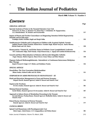
Fig 1 gives the algorithmic approach to a newborn
with suspected IEM. Disease category can be diagnosed
based on blood ammonia, blood gas analysis and urine
ketone testing. Hyperammonemia without acidosis is
caused by urea cycle defects. Metabolic acidosis with or
without hyperammonemia is a feature of organic
acidemias and fatty acid oxidation defects. Fig. 2 explains
the algorithmic approach to neonate with persistent
hypoglycemia and suspected underlying IEM. Table 2
explains the categorization of IEM based on simple
metabolic screening tests.
These tests need to be performed in a targeted manner,
based on presumptive diagnosis reached after first line
investigations:
• Gas chromatography mass spectrometry (GCMS) of
urine- for diagnosis of organic acidemias.
• Plasma amino acids and acyl carnitine profile: by
tandem mass spectrometry (TMS)- for diagnosis of
organic acidemias, urea cycle defects,
aminoacidopathies and fatty acid oxidation defects.
• High performance liquid chromatography (HPLC):
for quantitative analysis of amino acids in blood and
urine; required for diagnosis of organic acidemias
and aminoacidopathies.
• Lactate/pyruvate ratio- in cases with elevated
lactate.
• Urinary orotic acid- in cases with hyperammonemia
for classification of urea cycle defect.
• Enzyme assay: This is required for definitive
diagnosis, but not available for most IEM’s.
Available enzyme assays include: biotinidase assayin cases with suspected biotinidase deficiency
(intractable seizures, seborrheic rash, alopecia); and
. GALT (galactose 1-phosphate uridyl transferase )
assay- in cases with suspected galactosemia
Indian Journal of Pediatrics, Volume 75—March, 2008
5. 79
Approach to Inborn Error of Metabolism Presenting in the Neonate
Fig 1. Approach to newborn with suspected metabolic disorder Suspected Metabolic Disorder
(hypoglycemia, cataracts, reducing sugars in urine).
• Neuroimaging: MRI may provide helpful pointers
towards etiology while results of definitive
investigations are pending. Some IEM may be
associated with structural malformations e.g.,
Zellweger syndrome has diffuse cortical migration
and sulcation abnormalities. Agenesis of corpus
callosum has been reported in Menke’s disease,
pyruvate decarboxylase deficiency and nonketotic
hyperglycinemia.4 Examples of other neuroimaging
findings in IEM include:
• Maple syrup urine disease (MSUD): brainstem and
cerebellar edema
• Propionic and methylmalonic acidemia: basal
ganglia signal change
• Glutaric aciduria: frontotemporal atrophy, subdural
hematomas
Indian Journal of Pediatrics, Volume 75—March, 2008
• Magnetic resonance spectroscopy (MRS): may be
helpful in selected disorders e.g. lactate peak
elevated in mitochondrial disorders, leucine peak
elevated in MSUD.
• Electroencephalography (EEG): some EEG
abnormalities may be suggestive of particular IEM;
e.g., comb-like rhythm in MSUD, burst suppression
in NKH and holocarboxylase synthetase deficiency.5
Plasma very long chain fatty acid (VLCFA) levels:
elevated in peroxisomal disorders.
Mutation analysis when available.
CSF aminoacid analysis: CSF Glycine levels elevated in
NKH.
Precautions to be observed while collecting samples
Should be collected before specific treatment is started or
feeds are stopped, as may be falsely normal if the child is
off feeds.
273
6. 80
S. Sharma et al
Fig. 2. Approach to newborn with persistent hypoglycemia and suspected IEM
Samples for blood ammonia and lactate should be
transported in ice and immediately tested. Lactate
sample should be arterial and should be collected after 2
hrs fasting in a preheparinized syringe. Ammonia sample
is to be collected approximately after 2 hours of fasting in
EDTA vacutainer. Avoid air mixing. Sample should be
free flowing.
Detailed history including drug details should be
provided to the lab. (sodium valproate therapy may
increase ammonia levels).
Samples to be obtained in infant with suspected IEM
when diagnosis is uncertain and death seems inevitable
(Metabolic autopsy)6
Blood: 5-10 ml; frozen at -200C; both heparinized (for
chromosomal studies) and EDTA (for DNA studies)
samples to be taken
Urine: frozen at –20oC
CSF: store at –20oC
Skin biopsy: including dermis in culture medium or
saline with glucose. Store at 4-80C. Do not freeze.
Liver, muscle, kidney and heart biopsy: as indicated.
Clinical photograph (in cases with dysmorphism)
Infantogram (in cases with skeletal abnormalities)
TREATMENT
In most cases, treatment needs to be instituted empirically
without a specific diagnosis. The metabolic screen helps to
274
broadly categorize the patient’s IEM (e.g., urea cycle
defect, organic academia, congenital lactic acidosis etc), on
the basis of which, empirical treatment can be instituted.
Aims of treatment
1. To reduce the formation of toxic metabolites by
decreasing substrate availability (by stopping feeds
and preventing endogenous catabolism)
2. To provide adequate calories
3. To enhance the excretion of toxic metabolites.
4. To institute co-factor therapy for specific disease and
also empirically if diagnosis not established.
5. Supportive care- treatment of seizures (avoid
sodium valproate – may increase ammonia levels),
maintain euglycemia and normothermia, fluid,
electrolyte and acid-base balance, treatment of
infection, mechanical ventilation if required.
Management of hyperammonemia7,8
Discontinue all feeds. Provide adequate calories by
intravenous glucose and lipids. Maintain glucose infusion
rate 8-10mg/kg/min. Start intravenous lipid 0.5g/kg/
day (up to 3g/kg/day). After stabilization gradually add
protein 0.25 g/kg till 1.5 g/kg/day.
Dialysis is the only means for rapid removal of
ammonia, and hemodialysis is more effective and faster
than peritoneal dialysis, however peritoneal dialysis may
be more widely available and feasible. Exchange
transfusion is not useful.
Indian Journal of Pediatrics, Volume 75—March, 2008
7. 81
Approach to Inborn Error of Metabolism Presenting in the Neonate
Alternative pathways for nitrogen excretion:
Sodium benzoate (IV or oral)- loading dose 250 mg/kg
then 250-400 mg/kg/day in 4 divided doses. (Intravenous
preparation not available in India).
Sodium phenylbutyrate (not available in India)loading dose 250 mg/kg followed by 250-500 mg/kg/
day.
L-arginine (oral or IV)- 300 mg/kg/day (Intravenous
preparation not available in India)
L-carnitine (oral or IV)- 200 mg/kg/day
Supportive care: treatment of sepsis, seizures,
ventilation. Avoid sodium valproate.
Acute management of newborn with suspected organic
acidemia9
• The patient is kept nil per orally and intravenous
glucose is provided.
• Supportive care: hydration, treatment of sepsis,
seizures, ventilation.
• Carnitine: 100 mg/kg/day IV or oral.
• Treat acidosis: Sodium bicarbonate 0.35-0.5mEq/
kg/hr (max 1-2mEq/kg/hr)
• Start Biotin 10 mg/day orally.
• Start Vitamin B12 1-2 mg/day I/M (useful in B12
responsive forms of methylmalonic acidemias)
• Start Thiamine 300 mg/day (useful in Thiamineresponsive variants of MSUD).
• If hyperammonemia is present, treat as explained
above.
Management of congenital lactic acidosis
• Supportive care: hydration, treatment of sepsis,
seizures, ventilation. Avoid sodium valproate.
• Treat acidosis: sodium bicarbonate 0.35-0.5mEq/
kg/hr (max 1-2mEq/kg/hr)
• Thiamine: up to 300 mg/day in 4 divided doses.
• Riboflavin: 100 mg/day in 4 divided doses.
• Add co-enzyme Q: 5-15 mg/kg/day
• L-carnitine: 50-100 mg/kg orally.
Treatment of newborn with refractory seizures with no
obvious etiology (suspected metabolic etiology)10
• If patient persists to have seizures despite 2 or 3
antiepileptic drugs in adequate doses, consider trial
of pyridoxine 100 mg intravenously. If intravenous
preparation not available, oral pyridoxine can be
given (15 mg/Kg/day).
• If seizures persist despite pyridoxine, give trial of
biotin 10 mg/day and folinic acid 15 mg/day
(folinic acid responsive seizures).
• Rule out glucose transporter defect: measure CSF
Indian Journal of Pediatrics, Volume 75—March, 2008
and blood glucose. In glucose transporter defect,
CSF glucose level is equal to or less than 1/3rd of the
blood glucose level. This disorder responds to the
ketogenic diet.
Management of asymptomatic newborn with a history
of sibling death with suspected IEM:
• After baseline metabolic screen, start oral dextrose
feeds (10% dextrose).
• After 24 hours, repeat screen. If normal, start breast
feeds. Monitor sugar, blood gases and urine
ketones, blood ammonia 6 hourly.
• Some authorities recommend starting medium
chain triglycerides (MCT oil) before starting breast
feeds,3 however, this is not being followed in our
center (because of unpalatibility of MCT oil).
• After 48 hours, repeat metabolic screen. Obtain
samples for TMS and urine organic acid tests.
• The infant will need careful observation and followup for the first few months, as IEM may present in
different age groups in members of the same family.
Long term treatment of IEM
The following modalities are available
Dietary treatment: This is the mainstay of treatment in
phenylketonuria, maple syrup urine disease,
homocystinuria, galactosemia, and glycogen storage
disease Type I and III. Special diets for PKU and MSUD
are commercially available in the west. These are not
available in India, but can be imported. These special diets
are however very expensive, and cannot be afforded by
most Indian patients. Based on the amino acid content of
some common food products available in India, dietary
exchanges are calculated and a low phenylalanine diet for
PKU and diet low in branched chain amino acids for
MSUD are being used in our center. However, there are
no studies to document the efficacy of these indigenous
diets. Some disorders like urea cycle disorders and
organic acidurias require dietary modification (protein
restriction) in addition to other modalities.11
Enzyme replacement therapy (ERT): ERT is now
commercially available for some lysosomal storage
disorders.12 However, these disorders do not manifest in
the newborn period, an exception being Pompe’s disease
(Glycogen storage disorder Type II)which may present in
the newborn period and for which ERT is now available.
Cofactor replacement therapy: The catalytic properties of
many enzymes depend on the participation of non
protein prosthetic groups, such as vitamins and minerals,
as obligatory cofactors. The following co-factors may be
beneficial in certain IEM:13
• Thiamine: mitochondrial disorders, thiamine
responsive variants of MSUD, PDH deficiency &
complex I deficiency)
275
8. 82
S. Sharma et al
Appendix: Commercially Available Formulations Used in IEM
Co-factor
Trade name, formulation
Pyridoxine
Tab Benadon (40mg) (Nicholas Piramal), Inj Vitneurin (1 ampoule contains
50 mg pyridoxine)
Inj Trineurosol (1000mcg/ml) (Tridoss Laboratories)
Tab Benalgis (75 mg) (Franco India)
Tab Riboflavin (5 mg) (Shreya)
Tab Essvit (5mg, 10mg) (Ecopharma)
Syrup L-Carnitor (5ml=500 mg), Tab L-Carnitor (500 mg), Inj carnitor (1g/
5ml) (Elder)
Tab Leukorin (15 mg) (Samarth)
Satchet 20g (Hesh Co.)
ARG-9 Satchet (3g) (Noveau Medicament)
Tab CoQ 30 mg, 50 mg. (Universal Medicare)
Hydroxycobalamin (Vitamin B12)
Thiamine
Riboflavin
Biotin
Carnitine
Folinic acid
Sodium Benzoate
Arginine
Coenzyme Q
• Riboflavin: Glutaric aciduria Type I, Type II, mild
variants of ETF, ETF-DH, complex I deficiency
• Pyridoxine: 50% of cases of homocystinuria due to
cystathionine β-synthetase deficiency, pyridoxine
dependency with seizures, xanthurenic aciduria,
primary hyperoxaluria type I, Hyperornithemia
with gyrate atrophy
• Cobalamin: Methylmalonic academia (cblA, cblB),
Homocystinuria and
methylmalonic academia
(cblC, cblD, cblF)
• Folinic acid: Hereditary orotic aciduria, Methionine
synthase deficiency, Cerebral folate transporter
deficiency, hereditary folate malabsorption, KearnsSayre syndrome
• Biotin: Biotinidase deficiency, holocarboxylase
synthetase deficiency
PREVENTION
Neonatal screening: Tandem mass spectrometry is used in
some countries for neonatal screening for IEM. Disorders
which can be detected by TMS include aminoacidopathies
(phenylketonuria, MSUD, Homocystinuria, Citrullinemia,
Argininosuccinic aciduria, hepatorenal tyrosinemia), fatty
acid oxidation defects, organic acidemias (glutaric
aciduria, propionic acidemia, methylmalonic acidemia,
isovaleric acidemia). The cost of this procedure is high, a
potent dis-inventive for resource poor countries like India.
Also, the though the test is highly sensitive, the specificity
is relatively low; and there are difficulties in interpretation
of abnormal test results in apparently healthy infants.
Genetic counselling and prenatal diagnosis: Most of the IEM
are single gene defects, inherited in an autosomal
recessive manner, with a 25% recurrence risk. Therefore,
when the diagnosis is known and confirmed in the index
case, prenatal diagnosis can be offered, wherever
available for the subsequent pregnancies. The samples
required are chorionic villus tissue or amniotic fluid.
Modalities available are:14
• Substrate or metabolite detection: useful in
276
phenylketonuria, peroxisomal defects.
• Enzyme assay: useful in lysosomal storage
disorders like Niemann-Pick disease, Gaucher
disease.
• DNA based (molecular) diagnosis: Detection of
mutation in proband/ carrier parents is a
prerequisite.
REFERENCES
1. Childs B, Valle D, Jimenez-Sanchez. The Inborn error and
biochemical variability. In Scriver CR, Beaudet AL, Sly WS,
Valle D, eds. The metabolic and molecular basis of inherited
disease, 8 th ed, New York; McGraw-Hill, 2001: 155-166.
2. Clarke, JTR. A Clinical guide to inherited metabolic diseases. 3rd
Ed. Cambridge University Press, Cambridge; 2006.
3. Cataltepe SU, Levy HL. Inborn errors of metabolism. In
Cloherty JP, Eichenwald EC, Stark AR, eds. Manual of neonatal
care. 6th ed. Philadelphia; Lippincott Williams and Wilkins,
2008; 558-573.
4. Blaser S, Feigenbaum A. A neuroimaging approach to inborn
errors of metabolism. Neuroimag Clin N Am 2004; 14: 307-329.
5. Nordli DR, De Vivo DC. Classification of infantile seizures:
Implications for identification and treatment of inborn errors
of metabolism. J Child Neurol 2002; 17 (Suppl 3): 3S3-3S8.
6. Leonard JV, Morris AAM. Diagnosis and early management
of inborn errors of metabolism presenting around the time of
birth. Acta Pediatrica 2006; 95: 6-14.
7. Summar M. Current strategies for the management of
neonatal urea cycle disorders. J Pediatr 2001; 38: S30-S39.
8. Leonard JV, Morris AAM. Urea cycle disorders. Semin
Neonatol 2002; 7: 27-35.
9. de Baulny HO, Saudubray JM. Branched-chain organic
acidurias. Semin Neonatol 2002; 7: 65-74.
10. Wolf NI, Bast T, Surtees S. Epilepsy in inborn errors of
metabolism. Epileptic Disord 2005; 7 : 67-81.
11. Kabra M. Dietary management of Inborn errors of
metabolism. Indian J Pediatr 2002; 69: 421-426.
12. Brady RO, Schiffmann R. Enzyme-replacement therapy for
metabolic storage disorders. Lancet Neurol 2004; 3: 752-756.
13. Saudubray JM, Sedel F, Walter JH. Clinical approach to
treatable inborn metabolic diseases: an introduction. J Inherit
Metab Dis 2006; 29: 261-274.
14. Elias S, Simpson JL, Shulman LP. Techniques for prenatal
diagnosis. In Rimoin DL, Connor JH, Pyeritz RE, Korf BR, eds.
Emery and Rimoin’s Principles and practice of medical genetics.
London; Churchill-Livingstone, 2002: 802-825.
Indian Journal of Pediatrics, Volume 75—March, 2008