SlideShare a Scribd company logo
1 of 5
Download to read offline
NATURE|Vol 451|21 February 2008|doi:10.1038/nature06803                                                                                              INSIGHT PROGRESS


Imaging of atherosclerotic
cardiovascular disease
Javier Sanz1 & Zahi A. Fayad1,2

Atherosclerosis is characterized by thickening of the walls of the arteries, a process that occurs slowly and
‘silently’ over decades. This prolonged course of disease provides a window of opportunity for diagnosis
before symptoms occur. But, until recently, only advanced atherosclerotic disease could be observed. Now,
developments in imaging technology offer many enticing prospects, including detecting atherosclerosis early,
grouping individuals by the probability that they will develop symptoms of atherosclerosis, assessing the
results of treatment and improving the current understanding of the biology of atherosclerosis.

Despite considerable therapeutic advances over the past 50 years, cardio-                      and prognostic value, and their strengths and limitations have been
vascular disease is the leading cause of death worldwide. This is mainly                       reviewed recently2.
a result of the increasing prevalence of atherosclerosis, owing to the age-                       MRI, in particular, has emerged as a versatile technique that can be
ing population, the improved survival of patients with atherosclerotic                         used to assess multiple cardiac parameters non-invasively in a single ses-
cardiovascular disease and, above all, the widespread under-recognition                        sion. These parameters include cardiac structure and function, metabolic
and undertreatment of individuals with risk factors for atherosclerosis.                       status, the presence of regions lacking sufficient blood flow (ischaemic
Atherosclerosis is characterized by the thickening of the arterial wall                        regions), and coronary artery stenosis3. At present, MRI is considered to
to form an atherosclerotic plaque, a process in which cholesterol depo-                        be the most accurate modality for assessing the volume, mass and ejection
sition, inflammation, extracellular-matrix formation and thrombosis                            fraction of both the left ventricle and the right ventricle, parameters with
have important roles1 (Fig. 1) (see pages 904 and 914). Symptoms occur                         important prognostic implications. MRI can also detect changes in the
late in the course of disease and are usually caused by the narrowing                          magnetic properties of the tissue that are associated with increased water
of the lumen of the artery, which can happen gradually (as a result of                         content; this allows imaging of myocardial oedema, which occurs in acute
progressive plaque growth) or suddenly (as a result of plaque rupture                          ischaemic injury. In addition, MRI can capture the accumulation of gado-
and, subsequently, thrombosis). The resultant decrease in blood supply                         linium ion (Gd3+)-based contrast agents that occurs in areas of myocardial
can affect almost any organ, although coronary heart disease and stroke                        scarring and/or necrosis within a few minutes of administration (referred
are the most common consequences.                                                              to as delayed enhancement), allowing myocardial infarction to be imaged
   Traditionally, diagnosis of atherosclerosis was possible only at                            with unsurpassed resolution. The proportion of the myocardium showing
advanced stages of disease, either by directly revealing the narrowing                         delayed enhancement inversely correlates with the likelihood of dysfunc-
of the arterial lumen (stenosis) or by evaluating the effect of arterial                       tional myocardial segments recovering contractility. Recovery can occur
stenosis on organ perfusion. However, new imaging approaches allow                             spontaneously or through revascularization, processes that are indica-
the assessment not only of the morphology of blood vessels but also                            tive of heart injury known as ‘stunning’ and ‘hibernation’, respectively4.
of the composition of the vessel walls, enabling atherosclerosis-associ-                       In a recent study, the detection of even small amounts of myocardium
ated abnormalities in the arteries (including the coronary arteries) to                        showing delayed enhancement in patients without known myocardial
be observed, down to the cellular and molecular level in some cases.                           infarction was identified as the best predictor of future adverse cardiac
Some of these approaches are now in clinical use or are being tested in                        events and death, in comparison with other commonly used clinical indi-
clinical trials, whereas others are better suited to basic and translational                   ces5. Moreover, because MRI provides highly reproducible results and does
research. Here, we discuss recent advances in imaging cardiovascular                           not involve ionizing radiation, it can be used serially in animal or human
atherosclerotic disease, including revealing both the primary changes,                         studies to test the effects of therapeutic interventions on the myocardium
in the blood vessel wall, and the secondary changes, in the structure                          in vivo; such testing therefore requires fewer individuals than for other
and function of the heart. We focus first on advances in computed                              imaging techniques6. On the basis of these capabilities, MRI of the heart,
tomography (CT) and magnetic resonance imaging (MRI) and then                                  either alone or in combination with other imaging modalities, could be
discuss the growing field of molecular imaging.                                                important for assessing the potential benefits of myocardial regenerative
                                                                                               therapy (see page 937).
The heart
Cardiac function, perfusion and contractility can be assessed non-inva-                        The coronary arteries
sively by using various techniques: ultrasound, single-photon-emission                         The narrowing of non-cardiac arteries has traditionally been detected
CT (SPECT), positron-emission tomography (PET) and, more recently,                             non-invasively by using techniques such as ultrasound, CT or MRI. CT
MRI. These imaging techniques all provide information with diagnostic                          is well suited to studying all vascular regions, although it requires the use

1
 The Zena and Michael A. Wiener Cardiovascular Institute and Marie-Josee and Henry R. Kravis Center for Cardiovascular Health, Mount Sinai School of Medicine, One Gustave L. Levy Place,
New York, New York 10029, USA. 2Translational and Molecular Imaging Institute, Imaging Science Laboratories, Departments of Radiology and Medicine, Mount Sinai School of Medicine, One
Gustave L. Levy Place, New York, New York 10029, USA.

                                                                                                                                                                                     953
INSIGHT PROGRESS                                                                                                                          NATURE|Vol 451|21 February 2008


of potentially nephrotoxic contrast agents and ionizing radiation. These                    The arterial walls
limitations can now be largely overcome by using whole-body magnetic                        Atherosclerosis is a disease of the blood vessel wall, so the ability to
resonance angiography, which can be carried out in less than 90 s. This                     identify plaques before luminal stenosis develops is the cornerstone of
technique allows stenoses to be detected in the entire arterial system                      early disease detection. However, the thinness of the normal vessel wall
— except the coronary circulation — in a single examination7.                               (< 1 mm for most arteries) presents a huge challenge for imaging. Several
   Until recently, imaging of coronary stenoses required the insertion of                   invasive (catheter-based) techniques have been used to evaluate the mor-
a catheter into the coronary artery during X-ray angiography. In the past                   phology of plaques and other features of the vessel wall. These techniques
decade, however, it has become possible to image the coronary arter-                        include angioscopy (direct visualization of the inner surface of the ves-
ies non-invasively, by using contrast-enhanced CT. CT technology has                        sel wall by using fibre-optic technology), optical coherence tomography,
evolved from machines that needed about 300 s to obtain a single image                      thermography, near-infrared spectroscopy, intravascular MRI and, most
to multidetector CT (MDCT) scanners that can simultaneously acquire                         extensively, intravascular ultrasound11. These modalities are suitable for
256 ‘slices’ in less than 250 ms, providing a complete coronary angiogram                   evaluating the coronary arteries and — because of the proximity of the
in less than 15 s. In selected patients with stable disease and a normal                    imaging probe to the vessel wall — provide high spatial resolution (for
cardiac rhythm, MDCT has a sensitivity of 96% and a specificity of 74%                      example, < 15 μm with optical coherence tomography). However, the
for detecting significant coronary stenoses (defined as more than 50%                       requirement for catheterization is a definite limitation. Ultrasound can
narrowing of the diameter of the artery) compared with the traditional,                     also be used non-invasively to measure the intima-media thickness of the
invasive technique, catheterization, which is the gold standard8. From a                    carotid arteries, because these arteries are located superficially. Increased
clinical perspective, the most important advantage of MDCT is its high                      carotid intima-media thickness provides some additive information to
negative predictive value: that is, a normal result on an MDCT exam can                     conventional risk factors in determining the risk of future myocardial
convincingly rule out the possibility that significant coronary disease is                  infarction or stroke12.
present9. One limitation is that the heart rate must be slow for the image                     With recently developed CT technology, the coronary arteries can now
to be of adequate quality, but this might be overcome by using the newest                   be imaged non-invasively (as described earlier). CT has, however, long
generation of CT equipment, in which two X-ray sources and detectors are                    been used for the non-invasive detection of coronary calcium deposits
present in a single scanner (known as dual-source CT), thereby improving                    (yielding a ‘calcium score’), a specific indicator of atherosclerosis that
the temporal resolution of images10.                                                        has prognostic value in asymptomatic individuals. Depending on the

 Process        Endothelial-       Endothelial-       Inflammation            Proteolysis         Lipid-core         Angiogenesis         Thrombosis
                cell dysfunction   cell activation                            Apoptosis           and fibrous-
                                                                                                  cap formation

 Target         Flow-mediated      Adhesion           Macrophages             MMPs                Lipid core         αvβ3-Integrin        Fibrin
                vasodilation       molecules                                  Cathepsins          Fibrous cap                             Platelets (αIIbβ3-integrin)
                                                                                                                                          Tissue factor


                                                                                                                      Fibrin
                                                                                                      Fibrous cap
                                                                                                                     Platelet                                 Thrombus
                                                              Monocyte
   Blood
                                                              recruitment
                                   VCAM1, ICAM1
                                   and selectins
                Endothelial cell
                                                                                                 Smooth
                                                                                                 muscle
                                                                                                 cell
               ↓Nitric-oxide                                                 Apoptotic cell
               production
                                                                                                                      Tissue factor

                LDL                                                                                                                                            Cholesterol
                                                                            MMP                                                                        Lipid-rich
                                   Collagen fibril                                                                                                     necrotic core
                                                     T cell


                                                               Foam cell

   Tissue
                                                                                                                                                   Internal elastic lamina
                                                                                                     αvβ3-Integrin


                                                                                                 New blood vessel


 Approximate             I                                      II          III                                 IV                    V              VI
 AHA lesion
 stage
Figure 1 | The development of an atherosclerotic lesion. The progression of                 at each stage are also listed. AHA, American Heart Association; ICAM1,
an atherosclerotic lesion is shown in a simplified form, developing from a                  intercellular adhesion molecule 1; LDL, low-density lipoprotein; MMP,
normal blood vessel (far left) to a vessel with an atherosclerotic plaque and               matrix metalloproteinase; VCAM1, vascular cell-adhesion molecule 1.
superimposed thrombus (far right). Potential targets for molecular imaging                  Figure adapted, with permission, from ref. 25.
954
NATURE|Vol 451|21 February 2008                                                                                                                                             INSIGHT PROGRESS


 Table 1 | Targets and contrast agents for molecular imaging of atherosclerosis
 Target                Biological roles                                                   Contrast agent*                            Rationale for use                                               Modality
 Inflammatory          Crucial in early plaque development, lipid                         Ultrasmall superparamagnetic               Phagocytosed by macrophages                                     MRI
 cells                 modification, smooth muscle cell proliferation,                    iron-oxide particles29
 (including            extracellular-matrix formation, angiogenesis,                      [18F]Fluorodeoxyglucose32,33               Taken up by metabolically active cells                          PET
 macrophages)          plaque rupture and thrombosis
                                                                                          N1177 (iodinated particle)31               Accumulates in macrophages                                      CT
                                                                                          CD204-specific-antibody-                   Target the macrophage scavenger receptor                        MRI
                                                                                          carrying, Gd3+-loaded                      (CD204)
                                                                                          micelles30
                                                                                          99m
                                                                                                Tc-labelled interleukin-2            Binds to activated T cells                                      SPECT
                                                                                          99m
 Apoptotic cells Release pro-coagulant and pro-oxidative stimuli                                Tc-labelled annexin V                High affinity for phosphatidylserine, a molecule                SPECT
                 that contribute to plaque destabilization and                            Annexin-V-crosslinked iron                 present at the surface of apoptotic cells                       MRI and
                 myocardial injury                                                        oxide–Cy5.5                                                                                                NIRF
 Proteases             Participate in plaque remodelling and rupture, as                  Gelatinase probe                           Fluorescent substrate for MMP2 and MMP9                         NIRF
                       well as in infarct remodelling and healing                         Prosense37                                 Fluorescent substrate for cathepsins                            NIRF
                                                                                          P947                                       Contains an MMP-inhibitory peptide                              MRI
 Vascular              Mediate the adhesion of inflammatory cells to                      VINP-28 (ref. 35)                          Contains a peptide with high affinity for VCAM1                 MRI and
 cell-adhesion         the endothelium and the recruitment of these                                                                                                                                  NIRF
 molecules             cells into atherosclerotic lesions and injured                     Microbubbles containing sialyl- Bind to selectins                                                          Ultrasound
                       myocardium                                                         LewisX (ref. 36)

 Extracellular         An important component of plaques, particularly                    Gadofluorine                               Binds to extracellular-matrix components                        MRI
 matrix                abundant in advanced lesions                                                                                  (such as tenascin, proteoglycans and collagen)
                                                                                          99m
 Lipoproteins          Involved in the trafficking of cholesterol between                       Tc-labelled MDA2                     Binds to oxidized LDL, a potent intraplaque                     SPECT
                       the blood and atherosclerotic plaques                                                                         pro-inflammatory stimulus
                                                                                          HDL-like nanoparticles                     HDL removes cholesterol from plaques                            MRI
                                                                                                                                     (known as reverse cholesterol transport)
 New blood             Contribute to intraplaque haemorrhage, plaque      Paramagnetic nanoparticles38                               Target αVβ3-integrin, a key mediator of                         MRI
 vessels               growth and destabilization, and myocardial healing 99mTc-labelled NC100692                                    angiogenesis                                                    SPECT
                       and remodelling
 Thrombi               A hallmark of acute vascular syndromes, and                        EP-2104R (ref. 34)                         Contains a peptide with a high affinity for fibrin              MRI
                       promote plaque growth                                              99m
                                                                                                Tc-labelled apcitide                 Contains a peptide with a high affinity for the                 SPECT
                                                                                                                                     platelet cell-surface molecule αIIbβ3-integrin
                                                                                          IR-786-labelled platelets40                Incorporated into thrombi                                       NIRF
 *For agents for which no reference is given, and for discussion of other potential targets, see refs 22, 24–28. HDL, high-density lipoprotein; LDL, low-density lipoprotein; MDA2, monoclonal antibody specific for
 malondialdehyde; MMP, matrix metalloproteinase; NIRF, near-infrared fluorescence; Tc, technetium; VCAM1, vascular cell-adhesion molecule 1.


individual studied (in terms of age, ethnicity, baseline risk of cardiovas-                                  wall, this remains challenging because of the small diameter, the tortuos-
cular disease, and so on) and the thresholds used, the risk of subsequent                                    ity (twistedness) and the continuous movement of the coronary arteries.
death or myocardial infarction associated with a high calcium score                                          Therefore, MRI is mainly used to study extra-cardiac vessels. MRI can
increases up to 12-fold after adjusting for conventional risk factors13. As                                  also be used to provide insight into the composition and biological activ-
a result, it has been proposed that the coronary calcium score, alone or                                     ity of different types of atherosclerotic lesion, one of the most important
in combination with the carotid intima-media thickness, could be used                                        goals of imaging. The probability of plaque rupture and the subsequent
for initial stratification of cardiovascular risk in the general population14.                               clinical complications differ substantially between plaque types. Features
Nonetheless, this approach is not without controversy, largely because                                       of higher rupture risk include the following: active inflammation, a thin
of the required X-ray exposure and the financial cost15. Although coro-                                      fibrous cap with a large lipid core, erosion or fissure of the plaque surface,
nary calcifications are easily detected by CT, about three-quarters of                                       a superimposed thrombus, a stenosis that narrows the luminal diameter
all coronary lesions are non-calcified plaques. Such plaques can now                                         by more than 90%, superficial calcified nodules, intraplaque haemorrhage
be detected with modern CT scanners after contrast agents have been                                          and outward remodelling20. By combining images acquired with different
administered to patients. Moreover, in patients with chest pain, it was                                      parameters, MRI can reliably detect and quantify plaque components such
recently shown that the extent of non-calcified atherosclerosis in a coro-                                   as lipids, fibro-cellular tissue, calcium and intraplaque haemorrhage, and
nary CT angiogram is correlated with increasing mortality16. CT can                                          can detect and characterize a superimposed thrombus21,22. The clinical
also provide reasonably accurate quantification of plaque size and crude                                     implications of these capabilities were highlighted in a recent study of
characterization of plaque composition, on the basis of lipid-rich tissue                                    asymptomatic patients with moderate carotid stenosis (50–79% luminal
attenuating X-rays to a smaller extent than fibrous tissue17.                                                narrowing) in which several high-risk features of the plaques observed by
   MRI has also developed into an excellent modality for non-invasively                                      using MRI predicted subsequent cerebrovascular events23.
evaluating the blood vessel wall, and it has the advantage over CT of not
exposing the patient to ionizing radiation. ‘Black-blood’ techniques (an                                     Molecular imaging
imaging approach in which the blood appears black and the arterial wall                                      Not only has the ability to image cardiovascular anatomy and physi-
can be seen) accurately depict plaque presence, size and morphology with                                     ology on a macroscopic scale (as has been discussed so far) improved
submillimetre resolution and high reproducibility, providing new indi-                                       markedly in the past decade, but it has also become increasingly
ces of atherosclerotic burden that can be applied to large populations18.                                    possible to detect biological processes at the cellular or even molecular
Using this technique, with serial testing of an individual, it is possible to                                level. Molecular imaging relies on the use of contrast agents that target
track changes in arterial disease and to test the effects of therapies for                                   specific cells or molecular pathways of relevance to disease. In addition
atherosclerosis in a completely non-invasive manner19. Although prelimi-                                     to the various imaging techniques being developed, contrast agents for
nary data show that it is feasible to use MRI to evaluate the coronary artery                                tracking potentially important components of atherosclerotic disease
                                                                                                                                                                                                                 955
INSIGHT PROGRESS                                                                                                          NATURE|Vol 451|21 February 2008


 a                                                                                   The choice of target for imaging is also clearly important. Because
                             b                        c
                                                                                  inflammation has a crucial role at all stages of atherosclerosis (Fig. 1),
                                                                                  macrophages are currently one of the most appealing targets. Ultrasmall
                                                                                  paramagnetic iron-oxide particles are engulfed by macrophages in vivo,
                                                                                  and this causes a detectable decrease in the MRI signal in proportion to the
                                                                                  degree of atherosclerotic plaque inflammation, as shown in human stud-
                                                                                  ies29. A strong correlation between macrophage density and MRI signal
                                                                                  was also found recently in a mouse model of atherosclerosis, by using a
                                                                                  contrast agent consisting of Gd3+-loaded micelles targeted to the mac-
                                                      d                           rophage scavenger receptor30. Similarly, in rabbits, specific uptake of an
                                                                                  iodine-containing contrast agent by macrophages allows atherosclerotic
                                                                                  lesions to be detected by using CT31 (Fig. 2a, b). Also, with PET, the sig-
                                                                                  nal from [18F]fluorodeoxyglucose correlates with the concentration of
                                                                                  macrophages in human atherosclerotic plaques32. Moreover, by using
                                                                                  specialized equipment, several imaging techniques can be used concur-
                                                                                  rently — for example, PET together with CT or, recently, MRI (Fig. 2c, d)
Figure 2 | Multimodal imaging of inflammation and atherosclerosis.
a, Molecular imaging of macrophages by using CT, with the iodinated
                                                                                  — for the sensitive and reproducible detection of vascular inflammation33.
contrast agent N1177. After in vitro incubation of mouse macrophages              This combination approach allows the most appropriate technique(s) for
with N1177, light microscopy in a phase-contrast mode shows the presence          a particular patient, vascular region and/or disease stage to be chosen and
of multiple cytoplasmic granules (red arrow), confirming uptake of the            takes advantage of the particular strengths of each modality.
contrast agent by macrophages. b, Three-dimensional reconstruction                   Thrombi are another attractive target for imaging, because acute
of a rabbit’s abdominal aorta at 2 h after intravenous administration of          clinical events often occur as a result of thrombosis triggered by plaque
N1177. A false-colour image of N1177 staining is shown overlaid on an             rupture (Fig. 1). In animal models, thrombi of different ages and in dif-
angiogram. Intense red spots indicate areas of N1177 accumulation in              ferent vascular regions have been detected with MRI34, and this approach
aortic plaques, which are rich in macrophages (white arrows). Organs with         is now being investigated in humans26. At the other end of the timeline
high macrophage density are also visible, including the spleen (top right).
                                                                                  of atherosclerotic-plaque progression (Fig. 1), cell-adhesion molecules
c, A combined PET and CT image of a human neck (axial view).
Atherosclerotic pathology is present at the bifurcation of the right              participate in the early development of lesions by facilitating the recruit-
common carotid artery (red arrows), as determined by the presence of              ment of leukocytes into the vessel wall. In an animal model, increased
heavy calcification and large amounts of [18F]fluorodeoxyglucose, which           amounts of vascular cell-adhesion molecule 1 (VCAM1) were found in
is indicative of inflammatory activity. d, Black-blood MRI of the same            aortic plaques by using a dual contrast agent detectable by both MRI and
artery. Carotid arterial wall thickening is evident, as are two areas of signal   optical imaging35. A similar approach, which used ultrasound detection
drop (red arrows), which correspond to calcified regions. (Panels a and b         of microbubbles, found increased expression of endothelial selectins in
reproduced, with permission, from ref. 31. Panels c and d reproduced, with        the heart of rats that had been subjected to transient myocardial ischaemia
permission, from ref. 26.)                                                        followed by reperfusion36. Development of such probes for clinical use
                                                                                  could allow the identification of atherosclerosis at early stages and the
are at various stages of development22,24–28 (Fig. 1 and Table 1). Most of        detection of plaque rupture (which, even when clinically silent, indicates
the available probes are in experimental testing, although some have              disease instability).
already advanced to clinical evaluation. Imaging probes typically include            Another possibility is to use probes that emit a detectable signal only
a moiety (such as an antibody or specific ligand) with high affinity for          after they have been activated by the target. For example, in a recent study
the desired target molecule. Alternatively, the probe can be modified             of an experimental model of atherosclerosis, a fluorescent probe activated
to facilitate uptake by specific cells. In addition, probes are designed to       by enzymatic degradation was used to reveal intraplaque protease activity
be detected by various modalities, including ultrasound (which detects            with near-infrared fluorescence37.
microbubbles), SPECT and PET (radioactive isotopes), MRI (paramag-                   In addition to being diagnostic and prognostic indicators, probes
netic and superparamagnetic compounds), CT (iodinated compounds)                  could also be used for therapeutic purposes, to deliver drugs in a targeted
or optical imaging (fluorochromes). Many of the targets of interest are           manner. An example of this is a study in which rabbits were adminis-
located in deep organs and are present at very low (nanomolar) concen-            tered paramagnetic nanoparticles loaded with an antiangiogenic drug,
trations; imaging modalities therefore need to be highly sensitive, as well       resulting in a reduction in the extent of blood vessels in atherosclerotic
as safe and economically viable.                                                  plaques, as observed by non-invasive tracking with MRI38. In addition,
   Ultrasound is widely available, safe and inexpensive, but it has insuf-        haematopoietic or cardiac stem cells for use in cell-based therapy could
ficient penetration for the non-invasive imaging of deep vessels (including       be tagged with appropriate imaging probes, providing insights into the
the coronary arteries) with high spatial resolution or sensitivity. SPECT         role and fate of these cells after their administration39.
and PET have a high sensitivity, but they also have limited spatial resolu-
tion and the additional disadvantage of requiring the use of radioactive          Future directions
agents. By contrast, MRI has a somewhat lower sensitivity than SPECT              Rapid technological progress is transforming the imaging of atheroscle-
and PET and requires prolonged imaging times, but it is safe and provides         rotic cardiovascular disease from a method of diagnosis in symptom-
excellent resolution (~10 μm with high-field magnets). CT, conversely,            atic patients to a tool for the non-invasive detection of early subclinical
offers the advantages of fast scanning times and superior performance for         abnormalities. In addition, a new generation of hybrid technology is
coronary angiography, at the expense of limited sensitivity and the use of        now becoming available; this technology combines multiple imaging
nephrotoxic agents and ionizing radiation. Optical imaging techniques —           modalities in a single platform, using one machine for more than one
for example, near-infrared fluorescence reflectance or fluorescence molec-        type of imaging (Fig. 2c). And new probes designed to be detected by
ular tomography — have excellent sensitivity and temporal resolution and          several modalities can take advantage of the strengths of each.
allow the tissue distribution of the probe to be precisely determined with           The availability of more powerful imaging techniques has the potential
ex vivo fluorescence microscopy. So far, however, such techniques can             to improve our understanding of the biology of atherosclerosis. Much of
be used non-invasively only to monitor superficial structures because of          the current knowledge has been inferred from static histopathological
the limited ability of light to penetrate tissue. Optical imaging techniques      observations of animal models or human tissue samples studied at dif-
and some SPECT and MRI techniques have the advantage of being able                ferent disease stages or after various therapeutic interventions. By using
to detect more than one molecular signature at a time.                            molecular imaging, it is now becoming increasingly possible to obtain
956
NATURE|Vol 451|21 February 2008                                                                                                                                 INSIGHT PROGRESS


non-invasively — from living experimental animals and, even, humans                                     Committee to Update the 2000 Expert Consensus Document on Electron Beam
— the type of information that was previously available only through                                    Computed Tomography). Circulation 115, 402–426 (2007).
                                                                                                  14.   Naghavi, M. et al. From vulnerable plaque to vulnerable patient — part III: executive
immunohistochemistry.                                                                                   summary of the Screening for Heart Attack Prevention and Education (SHAPE) Task Force
   Improved imaging technologies also hold promise for aiding drug                                      report. Am. J. Cardiol. 98, 2H–15H (2006).
development. Rather than relying on the plasma concentrations of a                                15.   Clouse, M. E. et al. How useful is computed tomography for screening for coronary artery
                                                                                                        disease? Circulation 113, 125–146 (2006).
specific therapeutic agent to infer that it has been delivered to the target                      16.   Min, J. K. et al. Prognostic value of multidetector coronary computed tomographic
organ, imaging might be able to provide a direct read-out of the agent’s                                angiography for prediction of all-cause mortality. J. Am. Coll. Cardiol. 50, 1161–1170 (2007).
local concentration and activity. Such information could be enormously                            17.   Cordeiro, M. A. & Lima, J. A. Atherosclerotic plaque characterization by multidetector row
                                                                                                        computed tomography angiography. J. Am. Coll. Cardiol. 47, C40–C47 (2006).
helpful for deciding which therapies are the best candidates for proceed-                         18.   Khera, A. et al. Relationship between C-reactive protein and subclinical atherosclerosis: the
ing to clinical trials26.                                                                               Dallas Heart Study. Circulation 113, 38–43 (2006).
   Prospective studies that will provide information about the signifi-                           19.   Corti, R. et al. Effects of aggressive versus conventional lipid-lowering therapy by
                                                                                                        simvastatin on human atherosclerotic lesions: a prospective, randomized, double-blind
cance of currently available imaging data, considered either alone or in                                trial with high-resolution magnetic resonance imaging. J. Am. Coll. Cardiol. 46, 106–112
the context of conventional risk factors and emerging serum and genetic                                 (2005).
biomarkers (see page 949), are in progress. Examples are the Multi-                               20.   Naghavi, M. et al. From vulnerable plaque to vulnerable patient: a call for new definitions
                                                                                                        and risk assessment strategies: part I. Circulation 108, 1664–1672 (2003).
Ethnic Study of Atherosclerosis (MESA; http://www.mesa-nhlbi.org/)                                21.   Saam, T. et al. The vulnerable, or high-risk, atherosclerotic plaque: noninvasive MR imaging
and the High-Risk Plaque (HRP) Initiative (http://www.hrpinitiative.                                    for characterization and assessment. Radiology 244, 64–77 (2007).
com), both studies of asymptomatic individuals of various ethnic back-                            22.   Briley-Saebo, K. C. et al. Magnetic resonance imaging of vulnerable atherosclerotic
grounds. Another example is a subset of the FREEDOM trial (Future                                       plaques: current imaging strategies and molecular imaging probes. J. Magn. Reson. Imaging
                                                                                                        26, 460–479 (2007).
Revascularization Evaluation in Patients with Diabetes Mellitus: Optimal                          23.   Takaya, N. et al. Association between carotid plaque characteristics and subsequent
Management of Multivessel Disease trial), which involves diabetic individ-                              ischemic cerebrovascular events: a prospective assessment with MRI — initial results.
uals with proven coronary heart disease. With data from such investiga-                                 Stroke 37, 818–823 (2006).
                                                                                                  24.   Choudhury, R. P., Fuster, V. & Fayad, Z. A. Molecular, cellular and functional imaging of
tions, it might be possible to distinguish which patients would benefit from                            atherothrombosis. Nature Rev. Drug Discov. 3, 913–925 (2004).
therapeutic intervention. Preventing atherothrombotic events through                                    This review discusses different techniques for imaging the blood vessel wall, focusing on
early detection would have enormous medical impact, and imaging is                                      molecular imaging.
                                                                                                  25.   Wu, J. C., Bengel, F. M. & Gambhir, S. S. Cardiovascular molecular imaging. Radiology 244,
set to have a prominent role in making this a reality. In this regard, it will                          337–355 (2007).
be imperative to evaluate emerging imaging technologies rigorously to                             26.   Jaffer, F. A., Libby, P. & Weissleder, R. Molecular imaging of cardiovascular disease.
ensure that they are cost-effective.                                        ■                           Circulation 116, 1052–1061 (2007).
                                                                                                        This is an up-to-date review of cardiovascular molecular imaging, including a discussion
1.  Lusis, A. J. Atherosclerosis. Nature 407, 233–241 (2000).                                           of contrast agents that are in clinical testing.
2.  Gershlick, A. H. et al. Role of non-invasive imaging in the management of coronary artery     27.   Wickline, S. A., Neubauer, A. M., Winter, P. M., Caruthers, S. D. & Lanza, G. M. Molecular
    disease: an assessment of likely change over the next 10 years. A report from the British           imaging and therapy of atherosclerosis with targeted nanoparticles. J. Magn. Reson.
    Cardiovascular Society Working Group. Heart 93, 423–431 (2007).                                     Imaging 25, 667–680 (2007).
3. Hendel, R. C. et al. ACCF/ACR/SCCT/SCMR/ASNC/NASCI/SCAI/SIR 2006                               28.   Sosnovik, D. E., Nahrendorf, M. & Weissleder, R. Molecular magnetic resonance imaging in
    appropriateness criteria for cardiac computed tomography and cardiac magnetic                       cardiovascular medicine. Circulation 115, 2076–2086 (2007).
    resonance imaging: a report of the American College of Cardiology Foundation Quality          29.   Trivedi, R. A. et al. Identifying inflamed carotid plaques using in vivo USPIO-enhanced
    Strategic Directions Committee Appropriateness Criteria Working Group, American                     MR imaging to label plaque macrophages. Arterioscler. Thromb. Vasc. Biol. 26, 1601–1606
    College of Radiology, Society of Cardiovascular Computed Tomography, Society for                    (2006).
    Cardiovascular Magnetic Resonance, American Society of Nuclear Cardiology, North              30.   Amirbekian, V. et al. Detecting and assessing macrophages in vivo to evaluate
    American Society for Cardiac Imaging, Society for Cardiovascular Angiography and                    atherosclerosis noninvasively using molecular MRI. Proc. Natl Acad. Sci. USA 104, 961–966
    Interventions, and Society of Interventional Radiology. J. Am. Coll. Cardiol. 48, 1475–1497         (2007).
    (2006).                                                                                       31.   Hyafil, F. et al. Noninvasive detection of macrophages using a nanoparticulate contrast
4. Sakuma, H. Magnetic resonance imaging for ischemic heart disease. J. Magn. Reson.                    agent for computed tomography. Nature Med. 13, 636–641 (2007).
    Imaging 26, 3–13 (2007).                                                                            This paper was the first to show selective visualization of macrophages by using CT.
5. Kwong, R. Y. et al. Impact of unrecognized myocardial scar detected by cardiac magnetic        32.   Tawakol, A. et al. In vivo 18F-fluorodeoxyglucose positron emission tomography imaging
    resonance imaging on event-free survival in patients presenting with signs or symptoms of           provides a noninvasive measure of carotid plaque inflammation in patients. J. Am. Coll.
    coronary artery disease. Circulation 113, 2733–2743 (2006).                                         Cardiol. 48, 1818–1824 (2006).
6. Ibanez, B. et al. Early metoprolol administration before coronary reperfusion results in       33.   Rudd, J. H. et al. 18Fluorodeoxyglucose positron emission tomography imaging
    increased myocardial salvage: analysis of ischemic myocardium at risk using cardiac                 of atherosclerotic plaque inflammation is highly reproducible: implications for
    magnetic resonance. Circulation 115, 2909–2916 (2007).                                              atherosclerosis therapy trials. J. Am. Coll. Cardiol. 50, 892–896 (2007).
7. Fenchel, M. et al. Atherosclerotic disease: whole-body cardiovascular imaging with MR                This paper validates the reproducibility of concurrent PET and CT for the imaging of
    system with 32 receiver channels and total-body surface coil technology — initial clinical          vascular inflammation.
    results. Radiology 238, 280–291 (2006).                                                       34.   Sirol, M. et al. Chronic thrombus detection with in vivo magnetic resonance imaging and a
8. Hamon, M. et al. Diagnostic performance of multislice spiral computed tomography of                  fibrin-targeted contrast agent. Circulation 112, 1594–1600 (2005).
    coronary arteries as compared with conventional invasive coronary angiography: a meta-        35.   Nahrendorf, M. et al. Noninvasive vascular cell adhesion molecule-1 imaging identifies
    analysis. J. Am. Coll. Cardiol. 48, 1896–1910 (2006).                                               inflammatory activation of cells in atherosclerosis. Circulation 114, 1504–1511 (2006).
9. Budoff, M. J. et al. Assessment of coronary artery disease by cardiac computed                 36.   Villanueva, F. S. et al. Myocardial ischemic memory imaging with molecular
    tomography: a scientific statement from the American Heart Association Committee                    echocardiography. Circulation 115, 345–352 (2007).
    on Cardiovascular Imaging and Intervention, Council on Cardiovascular Radiology               37.   Jaffer, F. A. et al. Optical visualization of cathepsin K activity in atherosclerosis with a novel,
    and Intervention, and Committee on Cardiac Imaging, Council on Clinical Cardiology.                 protease-activatable fluorescence sensor. Circulation 115, 2292–2298 (2007).
    Circulation 114, 1761–1791 (2006).                                                            38.   Winter, P. M. et al. Endothelial αVβ3 integrin-targeted fumagillin nanoparticles inhibit
10. Weustink, A. C. et al. Reliable high-speed coronary computed tomography in symptomatic              angiogenesis in atherosclerosis. Arterioscler. Thromb. Vasc. Biol. 26, 2103–2109 (2006).
    patients. J. Am. Coll. Cardiol. 50, 786–794 (2007).                                           39.   Beeres, S. L. et al. Role of imaging in cardiac stem cell therapy. J. Am. Coll. Cardiol. 49,
11. MacNeill, B. D., Lowe, H. C., Takano, M., Fuster, V. & Jang, I.-K. Intravascular modalities         1137–1148 (2007).
    for detection of vulnerable plaque: current status. Arterioscler. Thromb. Vasc. Biol. 23,     40.   Flaumenhaft, R. et al. Localization and quantification of platelet-rich thrombi in large blood
    1333–1342 (2003).                                                                                   vessels with near-infrared fluorescence imaging. Circulation 115, 84–93 (2007).
12. Lorenz, M. W., Markus, H. S., Bots, M. L., Rosvall, M. & Sitzer, M. Prediction of clinical
    cardiovascular events with carotid intima-media thickness: a systematic review and meta-      Acknowledgements This work was partly funded by the National Institutes of
    analysis. Circulation 115, 459–467 (2007).                                                    Health and the National Heart, Lung, and Blood Institute.
13. Greenland, P. et al. ACCF/AHA 2007 clinical expert consensus document on coronary
    artery calcium scoring by computed tomography in global cardiovascular risk assessment        Author Information Reprints and permissions information is available at
    and in evaluation of patients with chest pain: a report of the American College of            npg.nature.com/reprints. The authors declare no competing financial interests.
    Cardiology Foundation Clinical Expert Consensus Task Force (ACCF/AHA Writing                  Correspondence should be addressed to Z.A.F. (zahi.fayad@mssm.edu).


                                                                                                                                                                                                     957

More Related Content

What's hot

Ccsvi and ms ameds centrum
Ccsvi and ms ameds centrumCcsvi and ms ameds centrum
Ccsvi and ms ameds centrumAmeds Centrum
 
Thoracic aorta mri nice
Thoracic aorta mri niceThoracic aorta mri nice
Thoracic aorta mri nicePraveen Kumar
 
Acs0609 Surgical Treatment Of Carotid Artery Disease
Acs0609 Surgical Treatment Of Carotid Artery DiseaseAcs0609 Surgical Treatment Of Carotid Artery Disease
Acs0609 Surgical Treatment Of Carotid Artery Diseasemedbookonline
 
Acs0617 Infrainguinal Arterial Procedures
Acs0617 Infrainguinal Arterial ProceduresAcs0617 Infrainguinal Arterial Procedures
Acs0617 Infrainguinal Arterial Proceduresmedbookonline
 
Acs0616 Repair Of Femoral And Popliteal Artery Aneurysms
Acs0616 Repair Of Femoral And Popliteal Artery AneurysmsAcs0616 Repair Of Femoral And Popliteal Artery Aneurysms
Acs0616 Repair Of Femoral And Popliteal Artery Aneurysmsmedbookonline
 
An approach to myocardial biopsy interpretation
An approach to myocardial biopsy interpretationAn approach to myocardial biopsy interpretation
An approach to myocardial biopsy interpretationSaurav Singh
 
Acs0611 Repair Of Infrarenal Abdominal Aortic Aneurysms
Acs0611 Repair Of Infrarenal Abdominal Aortic AneurysmsAcs0611 Repair Of Infrarenal Abdominal Aortic Aneurysms
Acs0611 Repair Of Infrarenal Abdominal Aortic Aneurysmsmedbookonline
 
Management of left ventricular aneurysm
Management of left ventricular aneurysmManagement of left ventricular aneurysm
Management of left ventricular aneurysmAbdulsalam Taha
 
Intracoronary Optical Coherence Tomography (2)
Intracoronary Optical Coherence Tomography (2)Intracoronary Optical Coherence Tomography (2)
Intracoronary Optical Coherence Tomography (2)Joseph Strakna, MSABE
 
PET scan in cardiac imaging
PET scan in cardiac imagingPET scan in cardiac imaging
PET scan in cardiac imagingMilan Silwal
 
Endovascular aneurysm repair (evar) market
Endovascular aneurysm repair (evar) marketEndovascular aneurysm repair (evar) market
Endovascular aneurysm repair (evar) marketAkshay Shinde
 
CT, MRI in vascular surgery
CT, MRI in vascular surgery CT, MRI in vascular surgery
CT, MRI in vascular surgery Tapish Sahu
 
Carotid Blowout Syndrome
Carotid Blowout SyndromeCarotid Blowout Syndrome
Carotid Blowout SyndromeHimanshu Soni
 
Mock test for imm radiology DR. Muhammad BIn Zulfiqar
Mock test for imm radiology DR. Muhammad BIn ZulfiqarMock test for imm radiology DR. Muhammad BIn Zulfiqar
Mock test for imm radiology DR. Muhammad BIn ZulfiqarDr. Muhammad Bin Zulfiqar
 
Technique of peripheral angiogram and complication
Technique of peripheral angiogram and complicationTechnique of peripheral angiogram and complication
Technique of peripheral angiogram and complicationMai Parachy
 
In stent restenosis
In stent restenosis In stent restenosis
In stent restenosis Sahar Gamal
 

What's hot (20)

Ccsvi and ms ameds centrum
Ccsvi and ms ameds centrumCcsvi and ms ameds centrum
Ccsvi and ms ameds centrum
 
Infrapopliteal pad
Infrapopliteal padInfrapopliteal pad
Infrapopliteal pad
 
Thoracic aorta mri nice
Thoracic aorta mri niceThoracic aorta mri nice
Thoracic aorta mri nice
 
Acs0609 Surgical Treatment Of Carotid Artery Disease
Acs0609 Surgical Treatment Of Carotid Artery DiseaseAcs0609 Surgical Treatment Of Carotid Artery Disease
Acs0609 Surgical Treatment Of Carotid Artery Disease
 
Acs0617 Infrainguinal Arterial Procedures
Acs0617 Infrainguinal Arterial ProceduresAcs0617 Infrainguinal Arterial Procedures
Acs0617 Infrainguinal Arterial Procedures
 
Acs0616 Repair Of Femoral And Popliteal Artery Aneurysms
Acs0616 Repair Of Femoral And Popliteal Artery AneurysmsAcs0616 Repair Of Femoral And Popliteal Artery Aneurysms
Acs0616 Repair Of Femoral And Popliteal Artery Aneurysms
 
An approach to myocardial biopsy interpretation
An approach to myocardial biopsy interpretationAn approach to myocardial biopsy interpretation
An approach to myocardial biopsy interpretation
 
Acs0611 Repair Of Infrarenal Abdominal Aortic Aneurysms
Acs0611 Repair Of Infrarenal Abdominal Aortic AneurysmsAcs0611 Repair Of Infrarenal Abdominal Aortic Aneurysms
Acs0611 Repair Of Infrarenal Abdominal Aortic Aneurysms
 
Management of left ventricular aneurysm
Management of left ventricular aneurysmManagement of left ventricular aneurysm
Management of left ventricular aneurysm
 
Intracoronary Optical Coherence Tomography (2)
Intracoronary Optical Coherence Tomography (2)Intracoronary Optical Coherence Tomography (2)
Intracoronary Optical Coherence Tomography (2)
 
Transcatheter therapy for mitral regurgitation
Transcatheter therapy for mitral regurgitationTranscatheter therapy for mitral regurgitation
Transcatheter therapy for mitral regurgitation
 
PET scan in cardiac imaging
PET scan in cardiac imagingPET scan in cardiac imaging
PET scan in cardiac imaging
 
Endovascular aneurysm repair (evar) market
Endovascular aneurysm repair (evar) marketEndovascular aneurysm repair (evar) market
Endovascular aneurysm repair (evar) market
 
In stent re stenosis
In stent re stenosisIn stent re stenosis
In stent re stenosis
 
CT, MRI in vascular surgery
CT, MRI in vascular surgery CT, MRI in vascular surgery
CT, MRI in vascular surgery
 
Carotid Blowout Syndrome
Carotid Blowout SyndromeCarotid Blowout Syndrome
Carotid Blowout Syndrome
 
PET,SPECT, IVUS
PET,SPECT, IVUSPET,SPECT, IVUS
PET,SPECT, IVUS
 
Mock test for imm radiology DR. Muhammad BIn Zulfiqar
Mock test for imm radiology DR. Muhammad BIn ZulfiqarMock test for imm radiology DR. Muhammad BIn Zulfiqar
Mock test for imm radiology DR. Muhammad BIn Zulfiqar
 
Technique of peripheral angiogram and complication
Technique of peripheral angiogram and complicationTechnique of peripheral angiogram and complication
Technique of peripheral angiogram and complication
 
In stent restenosis
In stent restenosis In stent restenosis
In stent restenosis
 

Similar to 1 enfermedad cv ateroesclerótica figura 1 (1)

182 non invasive coronary angiography
182 non invasive coronary angiography182 non invasive coronary angiography
182 non invasive coronary angiographySHAPE Society
 
Radiology of artherosclreosis copy
Radiology of artherosclreosis   copyRadiology of artherosclreosis   copy
Radiology of artherosclreosis copyAmir Altaf Mir
 
The role of Echocardiography In coronary artery disease and Acute Myocardial...
The role of Echocardiography In  coronary artery disease and Acute Myocardial...The role of Echocardiography In  coronary artery disease and Acute Myocardial...
The role of Echocardiography In coronary artery disease and Acute Myocardial...Nizam Uddin
 
Correlation Between ECG Changes and 2D Speckle Tracking Echocardiography with...
Correlation Between ECG Changes and 2D Speckle Tracking Echocardiography with...Correlation Between ECG Changes and 2D Speckle Tracking Echocardiography with...
Correlation Between ECG Changes and 2D Speckle Tracking Echocardiography with...Premier Publishers
 
Management of carotid artery atherosclerosis
Management of carotid artery atherosclerosisManagement of carotid artery atherosclerosis
Management of carotid artery atherosclerosisabhilekh srivastava
 
Intracoronary optical coherence tomography
Intracoronary optical coherence tomographyIntracoronary optical coherence tomography
Intracoronary optical coherence tomographyRamachandra Barik
 
Thermal Imaging for the Diagnosis of Early Vascular Dysfunctions: A Case Report
Thermal Imaging for the Diagnosis of Early Vascular Dysfunctions: A Case ReportThermal Imaging for the Diagnosis of Early Vascular Dysfunctions: A Case Report
Thermal Imaging for the Diagnosis of Early Vascular Dysfunctions: A Case Reportasclepiuspdfs
 
Left ventricular function evaluation
Left ventricular function evaluationLeft ventricular function evaluation
Left ventricular function evaluationMostafa Sayed
 
TRANS-CRANIAL DOPPLER
TRANS-CRANIAL DOPPLER  TRANS-CRANIAL DOPPLER
TRANS-CRANIAL DOPPLER RiyadhWaheed
 
Imaging of Renal Trauma A Comprehensive Review.pdf
Imaging of Renal Trauma A Comprehensive Review.pdfImaging of Renal Trauma A Comprehensive Review.pdf
Imaging of Renal Trauma A Comprehensive Review.pdfYukioTakeuchi2
 
Myocardial infarction with non-obstructive coronary arteries
Myocardial infarction with non-obstructive coronary arteriesMyocardial infarction with non-obstructive coronary arteries
Myocardial infarction with non-obstructive coronary arteriesAnwer Ghani
 
Dr Dake presentation ICCCV nov 2011
Dr Dake presentation ICCCV nov 2011Dr Dake presentation ICCCV nov 2011
Dr Dake presentation ICCCV nov 2011Sylvie Tuslanes
 

Similar to 1 enfermedad cv ateroesclerótica figura 1 (1) (20)

Esv2n41
Esv2n41Esv2n41
Esv2n41
 
182 non invasive coronary angiography
182 non invasive coronary angiography182 non invasive coronary angiography
182 non invasive coronary angiography
 
182 non invasive coronary angiography
182 non invasive coronary angiography182 non invasive coronary angiography
182 non invasive coronary angiography
 
Radiology of artherosclreosis copy
Radiology of artherosclreosis   copyRadiology of artherosclreosis   copy
Radiology of artherosclreosis copy
 
Issues in radiological pathology
Issues in radiological pathologyIssues in radiological pathology
Issues in radiological pathology
 
Coronary ectasia
Coronary ectasia Coronary ectasia
Coronary ectasia
 
The role of Echocardiography In coronary artery disease and Acute Myocardial...
The role of Echocardiography In  coronary artery disease and Acute Myocardial...The role of Echocardiography In  coronary artery disease and Acute Myocardial...
The role of Echocardiography In coronary artery disease and Acute Myocardial...
 
Correlation Between ECG Changes and 2D Speckle Tracking Echocardiography with...
Correlation Between ECG Changes and 2D Speckle Tracking Echocardiography with...Correlation Between ECG Changes and 2D Speckle Tracking Echocardiography with...
Correlation Between ECG Changes and 2D Speckle Tracking Echocardiography with...
 
Management of carotid artery atherosclerosis
Management of carotid artery atherosclerosisManagement of carotid artery atherosclerosis
Management of carotid artery atherosclerosis
 
Intracoronary optical coherence tomography
Intracoronary optical coherence tomographyIntracoronary optical coherence tomography
Intracoronary optical coherence tomography
 
An IoT Aware MEMS Cardio Care
An IoT Aware MEMS Cardio CareAn IoT Aware MEMS Cardio Care
An IoT Aware MEMS Cardio Care
 
Multi-modal CT scanning in the evaluation of cerebrovascular disease patients
Multi-modal CT scanning in the evaluation of cerebrovascular disease patientsMulti-modal CT scanning in the evaluation of cerebrovascular disease patients
Multi-modal CT scanning in the evaluation of cerebrovascular disease patients
 
Thermal Imaging for the Diagnosis of Early Vascular Dysfunctions: A Case Report
Thermal Imaging for the Diagnosis of Early Vascular Dysfunctions: A Case ReportThermal Imaging for the Diagnosis of Early Vascular Dysfunctions: A Case Report
Thermal Imaging for the Diagnosis of Early Vascular Dysfunctions: A Case Report
 
Left ventricular function evaluation
Left ventricular function evaluationLeft ventricular function evaluation
Left ventricular function evaluation
 
TRANS-CRANIAL DOPPLER
TRANS-CRANIAL DOPPLER  TRANS-CRANIAL DOPPLER
TRANS-CRANIAL DOPPLER
 
Imaging of Renal Trauma A Comprehensive Review.pdf
Imaging of Renal Trauma A Comprehensive Review.pdfImaging of Renal Trauma A Comprehensive Review.pdf
Imaging of Renal Trauma A Comprehensive Review.pdf
 
Myocardial infarction with non-obstructive coronary arteries
Myocardial infarction with non-obstructive coronary arteriesMyocardial infarction with non-obstructive coronary arteries
Myocardial infarction with non-obstructive coronary arteries
 
Tumours of the heart
Tumours of the heartTumours of the heart
Tumours of the heart
 
1512.full
1512.full1512.full
1512.full
 
Dr Dake presentation ICCCV nov 2011
Dr Dake presentation ICCCV nov 2011Dr Dake presentation ICCCV nov 2011
Dr Dake presentation ICCCV nov 2011
 

More from sagita28

6. Metabolismo del Hierro-VitB12-VitB9.pptx
6. Metabolismo del Hierro-VitB12-VitB9.pptx6. Metabolismo del Hierro-VitB12-VitB9.pptx
6. Metabolismo del Hierro-VitB12-VitB9.pptxsagita28
 
4-FISIOLOGIA DE LA TIROIDES-Dr. Alcantara.pptx
4-FISIOLOGIA DE LA TIROIDES-Dr. Alcantara.pptx4-FISIOLOGIA DE LA TIROIDES-Dr. Alcantara.pptx
4-FISIOLOGIA DE LA TIROIDES-Dr. Alcantara.pptxsagita28
 
Trasplante Renal-Isaias.pptx
Trasplante Renal-Isaias.pptxTrasplante Renal-Isaias.pptx
Trasplante Renal-Isaias.pptxsagita28
 
Fisiologia renal
Fisiologia renalFisiologia renal
Fisiologia renalsagita28
 
Fisiologia celular-Clase 1.pptx
Fisiologia celular-Clase 1.pptxFisiologia celular-Clase 1.pptx
Fisiologia celular-Clase 1.pptxsagita28
 
Tema 8-APARATO REPRODUCTOR MASCULINO.pdf
Tema 8-APARATO REPRODUCTOR MASCULINO.pdfTema 8-APARATO REPRODUCTOR MASCULINO.pdf
Tema 8-APARATO REPRODUCTOR MASCULINO.pdfsagita28
 
DESARROLLO PERSONAL.pdf
DESARROLLO PERSONAL.pdfDESARROLLO PERSONAL.pdf
DESARROLLO PERSONAL.pdfsagita28
 
complicacion aguda de DM-Rolo.pptx
complicacion aguda de DM-Rolo.pptxcomplicacion aguda de DM-Rolo.pptx
complicacion aguda de DM-Rolo.pptxsagita28
 
Esguinces Luxacion y Fracturas-CP1.pdf
Esguinces Luxacion y Fracturas-CP1.pdfEsguinces Luxacion y Fracturas-CP1.pdf
Esguinces Luxacion y Fracturas-CP1.pdfsagita28
 
Socrates-Expo.pdf
Socrates-Expo.pdfSocrates-Expo.pdf
Socrates-Expo.pdfsagita28
 
cavidades diafragmaticas-Dra. Antayhua.pptx
cavidades diafragmaticas-Dra. Antayhua.pptxcavidades diafragmaticas-Dra. Antayhua.pptx
cavidades diafragmaticas-Dra. Antayhua.pptxsagita28
 
Lesiones de tejidos blandos I. heridas-Dr. Olazabal.pdf
Lesiones de tejidos blandos I. heridas-Dr. Olazabal.pdfLesiones de tejidos blandos I. heridas-Dr. Olazabal.pdf
Lesiones de tejidos blandos I. heridas-Dr. Olazabal.pdfsagita28
 
hemoglobina.pdf
hemoglobina.pdfhemoglobina.pdf
hemoglobina.pdfsagita28
 
ALTERACION DEL ESTADO DE CONCIENCIA-Expo.pptx
ALTERACION DEL ESTADO DE CONCIENCIA-Expo.pptxALTERACION DEL ESTADO DE CONCIENCIA-Expo.pptx
ALTERACION DEL ESTADO DE CONCIENCIA-Expo.pptxsagita28
 
UNFV WEEK 2-INGLES I.pdf
UNFV WEEK 2-INGLES I.pdfUNFV WEEK 2-INGLES I.pdf
UNFV WEEK 2-INGLES I.pdfsagita28
 
La Celula-Dra. D´arrigo.pdf
La Celula-Dra. D´arrigo.pdfLa Celula-Dra. D´arrigo.pdf
La Celula-Dra. D´arrigo.pdfsagita28
 
Clase 2-Carbohidratos y Lipidos.pdf
Clase 2-Carbohidratos y Lipidos.pdfClase 2-Carbohidratos y Lipidos.pdf
Clase 2-Carbohidratos y Lipidos.pdfsagita28
 
Clase 2-COMPORTAMIENTO DE LOS GENES MEDICINA.pdf
Clase 2-COMPORTAMIENTO DE LOS GENES MEDICINA.pdfClase 2-COMPORTAMIENTO DE LOS GENES MEDICINA.pdf
Clase 2-COMPORTAMIENTO DE LOS GENES MEDICINA.pdfsagita28
 
Clase 1-ARBOL GENEALOGICO.pdf
Clase 1-ARBOL GENEALOGICO.pdfClase 1-ARBOL GENEALOGICO.pdf
Clase 1-ARBOL GENEALOGICO.pdfsagita28
 
TAREA 5-INTELIGENCIA MULTIPLES.pptx
TAREA 5-INTELIGENCIA MULTIPLES.pptxTAREA 5-INTELIGENCIA MULTIPLES.pptx
TAREA 5-INTELIGENCIA MULTIPLES.pptxsagita28
 

More from sagita28 (20)

6. Metabolismo del Hierro-VitB12-VitB9.pptx
6. Metabolismo del Hierro-VitB12-VitB9.pptx6. Metabolismo del Hierro-VitB12-VitB9.pptx
6. Metabolismo del Hierro-VitB12-VitB9.pptx
 
4-FISIOLOGIA DE LA TIROIDES-Dr. Alcantara.pptx
4-FISIOLOGIA DE LA TIROIDES-Dr. Alcantara.pptx4-FISIOLOGIA DE LA TIROIDES-Dr. Alcantara.pptx
4-FISIOLOGIA DE LA TIROIDES-Dr. Alcantara.pptx
 
Trasplante Renal-Isaias.pptx
Trasplante Renal-Isaias.pptxTrasplante Renal-Isaias.pptx
Trasplante Renal-Isaias.pptx
 
Fisiologia renal
Fisiologia renalFisiologia renal
Fisiologia renal
 
Fisiologia celular-Clase 1.pptx
Fisiologia celular-Clase 1.pptxFisiologia celular-Clase 1.pptx
Fisiologia celular-Clase 1.pptx
 
Tema 8-APARATO REPRODUCTOR MASCULINO.pdf
Tema 8-APARATO REPRODUCTOR MASCULINO.pdfTema 8-APARATO REPRODUCTOR MASCULINO.pdf
Tema 8-APARATO REPRODUCTOR MASCULINO.pdf
 
DESARROLLO PERSONAL.pdf
DESARROLLO PERSONAL.pdfDESARROLLO PERSONAL.pdf
DESARROLLO PERSONAL.pdf
 
complicacion aguda de DM-Rolo.pptx
complicacion aguda de DM-Rolo.pptxcomplicacion aguda de DM-Rolo.pptx
complicacion aguda de DM-Rolo.pptx
 
Esguinces Luxacion y Fracturas-CP1.pdf
Esguinces Luxacion y Fracturas-CP1.pdfEsguinces Luxacion y Fracturas-CP1.pdf
Esguinces Luxacion y Fracturas-CP1.pdf
 
Socrates-Expo.pdf
Socrates-Expo.pdfSocrates-Expo.pdf
Socrates-Expo.pdf
 
cavidades diafragmaticas-Dra. Antayhua.pptx
cavidades diafragmaticas-Dra. Antayhua.pptxcavidades diafragmaticas-Dra. Antayhua.pptx
cavidades diafragmaticas-Dra. Antayhua.pptx
 
Lesiones de tejidos blandos I. heridas-Dr. Olazabal.pdf
Lesiones de tejidos blandos I. heridas-Dr. Olazabal.pdfLesiones de tejidos blandos I. heridas-Dr. Olazabal.pdf
Lesiones de tejidos blandos I. heridas-Dr. Olazabal.pdf
 
hemoglobina.pdf
hemoglobina.pdfhemoglobina.pdf
hemoglobina.pdf
 
ALTERACION DEL ESTADO DE CONCIENCIA-Expo.pptx
ALTERACION DEL ESTADO DE CONCIENCIA-Expo.pptxALTERACION DEL ESTADO DE CONCIENCIA-Expo.pptx
ALTERACION DEL ESTADO DE CONCIENCIA-Expo.pptx
 
UNFV WEEK 2-INGLES I.pdf
UNFV WEEK 2-INGLES I.pdfUNFV WEEK 2-INGLES I.pdf
UNFV WEEK 2-INGLES I.pdf
 
La Celula-Dra. D´arrigo.pdf
La Celula-Dra. D´arrigo.pdfLa Celula-Dra. D´arrigo.pdf
La Celula-Dra. D´arrigo.pdf
 
Clase 2-Carbohidratos y Lipidos.pdf
Clase 2-Carbohidratos y Lipidos.pdfClase 2-Carbohidratos y Lipidos.pdf
Clase 2-Carbohidratos y Lipidos.pdf
 
Clase 2-COMPORTAMIENTO DE LOS GENES MEDICINA.pdf
Clase 2-COMPORTAMIENTO DE LOS GENES MEDICINA.pdfClase 2-COMPORTAMIENTO DE LOS GENES MEDICINA.pdf
Clase 2-COMPORTAMIENTO DE LOS GENES MEDICINA.pdf
 
Clase 1-ARBOL GENEALOGICO.pdf
Clase 1-ARBOL GENEALOGICO.pdfClase 1-ARBOL GENEALOGICO.pdf
Clase 1-ARBOL GENEALOGICO.pdf
 
TAREA 5-INTELIGENCIA MULTIPLES.pptx
TAREA 5-INTELIGENCIA MULTIPLES.pptxTAREA 5-INTELIGENCIA MULTIPLES.pptx
TAREA 5-INTELIGENCIA MULTIPLES.pptx
 

Recently uploaded

Call Girls In Andheri East Call 9920874524 Book Hot And Sexy Girls
Call Girls In Andheri East Call 9920874524 Book Hot And Sexy GirlsCall Girls In Andheri East Call 9920874524 Book Hot And Sexy Girls
Call Girls In Andheri East Call 9920874524 Book Hot And Sexy Girlsnehamumbai
 
Call Girls Service Chennai Jiya 7001305949 Independent Escort Service Chennai
Call Girls Service Chennai Jiya 7001305949 Independent Escort Service ChennaiCall Girls Service Chennai Jiya 7001305949 Independent Escort Service Chennai
Call Girls Service Chennai Jiya 7001305949 Independent Escort Service ChennaiNehru place Escorts
 
Call Girls Hebbal Just Call 7001305949 Top Class Call Girl Service Available
Call Girls Hebbal Just Call 7001305949 Top Class Call Girl Service AvailableCall Girls Hebbal Just Call 7001305949 Top Class Call Girl Service Available
Call Girls Hebbal Just Call 7001305949 Top Class Call Girl Service Availablenarwatsonia7
 
Kolkata Call Girls Services 9907093804 @24x7 High Class Babes Here Call Now
Kolkata Call Girls Services 9907093804 @24x7 High Class Babes Here Call NowKolkata Call Girls Services 9907093804 @24x7 High Class Babes Here Call Now
Kolkata Call Girls Services 9907093804 @24x7 High Class Babes Here Call NowNehru place Escorts
 
Call Girls Frazer Town Just Call 7001305949 Top Class Call Girl Service Avail...
Call Girls Frazer Town Just Call 7001305949 Top Class Call Girl Service Avail...Call Girls Frazer Town Just Call 7001305949 Top Class Call Girl Service Avail...
Call Girls Frazer Town Just Call 7001305949 Top Class Call Girl Service Avail...narwatsonia7
 
Call Girl Service Bidadi - For 7001305949 Cheap & Best with original Photos
Call Girl Service Bidadi - For 7001305949 Cheap & Best with original PhotosCall Girl Service Bidadi - For 7001305949 Cheap & Best with original Photos
Call Girl Service Bidadi - For 7001305949 Cheap & Best with original Photosnarwatsonia7
 
Asthma Review - GINA guidelines summary 2024
Asthma Review - GINA guidelines summary 2024Asthma Review - GINA guidelines summary 2024
Asthma Review - GINA guidelines summary 2024Gabriel Guevara MD
 
Call Girls Whitefield Just Call 7001305949 Top Class Call Girl Service Available
Call Girls Whitefield Just Call 7001305949 Top Class Call Girl Service AvailableCall Girls Whitefield Just Call 7001305949 Top Class Call Girl Service Available
Call Girls Whitefield Just Call 7001305949 Top Class Call Girl Service Availablenarwatsonia7
 
Call Girls Service Nandiambakkam | 7001305949 At Low Cost Cash Payment Booking
Call Girls Service Nandiambakkam | 7001305949 At Low Cost Cash Payment BookingCall Girls Service Nandiambakkam | 7001305949 At Low Cost Cash Payment Booking
Call Girls Service Nandiambakkam | 7001305949 At Low Cost Cash Payment BookingNehru place Escorts
 
Call Girl Lucknow Mallika 7001305949 Independent Escort Service Lucknow
Call Girl Lucknow Mallika 7001305949 Independent Escort Service LucknowCall Girl Lucknow Mallika 7001305949 Independent Escort Service Lucknow
Call Girl Lucknow Mallika 7001305949 Independent Escort Service Lucknownarwatsonia7
 
Ahmedabad Call Girls CG Road 🔝9907093804 Short 1500 💋 Night 6000
Ahmedabad Call Girls CG Road 🔝9907093804  Short 1500  💋 Night 6000Ahmedabad Call Girls CG Road 🔝9907093804  Short 1500  💋 Night 6000
Ahmedabad Call Girls CG Road 🔝9907093804 Short 1500 💋 Night 6000aliya bhat
 
VIP Call Girls Mumbai Arpita 9910780858 Independent Escort Service Mumbai
VIP Call Girls Mumbai Arpita 9910780858 Independent Escort Service MumbaiVIP Call Girls Mumbai Arpita 9910780858 Independent Escort Service Mumbai
VIP Call Girls Mumbai Arpita 9910780858 Independent Escort Service Mumbaisonalikaur4
 
call girls in Connaught Place DELHI 🔝 >༒9540349809 🔝 genuine Escort Service ...
call girls in Connaught Place  DELHI 🔝 >༒9540349809 🔝 genuine Escort Service ...call girls in Connaught Place  DELHI 🔝 >༒9540349809 🔝 genuine Escort Service ...
call girls in Connaught Place DELHI 🔝 >༒9540349809 🔝 genuine Escort Service ...saminamagar
 
Russian Call Girl Brookfield - 7001305949 Escorts Service 50% Off with Cash O...
Russian Call Girl Brookfield - 7001305949 Escorts Service 50% Off with Cash O...Russian Call Girl Brookfield - 7001305949 Escorts Service 50% Off with Cash O...
Russian Call Girl Brookfield - 7001305949 Escorts Service 50% Off with Cash O...narwatsonia7
 
College Call Girls Vyasarpadi Whatsapp 7001305949 Independent Escort Service
College Call Girls Vyasarpadi Whatsapp 7001305949 Independent Escort ServiceCollege Call Girls Vyasarpadi Whatsapp 7001305949 Independent Escort Service
College Call Girls Vyasarpadi Whatsapp 7001305949 Independent Escort ServiceNehru place Escorts
 
Call Girls ITPL Just Call 7001305949 Top Class Call Girl Service Available
Call Girls ITPL Just Call 7001305949 Top Class Call Girl Service AvailableCall Girls ITPL Just Call 7001305949 Top Class Call Girl Service Available
Call Girls ITPL Just Call 7001305949 Top Class Call Girl Service Availablenarwatsonia7
 
College Call Girls Pune Mira 9907093804 Short 1500 Night 6000 Best call girls...
College Call Girls Pune Mira 9907093804 Short 1500 Night 6000 Best call girls...College Call Girls Pune Mira 9907093804 Short 1500 Night 6000 Best call girls...
College Call Girls Pune Mira 9907093804 Short 1500 Night 6000 Best call girls...Miss joya
 
Call Girls Jp Nagar Just Call 7001305949 Top Class Call Girl Service Available
Call Girls Jp Nagar Just Call 7001305949 Top Class Call Girl Service AvailableCall Girls Jp Nagar Just Call 7001305949 Top Class Call Girl Service Available
Call Girls Jp Nagar Just Call 7001305949 Top Class Call Girl Service Availablenarwatsonia7
 
High Profile Call Girls Jaipur Vani 8445551418 Independent Escort Service Jaipur
High Profile Call Girls Jaipur Vani 8445551418 Independent Escort Service JaipurHigh Profile Call Girls Jaipur Vani 8445551418 Independent Escort Service Jaipur
High Profile Call Girls Jaipur Vani 8445551418 Independent Escort Service Jaipurparulsinha
 

Recently uploaded (20)

sauth delhi call girls in Bhajanpura 🔝 9953056974 🔝 escort Service
sauth delhi call girls in Bhajanpura 🔝 9953056974 🔝 escort Servicesauth delhi call girls in Bhajanpura 🔝 9953056974 🔝 escort Service
sauth delhi call girls in Bhajanpura 🔝 9953056974 🔝 escort Service
 
Call Girls In Andheri East Call 9920874524 Book Hot And Sexy Girls
Call Girls In Andheri East Call 9920874524 Book Hot And Sexy GirlsCall Girls In Andheri East Call 9920874524 Book Hot And Sexy Girls
Call Girls In Andheri East Call 9920874524 Book Hot And Sexy Girls
 
Call Girls Service Chennai Jiya 7001305949 Independent Escort Service Chennai
Call Girls Service Chennai Jiya 7001305949 Independent Escort Service ChennaiCall Girls Service Chennai Jiya 7001305949 Independent Escort Service Chennai
Call Girls Service Chennai Jiya 7001305949 Independent Escort Service Chennai
 
Call Girls Hebbal Just Call 7001305949 Top Class Call Girl Service Available
Call Girls Hebbal Just Call 7001305949 Top Class Call Girl Service AvailableCall Girls Hebbal Just Call 7001305949 Top Class Call Girl Service Available
Call Girls Hebbal Just Call 7001305949 Top Class Call Girl Service Available
 
Kolkata Call Girls Services 9907093804 @24x7 High Class Babes Here Call Now
Kolkata Call Girls Services 9907093804 @24x7 High Class Babes Here Call NowKolkata Call Girls Services 9907093804 @24x7 High Class Babes Here Call Now
Kolkata Call Girls Services 9907093804 @24x7 High Class Babes Here Call Now
 
Call Girls Frazer Town Just Call 7001305949 Top Class Call Girl Service Avail...
Call Girls Frazer Town Just Call 7001305949 Top Class Call Girl Service Avail...Call Girls Frazer Town Just Call 7001305949 Top Class Call Girl Service Avail...
Call Girls Frazer Town Just Call 7001305949 Top Class Call Girl Service Avail...
 
Call Girl Service Bidadi - For 7001305949 Cheap & Best with original Photos
Call Girl Service Bidadi - For 7001305949 Cheap & Best with original PhotosCall Girl Service Bidadi - For 7001305949 Cheap & Best with original Photos
Call Girl Service Bidadi - For 7001305949 Cheap & Best with original Photos
 
Asthma Review - GINA guidelines summary 2024
Asthma Review - GINA guidelines summary 2024Asthma Review - GINA guidelines summary 2024
Asthma Review - GINA guidelines summary 2024
 
Call Girls Whitefield Just Call 7001305949 Top Class Call Girl Service Available
Call Girls Whitefield Just Call 7001305949 Top Class Call Girl Service AvailableCall Girls Whitefield Just Call 7001305949 Top Class Call Girl Service Available
Call Girls Whitefield Just Call 7001305949 Top Class Call Girl Service Available
 
Call Girls Service Nandiambakkam | 7001305949 At Low Cost Cash Payment Booking
Call Girls Service Nandiambakkam | 7001305949 At Low Cost Cash Payment BookingCall Girls Service Nandiambakkam | 7001305949 At Low Cost Cash Payment Booking
Call Girls Service Nandiambakkam | 7001305949 At Low Cost Cash Payment Booking
 
Call Girl Lucknow Mallika 7001305949 Independent Escort Service Lucknow
Call Girl Lucknow Mallika 7001305949 Independent Escort Service LucknowCall Girl Lucknow Mallika 7001305949 Independent Escort Service Lucknow
Call Girl Lucknow Mallika 7001305949 Independent Escort Service Lucknow
 
Ahmedabad Call Girls CG Road 🔝9907093804 Short 1500 💋 Night 6000
Ahmedabad Call Girls CG Road 🔝9907093804  Short 1500  💋 Night 6000Ahmedabad Call Girls CG Road 🔝9907093804  Short 1500  💋 Night 6000
Ahmedabad Call Girls CG Road 🔝9907093804 Short 1500 💋 Night 6000
 
VIP Call Girls Mumbai Arpita 9910780858 Independent Escort Service Mumbai
VIP Call Girls Mumbai Arpita 9910780858 Independent Escort Service MumbaiVIP Call Girls Mumbai Arpita 9910780858 Independent Escort Service Mumbai
VIP Call Girls Mumbai Arpita 9910780858 Independent Escort Service Mumbai
 
call girls in Connaught Place DELHI 🔝 >༒9540349809 🔝 genuine Escort Service ...
call girls in Connaught Place  DELHI 🔝 >༒9540349809 🔝 genuine Escort Service ...call girls in Connaught Place  DELHI 🔝 >༒9540349809 🔝 genuine Escort Service ...
call girls in Connaught Place DELHI 🔝 >༒9540349809 🔝 genuine Escort Service ...
 
Russian Call Girl Brookfield - 7001305949 Escorts Service 50% Off with Cash O...
Russian Call Girl Brookfield - 7001305949 Escorts Service 50% Off with Cash O...Russian Call Girl Brookfield - 7001305949 Escorts Service 50% Off with Cash O...
Russian Call Girl Brookfield - 7001305949 Escorts Service 50% Off with Cash O...
 
College Call Girls Vyasarpadi Whatsapp 7001305949 Independent Escort Service
College Call Girls Vyasarpadi Whatsapp 7001305949 Independent Escort ServiceCollege Call Girls Vyasarpadi Whatsapp 7001305949 Independent Escort Service
College Call Girls Vyasarpadi Whatsapp 7001305949 Independent Escort Service
 
Call Girls ITPL Just Call 7001305949 Top Class Call Girl Service Available
Call Girls ITPL Just Call 7001305949 Top Class Call Girl Service AvailableCall Girls ITPL Just Call 7001305949 Top Class Call Girl Service Available
Call Girls ITPL Just Call 7001305949 Top Class Call Girl Service Available
 
College Call Girls Pune Mira 9907093804 Short 1500 Night 6000 Best call girls...
College Call Girls Pune Mira 9907093804 Short 1500 Night 6000 Best call girls...College Call Girls Pune Mira 9907093804 Short 1500 Night 6000 Best call girls...
College Call Girls Pune Mira 9907093804 Short 1500 Night 6000 Best call girls...
 
Call Girls Jp Nagar Just Call 7001305949 Top Class Call Girl Service Available
Call Girls Jp Nagar Just Call 7001305949 Top Class Call Girl Service AvailableCall Girls Jp Nagar Just Call 7001305949 Top Class Call Girl Service Available
Call Girls Jp Nagar Just Call 7001305949 Top Class Call Girl Service Available
 
High Profile Call Girls Jaipur Vani 8445551418 Independent Escort Service Jaipur
High Profile Call Girls Jaipur Vani 8445551418 Independent Escort Service JaipurHigh Profile Call Girls Jaipur Vani 8445551418 Independent Escort Service Jaipur
High Profile Call Girls Jaipur Vani 8445551418 Independent Escort Service Jaipur
 

1 enfermedad cv ateroesclerótica figura 1 (1)

  • 1. NATURE|Vol 451|21 February 2008|doi:10.1038/nature06803 INSIGHT PROGRESS Imaging of atherosclerotic cardiovascular disease Javier Sanz1 & Zahi A. Fayad1,2 Atherosclerosis is characterized by thickening of the walls of the arteries, a process that occurs slowly and ‘silently’ over decades. This prolonged course of disease provides a window of opportunity for diagnosis before symptoms occur. But, until recently, only advanced atherosclerotic disease could be observed. Now, developments in imaging technology offer many enticing prospects, including detecting atherosclerosis early, grouping individuals by the probability that they will develop symptoms of atherosclerosis, assessing the results of treatment and improving the current understanding of the biology of atherosclerosis. Despite considerable therapeutic advances over the past 50 years, cardio- and prognostic value, and their strengths and limitations have been vascular disease is the leading cause of death worldwide. This is mainly reviewed recently2. a result of the increasing prevalence of atherosclerosis, owing to the age- MRI, in particular, has emerged as a versatile technique that can be ing population, the improved survival of patients with atherosclerotic used to assess multiple cardiac parameters non-invasively in a single ses- cardiovascular disease and, above all, the widespread under-recognition sion. These parameters include cardiac structure and function, metabolic and undertreatment of individuals with risk factors for atherosclerosis. status, the presence of regions lacking sufficient blood flow (ischaemic Atherosclerosis is characterized by the thickening of the arterial wall regions), and coronary artery stenosis3. At present, MRI is considered to to form an atherosclerotic plaque, a process in which cholesterol depo- be the most accurate modality for assessing the volume, mass and ejection sition, inflammation, extracellular-matrix formation and thrombosis fraction of both the left ventricle and the right ventricle, parameters with have important roles1 (Fig. 1) (see pages 904 and 914). Symptoms occur important prognostic implications. MRI can also detect changes in the late in the course of disease and are usually caused by the narrowing magnetic properties of the tissue that are associated with increased water of the lumen of the artery, which can happen gradually (as a result of content; this allows imaging of myocardial oedema, which occurs in acute progressive plaque growth) or suddenly (as a result of plaque rupture ischaemic injury. In addition, MRI can capture the accumulation of gado- and, subsequently, thrombosis). The resultant decrease in blood supply linium ion (Gd3+)-based contrast agents that occurs in areas of myocardial can affect almost any organ, although coronary heart disease and stroke scarring and/or necrosis within a few minutes of administration (referred are the most common consequences. to as delayed enhancement), allowing myocardial infarction to be imaged Traditionally, diagnosis of atherosclerosis was possible only at with unsurpassed resolution. The proportion of the myocardium showing advanced stages of disease, either by directly revealing the narrowing delayed enhancement inversely correlates with the likelihood of dysfunc- of the arterial lumen (stenosis) or by evaluating the effect of arterial tional myocardial segments recovering contractility. Recovery can occur stenosis on organ perfusion. However, new imaging approaches allow spontaneously or through revascularization, processes that are indica- the assessment not only of the morphology of blood vessels but also tive of heart injury known as ‘stunning’ and ‘hibernation’, respectively4. of the composition of the vessel walls, enabling atherosclerosis-associ- In a recent study, the detection of even small amounts of myocardium ated abnormalities in the arteries (including the coronary arteries) to showing delayed enhancement in patients without known myocardial be observed, down to the cellular and molecular level in some cases. infarction was identified as the best predictor of future adverse cardiac Some of these approaches are now in clinical use or are being tested in events and death, in comparison with other commonly used clinical indi- clinical trials, whereas others are better suited to basic and translational ces5. Moreover, because MRI provides highly reproducible results and does research. Here, we discuss recent advances in imaging cardiovascular not involve ionizing radiation, it can be used serially in animal or human atherosclerotic disease, including revealing both the primary changes, studies to test the effects of therapeutic interventions on the myocardium in the blood vessel wall, and the secondary changes, in the structure in vivo; such testing therefore requires fewer individuals than for other and function of the heart. We focus first on advances in computed imaging techniques6. On the basis of these capabilities, MRI of the heart, tomography (CT) and magnetic resonance imaging (MRI) and then either alone or in combination with other imaging modalities, could be discuss the growing field of molecular imaging. important for assessing the potential benefits of myocardial regenerative therapy (see page 937). The heart Cardiac function, perfusion and contractility can be assessed non-inva- The coronary arteries sively by using various techniques: ultrasound, single-photon-emission The narrowing of non-cardiac arteries has traditionally been detected CT (SPECT), positron-emission tomography (PET) and, more recently, non-invasively by using techniques such as ultrasound, CT or MRI. CT MRI. These imaging techniques all provide information with diagnostic is well suited to studying all vascular regions, although it requires the use 1 The Zena and Michael A. Wiener Cardiovascular Institute and Marie-Josee and Henry R. Kravis Center for Cardiovascular Health, Mount Sinai School of Medicine, One Gustave L. Levy Place, New York, New York 10029, USA. 2Translational and Molecular Imaging Institute, Imaging Science Laboratories, Departments of Radiology and Medicine, Mount Sinai School of Medicine, One Gustave L. Levy Place, New York, New York 10029, USA. 953
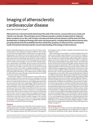
  • 2. INSIGHT PROGRESS NATURE|Vol 451|21 February 2008 of potentially nephrotoxic contrast agents and ionizing radiation. These The arterial walls limitations can now be largely overcome by using whole-body magnetic Atherosclerosis is a disease of the blood vessel wall, so the ability to resonance angiography, which can be carried out in less than 90 s. This identify plaques before luminal stenosis develops is the cornerstone of technique allows stenoses to be detected in the entire arterial system early disease detection. However, the thinness of the normal vessel wall — except the coronary circulation — in a single examination7. (< 1 mm for most arteries) presents a huge challenge for imaging. Several Until recently, imaging of coronary stenoses required the insertion of invasive (catheter-based) techniques have been used to evaluate the mor- a catheter into the coronary artery during X-ray angiography. In the past phology of plaques and other features of the vessel wall. These techniques decade, however, it has become possible to image the coronary arter- include angioscopy (direct visualization of the inner surface of the ves- ies non-invasively, by using contrast-enhanced CT. CT technology has sel wall by using fibre-optic technology), optical coherence tomography, evolved from machines that needed about 300 s to obtain a single image thermography, near-infrared spectroscopy, intravascular MRI and, most to multidetector CT (MDCT) scanners that can simultaneously acquire extensively, intravascular ultrasound11. These modalities are suitable for 256 ‘slices’ in less than 250 ms, providing a complete coronary angiogram evaluating the coronary arteries and — because of the proximity of the in less than 15 s. In selected patients with stable disease and a normal imaging probe to the vessel wall — provide high spatial resolution (for cardiac rhythm, MDCT has a sensitivity of 96% and a specificity of 74% example, < 15 μm with optical coherence tomography). However, the for detecting significant coronary stenoses (defined as more than 50% requirement for catheterization is a definite limitation. Ultrasound can narrowing of the diameter of the artery) compared with the traditional, also be used non-invasively to measure the intima-media thickness of the invasive technique, catheterization, which is the gold standard8. From a carotid arteries, because these arteries are located superficially. Increased clinical perspective, the most important advantage of MDCT is its high carotid intima-media thickness provides some additive information to negative predictive value: that is, a normal result on an MDCT exam can conventional risk factors in determining the risk of future myocardial convincingly rule out the possibility that significant coronary disease is infarction or stroke12. present9. One limitation is that the heart rate must be slow for the image With recently developed CT technology, the coronary arteries can now to be of adequate quality, but this might be overcome by using the newest be imaged non-invasively (as described earlier). CT has, however, long generation of CT equipment, in which two X-ray sources and detectors are been used for the non-invasive detection of coronary calcium deposits present in a single scanner (known as dual-source CT), thereby improving (yielding a ‘calcium score’), a specific indicator of atherosclerosis that the temporal resolution of images10. has prognostic value in asymptomatic individuals. Depending on the Process Endothelial- Endothelial- Inflammation Proteolysis Lipid-core Angiogenesis Thrombosis cell dysfunction cell activation Apoptosis and fibrous- cap formation Target Flow-mediated Adhesion Macrophages MMPs Lipid core αvβ3-Integrin Fibrin vasodilation molecules Cathepsins Fibrous cap Platelets (αIIbβ3-integrin) Tissue factor Fibrin Fibrous cap Platelet Thrombus Monocyte Blood recruitment VCAM1, ICAM1 and selectins Endothelial cell Smooth muscle cell ↓Nitric-oxide Apoptotic cell production Tissue factor LDL Cholesterol MMP Lipid-rich Collagen fibril necrotic core T cell Foam cell Tissue Internal elastic lamina αvβ3-Integrin New blood vessel Approximate I II III IV V VI AHA lesion stage Figure 1 | The development of an atherosclerotic lesion. The progression of at each stage are also listed. AHA, American Heart Association; ICAM1, an atherosclerotic lesion is shown in a simplified form, developing from a intercellular adhesion molecule 1; LDL, low-density lipoprotein; MMP, normal blood vessel (far left) to a vessel with an atherosclerotic plaque and matrix metalloproteinase; VCAM1, vascular cell-adhesion molecule 1. superimposed thrombus (far right). Potential targets for molecular imaging Figure adapted, with permission, from ref. 25. 954
  • 3. NATURE|Vol 451|21 February 2008 INSIGHT PROGRESS Table 1 | Targets and contrast agents for molecular imaging of atherosclerosis Target Biological roles Contrast agent* Rationale for use Modality Inflammatory Crucial in early plaque development, lipid Ultrasmall superparamagnetic Phagocytosed by macrophages MRI cells modification, smooth muscle cell proliferation, iron-oxide particles29 (including extracellular-matrix formation, angiogenesis, [18F]Fluorodeoxyglucose32,33 Taken up by metabolically active cells PET macrophages) plaque rupture and thrombosis N1177 (iodinated particle)31 Accumulates in macrophages CT CD204-specific-antibody- Target the macrophage scavenger receptor MRI carrying, Gd3+-loaded (CD204) micelles30 99m Tc-labelled interleukin-2 Binds to activated T cells SPECT 99m Apoptotic cells Release pro-coagulant and pro-oxidative stimuli Tc-labelled annexin V High affinity for phosphatidylserine, a molecule SPECT that contribute to plaque destabilization and Annexin-V-crosslinked iron present at the surface of apoptotic cells MRI and myocardial injury oxide–Cy5.5 NIRF Proteases Participate in plaque remodelling and rupture, as Gelatinase probe Fluorescent substrate for MMP2 and MMP9 NIRF well as in infarct remodelling and healing Prosense37 Fluorescent substrate for cathepsins NIRF P947 Contains an MMP-inhibitory peptide MRI Vascular Mediate the adhesion of inflammatory cells to VINP-28 (ref. 35) Contains a peptide with high affinity for VCAM1 MRI and cell-adhesion the endothelium and the recruitment of these NIRF molecules cells into atherosclerotic lesions and injured Microbubbles containing sialyl- Bind to selectins Ultrasound myocardium LewisX (ref. 36) Extracellular An important component of plaques, particularly Gadofluorine Binds to extracellular-matrix components MRI matrix abundant in advanced lesions (such as tenascin, proteoglycans and collagen) 99m Lipoproteins Involved in the trafficking of cholesterol between Tc-labelled MDA2 Binds to oxidized LDL, a potent intraplaque SPECT the blood and atherosclerotic plaques pro-inflammatory stimulus HDL-like nanoparticles HDL removes cholesterol from plaques MRI (known as reverse cholesterol transport) New blood Contribute to intraplaque haemorrhage, plaque Paramagnetic nanoparticles38 Target αVβ3-integrin, a key mediator of MRI vessels growth and destabilization, and myocardial healing 99mTc-labelled NC100692 angiogenesis SPECT and remodelling Thrombi A hallmark of acute vascular syndromes, and EP-2104R (ref. 34) Contains a peptide with a high affinity for fibrin MRI promote plaque growth 99m Tc-labelled apcitide Contains a peptide with a high affinity for the SPECT platelet cell-surface molecule αIIbβ3-integrin IR-786-labelled platelets40 Incorporated into thrombi NIRF *For agents for which no reference is given, and for discussion of other potential targets, see refs 22, 24–28. HDL, high-density lipoprotein; LDL, low-density lipoprotein; MDA2, monoclonal antibody specific for malondialdehyde; MMP, matrix metalloproteinase; NIRF, near-infrared fluorescence; Tc, technetium; VCAM1, vascular cell-adhesion molecule 1. individual studied (in terms of age, ethnicity, baseline risk of cardiovas- wall, this remains challenging because of the small diameter, the tortuos- cular disease, and so on) and the thresholds used, the risk of subsequent ity (twistedness) and the continuous movement of the coronary arteries. death or myocardial infarction associated with a high calcium score Therefore, MRI is mainly used to study extra-cardiac vessels. MRI can increases up to 12-fold after adjusting for conventional risk factors13. As also be used to provide insight into the composition and biological activ- a result, it has been proposed that the coronary calcium score, alone or ity of different types of atherosclerotic lesion, one of the most important in combination with the carotid intima-media thickness, could be used goals of imaging. The probability of plaque rupture and the subsequent for initial stratification of cardiovascular risk in the general population14. clinical complications differ substantially between plaque types. Features Nonetheless, this approach is not without controversy, largely because of higher rupture risk include the following: active inflammation, a thin of the required X-ray exposure and the financial cost15. Although coro- fibrous cap with a large lipid core, erosion or fissure of the plaque surface, nary calcifications are easily detected by CT, about three-quarters of a superimposed thrombus, a stenosis that narrows the luminal diameter all coronary lesions are non-calcified plaques. Such plaques can now by more than 90%, superficial calcified nodules, intraplaque haemorrhage be detected with modern CT scanners after contrast agents have been and outward remodelling20. By combining images acquired with different administered to patients. Moreover, in patients with chest pain, it was parameters, MRI can reliably detect and quantify plaque components such recently shown that the extent of non-calcified atherosclerosis in a coro- as lipids, fibro-cellular tissue, calcium and intraplaque haemorrhage, and nary CT angiogram is correlated with increasing mortality16. CT can can detect and characterize a superimposed thrombus21,22. The clinical also provide reasonably accurate quantification of plaque size and crude implications of these capabilities were highlighted in a recent study of characterization of plaque composition, on the basis of lipid-rich tissue asymptomatic patients with moderate carotid stenosis (50–79% luminal attenuating X-rays to a smaller extent than fibrous tissue17. narrowing) in which several high-risk features of the plaques observed by MRI has also developed into an excellent modality for non-invasively using MRI predicted subsequent cerebrovascular events23. evaluating the blood vessel wall, and it has the advantage over CT of not exposing the patient to ionizing radiation. ‘Black-blood’ techniques (an Molecular imaging imaging approach in which the blood appears black and the arterial wall Not only has the ability to image cardiovascular anatomy and physi- can be seen) accurately depict plaque presence, size and morphology with ology on a macroscopic scale (as has been discussed so far) improved submillimetre resolution and high reproducibility, providing new indi- markedly in the past decade, but it has also become increasingly ces of atherosclerotic burden that can be applied to large populations18. possible to detect biological processes at the cellular or even molecular Using this technique, with serial testing of an individual, it is possible to level. Molecular imaging relies on the use of contrast agents that target track changes in arterial disease and to test the effects of therapies for specific cells or molecular pathways of relevance to disease. In addition atherosclerosis in a completely non-invasive manner19. Although prelimi- to the various imaging techniques being developed, contrast agents for nary data show that it is feasible to use MRI to evaluate the coronary artery tracking potentially important components of atherosclerotic disease 955
  • 4. INSIGHT PROGRESS NATURE|Vol 451|21 February 2008 a The choice of target for imaging is also clearly important. Because b c inflammation has a crucial role at all stages of atherosclerosis (Fig. 1), macrophages are currently one of the most appealing targets. Ultrasmall paramagnetic iron-oxide particles are engulfed by macrophages in vivo, and this causes a detectable decrease in the MRI signal in proportion to the degree of atherosclerotic plaque inflammation, as shown in human stud- ies29. A strong correlation between macrophage density and MRI signal was also found recently in a mouse model of atherosclerosis, by using a contrast agent consisting of Gd3+-loaded micelles targeted to the mac- d rophage scavenger receptor30. Similarly, in rabbits, specific uptake of an iodine-containing contrast agent by macrophages allows atherosclerotic lesions to be detected by using CT31 (Fig. 2a, b). Also, with PET, the sig- nal from [18F]fluorodeoxyglucose correlates with the concentration of macrophages in human atherosclerotic plaques32. Moreover, by using specialized equipment, several imaging techniques can be used concur- rently — for example, PET together with CT or, recently, MRI (Fig. 2c, d) Figure 2 | Multimodal imaging of inflammation and atherosclerosis. a, Molecular imaging of macrophages by using CT, with the iodinated — for the sensitive and reproducible detection of vascular inflammation33. contrast agent N1177. After in vitro incubation of mouse macrophages This combination approach allows the most appropriate technique(s) for with N1177, light microscopy in a phase-contrast mode shows the presence a particular patient, vascular region and/or disease stage to be chosen and of multiple cytoplasmic granules (red arrow), confirming uptake of the takes advantage of the particular strengths of each modality. contrast agent by macrophages. b, Three-dimensional reconstruction Thrombi are another attractive target for imaging, because acute of a rabbit’s abdominal aorta at 2 h after intravenous administration of clinical events often occur as a result of thrombosis triggered by plaque N1177. A false-colour image of N1177 staining is shown overlaid on an rupture (Fig. 1). In animal models, thrombi of different ages and in dif- angiogram. Intense red spots indicate areas of N1177 accumulation in ferent vascular regions have been detected with MRI34, and this approach aortic plaques, which are rich in macrophages (white arrows). Organs with is now being investigated in humans26. At the other end of the timeline high macrophage density are also visible, including the spleen (top right). of atherosclerotic-plaque progression (Fig. 1), cell-adhesion molecules c, A combined PET and CT image of a human neck (axial view). Atherosclerotic pathology is present at the bifurcation of the right participate in the early development of lesions by facilitating the recruit- common carotid artery (red arrows), as determined by the presence of ment of leukocytes into the vessel wall. In an animal model, increased heavy calcification and large amounts of [18F]fluorodeoxyglucose, which amounts of vascular cell-adhesion molecule 1 (VCAM1) were found in is indicative of inflammatory activity. d, Black-blood MRI of the same aortic plaques by using a dual contrast agent detectable by both MRI and artery. Carotid arterial wall thickening is evident, as are two areas of signal optical imaging35. A similar approach, which used ultrasound detection drop (red arrows), which correspond to calcified regions. (Panels a and b of microbubbles, found increased expression of endothelial selectins in reproduced, with permission, from ref. 31. Panels c and d reproduced, with the heart of rats that had been subjected to transient myocardial ischaemia permission, from ref. 26.) followed by reperfusion36. Development of such probes for clinical use could allow the identification of atherosclerosis at early stages and the are at various stages of development22,24–28 (Fig. 1 and Table 1). Most of detection of plaque rupture (which, even when clinically silent, indicates the available probes are in experimental testing, although some have disease instability). already advanced to clinical evaluation. Imaging probes typically include Another possibility is to use probes that emit a detectable signal only a moiety (such as an antibody or specific ligand) with high affinity for after they have been activated by the target. For example, in a recent study the desired target molecule. Alternatively, the probe can be modified of an experimental model of atherosclerosis, a fluorescent probe activated to facilitate uptake by specific cells. In addition, probes are designed to by enzymatic degradation was used to reveal intraplaque protease activity be detected by various modalities, including ultrasound (which detects with near-infrared fluorescence37. microbubbles), SPECT and PET (radioactive isotopes), MRI (paramag- In addition to being diagnostic and prognostic indicators, probes netic and superparamagnetic compounds), CT (iodinated compounds) could also be used for therapeutic purposes, to deliver drugs in a targeted or optical imaging (fluorochromes). Many of the targets of interest are manner. An example of this is a study in which rabbits were adminis- located in deep organs and are present at very low (nanomolar) concen- tered paramagnetic nanoparticles loaded with an antiangiogenic drug, trations; imaging modalities therefore need to be highly sensitive, as well resulting in a reduction in the extent of blood vessels in atherosclerotic as safe and economically viable. plaques, as observed by non-invasive tracking with MRI38. In addition, Ultrasound is widely available, safe and inexpensive, but it has insuf- haematopoietic or cardiac stem cells for use in cell-based therapy could ficient penetration for the non-invasive imaging of deep vessels (including be tagged with appropriate imaging probes, providing insights into the the coronary arteries) with high spatial resolution or sensitivity. SPECT role and fate of these cells after their administration39. and PET have a high sensitivity, but they also have limited spatial resolu- tion and the additional disadvantage of requiring the use of radioactive Future directions agents. By contrast, MRI has a somewhat lower sensitivity than SPECT Rapid technological progress is transforming the imaging of atheroscle- and PET and requires prolonged imaging times, but it is safe and provides rotic cardiovascular disease from a method of diagnosis in symptom- excellent resolution (~10 μm with high-field magnets). CT, conversely, atic patients to a tool for the non-invasive detection of early subclinical offers the advantages of fast scanning times and superior performance for abnormalities. In addition, a new generation of hybrid technology is coronary angiography, at the expense of limited sensitivity and the use of now becoming available; this technology combines multiple imaging nephrotoxic agents and ionizing radiation. Optical imaging techniques — modalities in a single platform, using one machine for more than one for example, near-infrared fluorescence reflectance or fluorescence molec- type of imaging (Fig. 2c). And new probes designed to be detected by ular tomography — have excellent sensitivity and temporal resolution and several modalities can take advantage of the strengths of each. allow the tissue distribution of the probe to be precisely determined with The availability of more powerful imaging techniques has the potential ex vivo fluorescence microscopy. So far, however, such techniques can to improve our understanding of the biology of atherosclerosis. Much of be used non-invasively only to monitor superficial structures because of the current knowledge has been inferred from static histopathological the limited ability of light to penetrate tissue. Optical imaging techniques observations of animal models or human tissue samples studied at dif- and some SPECT and MRI techniques have the advantage of being able ferent disease stages or after various therapeutic interventions. By using to detect more than one molecular signature at a time. molecular imaging, it is now becoming increasingly possible to obtain 956
  • 5. NATURE|Vol 451|21 February 2008 INSIGHT PROGRESS non-invasively — from living experimental animals and, even, humans Committee to Update the 2000 Expert Consensus Document on Electron Beam — the type of information that was previously available only through Computed Tomography). Circulation 115, 402–426 (2007). 14. Naghavi, M. et al. From vulnerable plaque to vulnerable patient — part III: executive immunohistochemistry. summary of the Screening for Heart Attack Prevention and Education (SHAPE) Task Force Improved imaging technologies also hold promise for aiding drug report. Am. J. Cardiol. 98, 2H–15H (2006). development. Rather than relying on the plasma concentrations of a 15. Clouse, M. E. et al. How useful is computed tomography for screening for coronary artery disease? Circulation 113, 125–146 (2006). specific therapeutic agent to infer that it has been delivered to the target 16. Min, J. K. et al. Prognostic value of multidetector coronary computed tomographic organ, imaging might be able to provide a direct read-out of the agent’s angiography for prediction of all-cause mortality. J. Am. Coll. Cardiol. 50, 1161–1170 (2007). local concentration and activity. Such information could be enormously 17. Cordeiro, M. A. & Lima, J. A. Atherosclerotic plaque characterization by multidetector row computed tomography angiography. J. Am. Coll. Cardiol. 47, C40–C47 (2006). helpful for deciding which therapies are the best candidates for proceed- 18. Khera, A. et al. Relationship between C-reactive protein and subclinical atherosclerosis: the ing to clinical trials26. Dallas Heart Study. Circulation 113, 38–43 (2006). Prospective studies that will provide information about the signifi- 19. Corti, R. et al. Effects of aggressive versus conventional lipid-lowering therapy by simvastatin on human atherosclerotic lesions: a prospective, randomized, double-blind cance of currently available imaging data, considered either alone or in trial with high-resolution magnetic resonance imaging. J. Am. Coll. Cardiol. 46, 106–112 the context of conventional risk factors and emerging serum and genetic (2005). biomarkers (see page 949), are in progress. Examples are the Multi- 20. Naghavi, M. et al. From vulnerable plaque to vulnerable patient: a call for new definitions and risk assessment strategies: part I. Circulation 108, 1664–1672 (2003). Ethnic Study of Atherosclerosis (MESA; http://www.mesa-nhlbi.org/) 21. Saam, T. et al. The vulnerable, or high-risk, atherosclerotic plaque: noninvasive MR imaging and the High-Risk Plaque (HRP) Initiative (http://www.hrpinitiative. for characterization and assessment. Radiology 244, 64–77 (2007). com), both studies of asymptomatic individuals of various ethnic back- 22. Briley-Saebo, K. C. et al. Magnetic resonance imaging of vulnerable atherosclerotic grounds. Another example is a subset of the FREEDOM trial (Future plaques: current imaging strategies and molecular imaging probes. J. Magn. Reson. Imaging 26, 460–479 (2007). Revascularization Evaluation in Patients with Diabetes Mellitus: Optimal 23. Takaya, N. et al. Association between carotid plaque characteristics and subsequent Management of Multivessel Disease trial), which involves diabetic individ- ischemic cerebrovascular events: a prospective assessment with MRI — initial results. uals with proven coronary heart disease. With data from such investiga- Stroke 37, 818–823 (2006). 24. Choudhury, R. P., Fuster, V. & Fayad, Z. A. Molecular, cellular and functional imaging of tions, it might be possible to distinguish which patients would benefit from atherothrombosis. Nature Rev. Drug Discov. 3, 913–925 (2004). therapeutic intervention. Preventing atherothrombotic events through This review discusses different techniques for imaging the blood vessel wall, focusing on early detection would have enormous medical impact, and imaging is molecular imaging. 25. Wu, J. C., Bengel, F. M. & Gambhir, S. S. Cardiovascular molecular imaging. Radiology 244, set to have a prominent role in making this a reality. In this regard, it will 337–355 (2007). be imperative to evaluate emerging imaging technologies rigorously to 26. Jaffer, F. A., Libby, P. & Weissleder, R. Molecular imaging of cardiovascular disease. ensure that they are cost-effective. ■ Circulation 116, 1052–1061 (2007). This is an up-to-date review of cardiovascular molecular imaging, including a discussion 1. Lusis, A. J. Atherosclerosis. Nature 407, 233–241 (2000). of contrast agents that are in clinical testing. 2. Gershlick, A. H. et al. Role of non-invasive imaging in the management of coronary artery 27. Wickline, S. A., Neubauer, A. M., Winter, P. M., Caruthers, S. D. & Lanza, G. M. Molecular disease: an assessment of likely change over the next 10 years. A report from the British imaging and therapy of atherosclerosis with targeted nanoparticles. J. Magn. Reson. Cardiovascular Society Working Group. Heart 93, 423–431 (2007). Imaging 25, 667–680 (2007). 3. Hendel, R. C. et al. ACCF/ACR/SCCT/SCMR/ASNC/NASCI/SCAI/SIR 2006 28. Sosnovik, D. E., Nahrendorf, M. & Weissleder, R. Molecular magnetic resonance imaging in appropriateness criteria for cardiac computed tomography and cardiac magnetic cardiovascular medicine. Circulation 115, 2076–2086 (2007). resonance imaging: a report of the American College of Cardiology Foundation Quality 29. Trivedi, R. A. et al. Identifying inflamed carotid plaques using in vivo USPIO-enhanced Strategic Directions Committee Appropriateness Criteria Working Group, American MR imaging to label plaque macrophages. Arterioscler. Thromb. Vasc. Biol. 26, 1601–1606 College of Radiology, Society of Cardiovascular Computed Tomography, Society for (2006). Cardiovascular Magnetic Resonance, American Society of Nuclear Cardiology, North 30. Amirbekian, V. et al. Detecting and assessing macrophages in vivo to evaluate American Society for Cardiac Imaging, Society for Cardiovascular Angiography and atherosclerosis noninvasively using molecular MRI. Proc. Natl Acad. Sci. USA 104, 961–966 Interventions, and Society of Interventional Radiology. J. Am. Coll. Cardiol. 48, 1475–1497 (2007). (2006). 31. Hyafil, F. et al. Noninvasive detection of macrophages using a nanoparticulate contrast 4. Sakuma, H. Magnetic resonance imaging for ischemic heart disease. J. Magn. Reson. agent for computed tomography. Nature Med. 13, 636–641 (2007). Imaging 26, 3–13 (2007). This paper was the first to show selective visualization of macrophages by using CT. 5. Kwong, R. Y. et al. Impact of unrecognized myocardial scar detected by cardiac magnetic 32. Tawakol, A. et al. In vivo 18F-fluorodeoxyglucose positron emission tomography imaging resonance imaging on event-free survival in patients presenting with signs or symptoms of provides a noninvasive measure of carotid plaque inflammation in patients. J. Am. Coll. coronary artery disease. Circulation 113, 2733–2743 (2006). Cardiol. 48, 1818–1824 (2006). 6. Ibanez, B. et al. Early metoprolol administration before coronary reperfusion results in 33. Rudd, J. H. et al. 18Fluorodeoxyglucose positron emission tomography imaging increased myocardial salvage: analysis of ischemic myocardium at risk using cardiac of atherosclerotic plaque inflammation is highly reproducible: implications for magnetic resonance. Circulation 115, 2909–2916 (2007). atherosclerosis therapy trials. J. Am. Coll. Cardiol. 50, 892–896 (2007). 7. Fenchel, M. et al. Atherosclerotic disease: whole-body cardiovascular imaging with MR This paper validates the reproducibility of concurrent PET and CT for the imaging of system with 32 receiver channels and total-body surface coil technology — initial clinical vascular inflammation. results. Radiology 238, 280–291 (2006). 34. Sirol, M. et al. Chronic thrombus detection with in vivo magnetic resonance imaging and a 8. Hamon, M. et al. Diagnostic performance of multislice spiral computed tomography of fibrin-targeted contrast agent. Circulation 112, 1594–1600 (2005). coronary arteries as compared with conventional invasive coronary angiography: a meta- 35. Nahrendorf, M. et al. Noninvasive vascular cell adhesion molecule-1 imaging identifies analysis. J. Am. Coll. Cardiol. 48, 1896–1910 (2006). inflammatory activation of cells in atherosclerosis. Circulation 114, 1504–1511 (2006). 9. Budoff, M. J. et al. Assessment of coronary artery disease by cardiac computed 36. Villanueva, F. S. et al. Myocardial ischemic memory imaging with molecular tomography: a scientific statement from the American Heart Association Committee echocardiography. Circulation 115, 345–352 (2007). on Cardiovascular Imaging and Intervention, Council on Cardiovascular Radiology 37. Jaffer, F. A. et al. Optical visualization of cathepsin K activity in atherosclerosis with a novel, and Intervention, and Committee on Cardiac Imaging, Council on Clinical Cardiology. protease-activatable fluorescence sensor. Circulation 115, 2292–2298 (2007). Circulation 114, 1761–1791 (2006). 38. Winter, P. M. et al. Endothelial αVβ3 integrin-targeted fumagillin nanoparticles inhibit 10. Weustink, A. C. et al. Reliable high-speed coronary computed tomography in symptomatic angiogenesis in atherosclerosis. Arterioscler. Thromb. Vasc. Biol. 26, 2103–2109 (2006). patients. J. Am. Coll. Cardiol. 50, 786–794 (2007). 39. Beeres, S. L. et al. Role of imaging in cardiac stem cell therapy. J. Am. Coll. Cardiol. 49, 11. MacNeill, B. D., Lowe, H. C., Takano, M., Fuster, V. & Jang, I.-K. Intravascular modalities 1137–1148 (2007). for detection of vulnerable plaque: current status. Arterioscler. Thromb. Vasc. Biol. 23, 40. Flaumenhaft, R. et al. Localization and quantification of platelet-rich thrombi in large blood 1333–1342 (2003). vessels with near-infrared fluorescence imaging. Circulation 115, 84–93 (2007). 12. Lorenz, M. W., Markus, H. S., Bots, M. L., Rosvall, M. & Sitzer, M. Prediction of clinical cardiovascular events with carotid intima-media thickness: a systematic review and meta- Acknowledgements This work was partly funded by the National Institutes of analysis. Circulation 115, 459–467 (2007). Health and the National Heart, Lung, and Blood Institute. 13. Greenland, P. et al. ACCF/AHA 2007 clinical expert consensus document on coronary artery calcium scoring by computed tomography in global cardiovascular risk assessment Author Information Reprints and permissions information is available at and in evaluation of patients with chest pain: a report of the American College of npg.nature.com/reprints. The authors declare no competing financial interests. Cardiology Foundation Clinical Expert Consensus Task Force (ACCF/AHA Writing Correspondence should be addressed to Z.A.F. (zahi.fayad@mssm.edu). 957